. 2015 Nov 11;35(45):15073-81.
doi: 10.1523/JNEUROSCI.1087-15.2015.

Convergence of Hippocampal Pathophysiology in Syngap+/- and Fmr1-/y Mice

Affiliations

Convergence of Hippocampal Pathophysiology in Syngap+/- and Fmr1-/y Mice

Stephanie A Barnes et al. J Neurosci. .

Abstract

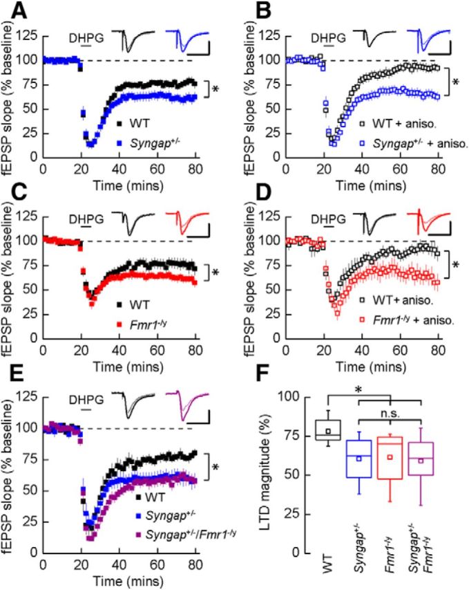
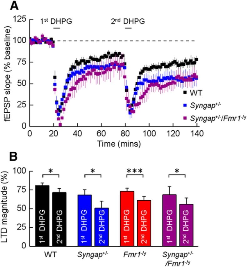
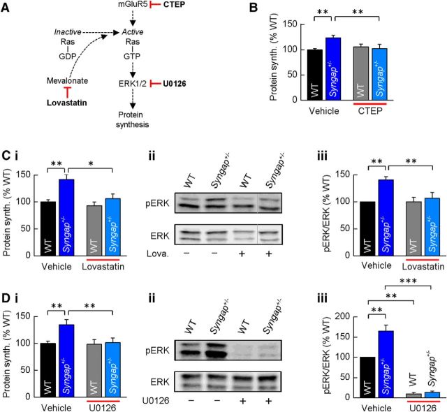
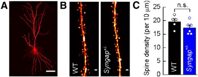
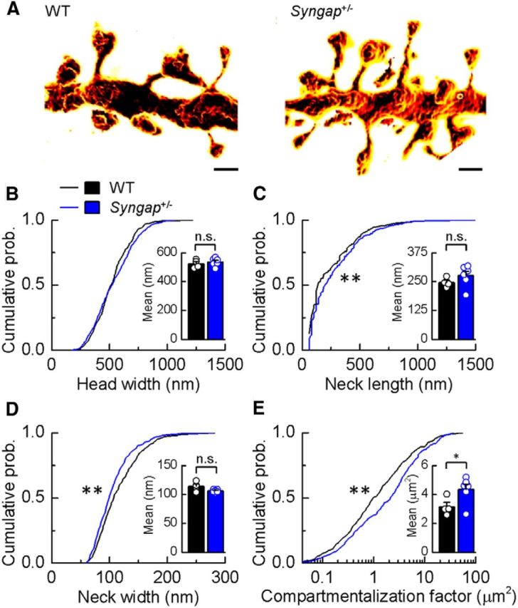
Previous studies have hypothesized that diverse genetic causes of intellectual disability (ID) and autism spectrum disorders (ASDs) converge on common cellular pathways. Testing this hypothesis requires detailed phenotypic analyses of animal models with genetic mutations that accurately reflect those seen in the human condition (i.e., have structural validity) and which produce phenotypes that mirror ID/ASDs (i.e., have face validity). We show that SynGAP haploinsufficiency, which causes ID with co-occurring ASD in humans, mimics and occludes the synaptic pathophysiology associated with deletion of the Fmr1 gene. Syngap(+/-) and Fmr1(-/y) mice show increases in basal protein synthesis and metabotropic glutamate receptor (mGluR)-dependent long-term depression that, unlike in their wild-type controls, is independent of new protein synthesis. Basal levels of phosphorylated ERK1/2 are also elevated in Syngap(+/-) hippocampal slices. Super-resolution microscopy reveals that Syngap(+/-) and Fmr1(-/y) mice show nanoscale alterations in dendritic spine morphology that predict an increase in biochemical compartmentalization. Finally, increased basal protein synthesis is rescued by negative regulators of the mGlu subtype 5 receptor and the Ras-ERK1/2 pathway, indicating that therapeutic interventions for fragile X syndrome may benefit patients with SYNGAP1 haploinsufficiency.

Significance statement: As the genetics of intellectual disability (ID) and autism spectrum disorders (ASDs) are unraveled, a key issue is whether genetically divergent forms of these disorders converge on common biochemical/cellular pathways and hence may be amenable to common therapeutic interventions. This study compares the pathophysiology associated with the loss of fragile X mental retardation protein (FMRP) and haploinsufficiency of synaptic GTPase-activating protein (SynGAP), two prevalent monogenic forms of ID. We show that Syngap(+/-) mice phenocopy Fmr1(-/y) mice in the alterations in mGluR-dependent long-term depression, basal protein synthesis, and dendritic spine morphology. Deficits in basal protein synthesis can be rescued by pharmacological interventions that reduce the mGlu5 receptor-ERK1/2 signaling pathway, which also rescues the same deficit in Fmr1(-/y) mice. Our findings support the hypothesis that phenotypes associated with genetically diverse forms of ID/ASDs result from alterations in common cellular/biochemical pathways.

Keywords: STED; SynGAP; fragile X syndrome; long-term depression; mGluR; neurodevelopmental disorder.


Figures

Figure 1.
Figure 1.
Syngap+/− phenocopies the hippocampal synaptic pathophysiology observed in Fmr1−/y mice. A, application of DHPG (50 μm) induced LTD that was significantly increased in Syngap+/− mice (62 ± 5%, n = 15; t test, p = 0.01) versus WT littermate controls (77 ± 3%, n = 18). Representative average fEPSPs before and after DHPG application are illustrated. B, In the presence of protein synthesis inhibitor anisomycin (aniso.; 20 μm), DHPG-induced LTD was not sustained in WT mice (91 ± 4%, n = 12), while remaining intact in Syngap+/− mice (64 ± 4%, n = 14). C, DHPG-induced mGluR-LTD was also significantly enhanced in Fmr1−/y mice (63 ± 4%, n = 17; t test, p = 0.04) versus WT littermate controls (77 ± 5%, n = 17). D, In the presence of anisomycin, mGluR-LTD remained at a similar magnitude in the Fmr1−/y (65 ± 7%, n = 9), whereas LTD could not be sustained in WT mice (91 ± 6%, n = 8). E, The magnitude of LTD was significantly enhanced in the Syngap+/−/Fmr1−/y double mutant mice (59 ± 4%, n = 12; ANOVA, p = 0.02) relative to WT littermate controls (78 ± 3%; n = 9). No significant differences were observed in LTD magnitude between the Syngap+/−/Fmr1−/y double mutant mice and either Syngap+/− (60 ± 4%, n = 10) or Fmr1−/y (62 ± 5%, n = 10; data not illustrated) single mutant mice. F, Summary of DHPG-induced LTD for each of the four genotypes generated from the Syngap+/− × Fmr1−/y cross. Box plots illustrate minima and maxima (whiskers), median (line), mean (square symbol), and interquartile range (box). Calibrations: A, D, 250 μV, 10 ms; B, C, 500 μV, 10 ms; E, 350 μV, 10 ms. n.s., Not significant. *p < 0.05.
Figure 2.
Figure 2.
A single application of DHPG does not “saturate” LTD in Syngap+/−/Fmr1−/y double mutant mice. A, Mean time courses illustrating fEPSP slope in experiments on slices prepared from WT, Syngap+/−, and Syngap+/−/Fmr1−/y mice where two applications of DHPG (50 μm) were applied to determine whether the magnitude of LTD elicited by the first application could be augmented by the second. B, Bar graphs illustrating the magnitude of mGluR-mediated LTD produced by a first and then a second application of DHPG in slices prepared from WT (first, 81 ± 3%; second, 71 ± 5%; n = 8; p < 0.05, paired t test), Syngap+/− (first, 68 ± 7%; second, 51 ± 9%; n = 5; p < 0.05), Fmr1−/y (first, 73 ± 4%; second, 61 ± 5%; n = 3; p < 0.001), and Syngap+/−/Fmr1−/y (first, 69 ± 11%; second, 56 ± 8%; n = 4; p < 0.05) mice. *p < 0.05; ***p < 0.001.
Figure 3.
Figure 3.
Basal protein synthesis and phosphorylated ERK1/2 levels are elevated in the hippocampus of Syngap+/− mice. A, Schematic of the experimental timeline for 35[S]-Met/Cys metabolic labeling. B, Basal protein synthesis levels were significantly elevated in dorsal hippocampal slices from Syngap+/− versus WT mice (WT, 100 ± 3%; Syngap+/−, 140 ± 12%; t test, p = 0.0007; n = 9). Increased protein synthesis rates were also observed in the Fmr1−/y versus WT mice (WT, 100 ± 2%; Fmr1−/y, 121 ± 7%; t test, p = 0.01; n = 6). Ci, Cii, Basal activation state of ERK1/2 in hippocampal slices was significantly increased in Syngap+/− mice versus WT controls (phosphorylated/total ERK1/2, 137 ± 7%; n = 23; p = 1.5 × 10⁻⁵). D, Protein synthesis levels were significantly elevated in vehicle-treated dorsal hippocampal slices from Syngap+/− versus WT mice (WT, 100 ± 5%; Syngap+/−, 135 ± 7%; n = 6; ANOVA, p = 0.0038). DHPG treatment (100 μm, 5 min) significantly increased protein synthesis rates in WT slices, but did not further increase 35[S]-Met/Cys incorporation in Syngap+/− slices (WT DHPG, 138 ± 14%; Syngap+/− DHPG, 148 ± 15%; ANOVA, WT treatment, p = 0.0028; n = 6). Ei, Eii, Western blot analysis of vehicle/DHPG-treated hippocampal slices revealed DHPG significantly increases the phosphorylation status of ERK1/2 in both Syngap+/− and WT mice (phosphorylated/total ERK1/2, WT vehicle, 100%; Syngap+/− vehicle, 130 ± 6%; WT DHPG, 180 ± 16%; Syngap+/− DHPG, 173 ± 21%; n = 7; ANOVA, genotype × treatment, p < 0.05). *p < 0.05; **p < 0.01; ***p < 0.001.
Figure 4.
Figure 4.
Elevated protein synthesis in Syngap+/− hippocampus is corrected by inhibitors of mGluR5 and Ras–ERK1/2 signaling. A, schematic of the drug targets for CTEP, lovastatin, and U0126, each shown previously to reduce Ras–ERK1/2 activation and correct basal protein synthesis in Fmr1−/y mice. B, CTEP (10 μm) reverses elevated protein synthesis in Syngap+/− mice (WT, vehicle, 100 ± 3%; CTEP, 106 ± 5%; Syngap+/−, vehicle, 124 ± 5%; CTEP, 102 ± 8%; n = 9; ANOVA, genotype, p = 0.0007; KO treatment, p = 0.0014). Ci–Ciii, Lovastatin (100 μm) reverses elevated protein synthesis (Ci) in Syngap+/− mice (WT, vehicle, 100 ± 4%; lovastatin, 93 ± 7; Syngap+/−, vehicle, 142 ± 9%; lovastatin, 106 ± 8%; n = 7; ANOVA, genotype, p = 0.0061; KO treatment, p = 0.013). Example Western blot (Cii) and quantification of ERK1/2 activity (Ciii) in vehicle- and lovastatin-treated hippocampal slices revealed lovastatin significantly reduces phosphorylation levels of ERK1/2 in Syngap+/− mice (phosphorylated/total ERK1/2, WT, vehicle, 100%; Syngap+/−, vehicle, 140 ± 6%; WT, lovastatin, 100 ± 8%; Syngap+/−, lovastatin, 106 ± 11%; n = 10; ANOVA, genotype, *p = 0.0086; KO treatment, p = 0.0057). The gray line to the left of the lovastatin-treated Syngap+/− Western blot indicates that this lane was not adjacent to each of the other three lanes, but was run on the same gel, and hence was processed identically. D, U0126 (5 μm) reverses elevated protein synthesis (Di) in Syngap+/− mice (WT, vehicle, 100 ± 4%; U0126, 98 ± 8%; Syngap+/−, vehicle, 134 ± 9%; U0126, 102 ± 9%; n = 9; ANOVA, genotype, p = 0.003; KO treatment, p = 0.004). Example Western blot (Dii) and quantification (Diii) of ERK1/2 activity in vehicle- and U0126-treated hippocampal slices show U0126 abolishes ERK1/2 activity in both WT and Syngap+/− mice (phosphorylated/total ERK1/2, WT, vehicle, 100%; Syngap+/−, vehicle, 165 ± 15%; WT, lovastatin, 10 ± 3%; Syngap+/−, lovastatin, 13 ± 3%; n = 5; ANOVA, genotype, p = 0.0054; WT treatment, p = 0.005; KO treatment, p = 0.0002). *p < 0.05; **p < 0.01; ***p < 0.001.
Figure 5.
Figure 5.
CA1 pyramidal neuron dendritic spine density in Syngap+/− mice. A, Composite image of a representative CA1 pyramidal neuron labeled by intracellular filling with Alexa Fluor 568 in lightly fixed section through CA1 of hippocampus. Scale bar, 50 μm. B, Representative composite images of apical oblique dendritic segments from WT and Syngap+/− mice. Scale bars, 1 μm. C, Mean spine density of apical dendrites for WT (n = 5) and Syngap+/− (n = 6) animals indicating no difference in dendritic spine density (WT, 19.58 ± 1.03 spines/10 μm; Syngap+/−, 17.34 ± 0.94 spines/10 μm; p = 0.1430, two-tailed independent t test with Welch's correction). n.s., Not significant.
Figure 6.
Figure 6.
CA1 pyramidal neuron dendritic spine morphology in Syngap+/− mice. A, Surface-rendered STED images of apical dendritic segments from pyramidal neurons in the CA1 of the hippocampus of WT and Syngap+/− mice revealing morphological details that are invisible to conventional light microscopy. Scale bars, 500 nm. B–D, Kolmogorov–Smirnov tests of the cumulative frequency distributions show that the distribution profiles of spine head widths (B) are not significantly different between genotypes (p = 0.11), whereas distribution profiles of spine neck lengths and widths show that Syngap+/− mice (n = 424 spines) have significantly more spines with longer (C; p < 0.005) and narrower necks (D; p < 0.0008) compared to WT mice (n = 393 spines). The mean values for these parameters are, however, not significantly different (WT, 523 ± 14 nm, 246 ± 10 nm, 114 ± 4 nm, n = 4; Syngap+/−, 536 ± 11 nm, 276 ± 19 nm, 106 ± 1 nm, n = 6, for head width, neck length, and neck width, respectively). E, To predict the impact of these spine morphology changes on diffusional coupling, a morphological compartmentalization factor was calculated. The cumulative frequency distributions of the compartmentalization factor significantly differ between genotypes (Kolmogorov–Smirnov test, p < 0.0001), and this is also reflected in the mean values for each genotype (two-tailed independent t test with Welch's correction, p = 0.04; WT, 3.16 ± 0.30 μm2; Syngap+/−, 4.35 ± 0.37 μm2). n.s., Not significant; *p < 0.05; **p < 0.01.

References

    1. Auerbach BD, Osterweil EK, Bear MF. Mutations causing syndromic autism define an axis of synaptic pathophysiology. Nature. 2011;480:63–68. doi: 10.1038/nature10658. - DOI - PMC - PubMed
    1. Barnett MW, Watson RF, Vitalis T, Porter K, Komiyama NH, Stoney PN, Gillingwater TH, Grant SG, Kind PC. Synaptic Ras GTPase activating protein regulates pattern formation in the trigeminal system of mice. J Neurosci. 2006;26:1355–1365. doi: 10.1523/JNEUROSCI.3164-05.2006. - DOI - PMC - PubMed
    1. Berryer MH, Hamdan FF, Klitten LL, Møller RS, Carmant L, Schwartzentruber J, Patry L, Dobrzeniecka S, Rochefort D, Neugnot-Cerioli M, Lacaille JC, Niu Z, Eng CM, Yang Y, Palardy S, Belhumeur C, Rouleau GA, Tommerup N, Immken L, Beauchamp MH, et al. Mutations in SYNGAP1 cause intellectual disability, autism, and a specific form of epilepsy by inducing haploinsufficiency. Hum Mutat. 2013;34:385–394. doi: 10.1002/humu.22248. - DOI - PubMed
    1. Carlisle HJ, Manzerra P, Marcora E, Kennedy MB. SynGAP regulates steady-state and activity-dependent phosphorylation of cofilin. J Neurosci. 2008;28:13673–13683. doi: 10.1523/JNEUROSCI.4695-08.2008. - DOI - PMC - PubMed
    1. Clement JP, Aceti M, Creson TK, Ozkan ED, Shi Y, Reish NJ, Almonte AG, Miller BH, Wiltgen BJ, Miller CA, Xu X, Rumbaugh G. Pathogenic SYNGAP1 mutations impair cognitive development by disrupting maturation of dendritic spine synapses. Cell. 2012;151:709–723. doi: 10.1016/j.cell.2012.08.045. - DOI - PMC - PubMed

Publication types

MeSH terms

Substances