Review
. 2016 Feb;39(1):98-106.
doi: 10.1097/COC.0000000000000239.

CTLA-4 and PD-1 Pathways: Similarities, Differences, and Implications of Their Inhibition

Affiliations
Review

CTLA-4 and PD-1 Pathways: Similarities, Differences, and Implications of Their Inhibition

Elizabeth I Buchbinder et al. Am J Clin Oncol. 2016 Feb.

Abstract

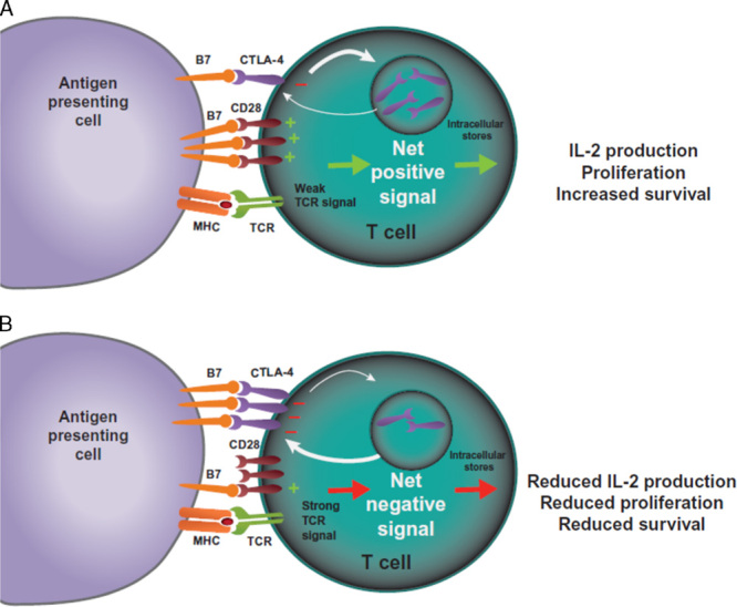
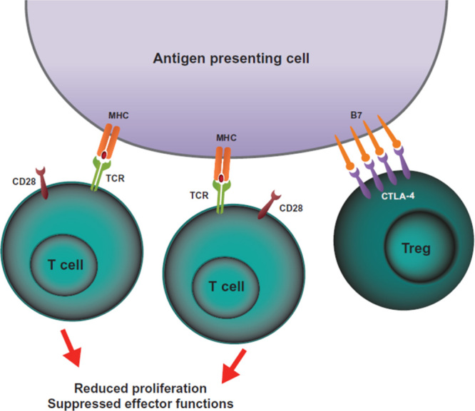
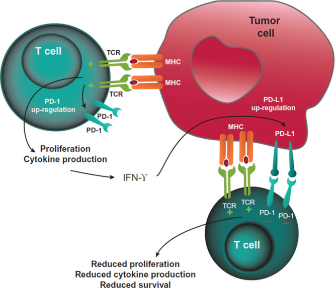
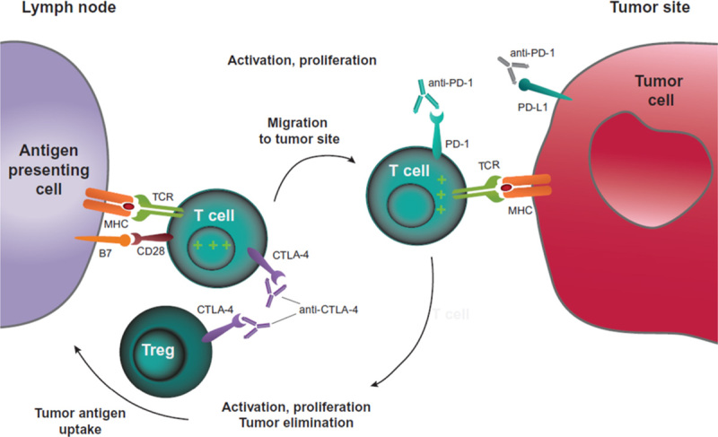
The cytotoxic T-lymphocyte-associated antigen 4 (CTLA-4) and programmed death 1 (PD-1) immune checkpoints are negative regulators of T-cell immune function. Inhibition of these targets, resulting in increased activation of the immune system, has led to new immunotherapies for melanoma, non-small cell lung cancer, and other cancers. Ipilimumab, an inhibitor of CTLA-4, is approved for the treatment of advanced or unresectable melanoma. Nivolumab and pembrolizumab, both PD-1 inhibitors, are approved to treat patients with advanced or metastatic melanoma and patients with metastatic, refractory non-small cell lung cancer. In addition the combination of ipilimumab and nivolumab has been approved in patients with BRAF WT metastatic or unresectable melanoma. The roles of CTLA-4 and PD-1 in inhibiting immune responses, including antitumor responses, are largely distinct. CTLA-4 is thought to regulate T-cell proliferation early in an immune response, primarily in lymph nodes, whereas PD-1 suppresses T cells later in an immune response, primarily in peripheral tissues. The clinical profiles of immuno-oncology agents inhibiting these 2 checkpoints may vary based on their mechanistic differences. This article provides an overview of the CTLA-4 and PD-1 pathways and implications of their inhibition in cancer therapy.


Conflict of interest statement

E.I.B. received research funding from Bristol-Myers Squibb, Genentech, and Merck; spouse previously employed by Merck. A.D. declares no conflicts of interest.

Figures

FIGURE 1
FIGURE 1
CTLA-4-mediated inhibition of T cells. T cells are activated when TCRs bind antigen displayed in the MHC on antigen-presenting cells in concert with CD28:B7-mediated costimulation. A, In the case of a weak TCR stimulus, CD28:B7 binding predominates, resulting in a net positive activating signal and IL-2 production, proliferation, and increased survival. B, In the case of a strong TCR stimulus, CTLA-4 expression is upregulated by increased transport to the cell surface from intracellular stores and decreased internalization. CTLA-4 competes with CD28 for binding of B7 molecules. Increased CTLA-4:B7 binding can result in a net negative signal, which limits IL-2 production and proliferation, and limits survival of the T cell. CTLA-4 indicates cytotoxic T-lymphocyte–associated antigen 4; IL-2, interleukin-2; MHC, major histocompatibility complex; TCR, T-cell receptor.
FIGURE 2
FIGURE 2
CTLA-4-mediated inhibition of Tregs. One hypothesis of how CTLA-4 expression on Tregs can inhibit T-cell activation is depicted. Constitutive expression of CTLA-4 on Tregs can sequester or cause internalization (not depicted) of B7 molecules on antigen-presenting cells. The lack of CD28:B7-mediated costimulation leads to reduced T-cell proliferation and reduced effector functions. CTLA-4 indicates cytotoxic T-lymphocyte–associated antigen 4; MHC, major histocompatibility complex; TCR, T-cell receptor; Tregs, regulatory T cells.
FIGURE 3
FIGURE 3
PD-1-mediated inhibition of T cells. T cells recognizing tumor antigens can be activated to proliferate, secrete inflammatory cytokines, and resist cell death. Prolonged TCR stimulation during an ongoing immune response can cause upregulated PD-1 expression. Tumor cells can express PD-L1 (and PD-L2, not shown) as a consequence of inflammatory cytokines and/or oncogenic signaling pathways. PD-1:PD-L1 binding inhibits TCR-mediated positive signaling, leading to reduced proliferation, reduced cytokine secretion, and reduced survival. IFN-γ indicates interferon-γ; MHC, major histocompatibility complex; PD-1, programmed death protein 1; PD-L1, programmed death ligand 1; PD-L2, programmed death ligand 2; TCR, T-cell receptor.
FIGURE 4
FIGURE 4
CTLA-4 and PD-1 pathway blockade. CTLA-4 blockade allows for activation and proliferation of more T-cell clones, and reduces Treg-mediated immunosuppression. PD-1 pathway blockade restores the activity of antitumor T cells that have become quiescent. A dual pathway blockade could have a synergistic effect, resulting in a larger and longer lasting antitumor immune response. CTLA-4 indicates cytotoxic T-lymphocyte–associated antigen 4; MHC, major histocompatibility complex; PD-1, programmed death 1; PD-L1, programmed death ligand 1; TCR, T-cell receptor; Treg, regulatory T cell.

Similar articles

Cited by

References

    1. Goldrath AW, Bevan MJ. Selecting and maintaining a diverse T-cell repertoire. Nature. 1999;402:255–262. - PubMed
    1. Fife BT, Bluestone JA. Control of peripheral T-cell tolerance and autoimmunity via the CTLA-4 and PD-1 pathways. Immunol Rev. 2008;224:166–182. - PubMed
    1. Greenwald RJ, Freeman GJ, Sharpe AH. The B7 family revisited. Annu Rev Immunol. 2005;23:515–548. - PubMed
    1. Krummel MF, Allison JP. CD28 and CTLA-4 have opposing effects on the response of T cells to stimulation. J Exp Med. 1995;182:459–465. - PMC - PubMed
    1. Dunn GP, Old LJ, Schreiber RD. The immunobiology of cancer immunosurveillance and immunoediting. Immunity. 2004;21:137–148. - PubMed

MeSH terms