

Review
doi: 10.1021/acs.analchem.5b04139.
Epub 2015 Nov 20.
Discovery in Droplets
Affiliations
- PMID: 26558892
- PMCID: PMC4865373
- DOI: 10.1021/acs.analchem.5b04139
Item in Clipboard
Review
Discovery in Droplets
Anal Chem.
.
No abstract available
Figures
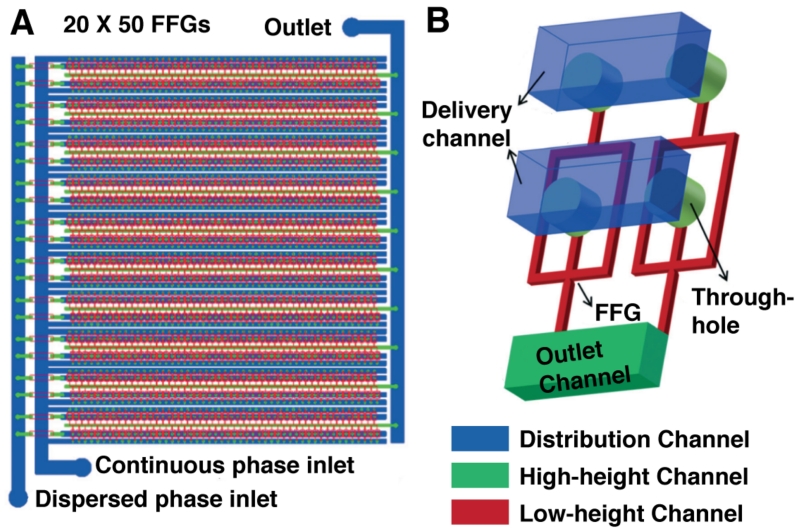
Ultra-high-throughput droplet generation. (A) The three-dimensional monolithic elastomer device (3D MED) consists of an array of 20 × 50 flow-focusing generators (FFGs). (B) Through-hole-containing manifolds enable parallelization of a conventional droplet generation mode for unparalleled scales of production. Adapted from Jeong, H.-H.; Yelleswarapu, V. R.; Yadavali, S.; Issadore, D.; Lee, D. Lab Chip
2015, Advance Article, DOI:10.1039/C5LC01025J (ref 9) with permission of The Royal Society of Chemistry.

Enabling technology for sorting. Flow-focusing droplet generation using an agarose/alginate aqueous phase in oil followed by cooling and oil removal generated particles with a thin, semi-porous polyelectrolyte gel. The particles are suitable for conventional FACS-based analysis. The gel-shell beads (GSB) shells could be disassembled at elevated pH to access contents. Reprinted by permission from Macmillan Publishers Ltd: Nature Chemistry (ref 20), copyright 2014. (B) Ultra-high-throughput sorting at 30 kHz is possible with a redesigned sorting junction. Gapped dividers prevent droplet scission and close positioning of waste and collection streams provide for rapid electrokinetic droplet mobilization after laser-induced fluorescence detection signals a positive event. Scale = 50 μm Adapted from Sciambi, A.; Abate, A. R. Lab Chip
2015, 15, 47–51 (ref 21) with permission of The Royal Society of Chemistry.
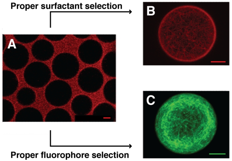
Retention as a function of dye and surfactant selection. (A) Microtubules stained with ATTO633 immediately partition to the oil phase in triblock co-polymer TRI2500 surfactant. (B) Selection of a higher molecular weight triblock copolymer surfactant TRI7000 properly compartmentalizes the ATTO633-stained protein. (C) Microtubules stained with ATTO488, a more hydrophilic dye, retain properly in TRI2500-stabilized droplets. Scale = 10 μm Adapted with permission from Janiesch, J.-W.; Weiss, M.; Kannenberg, G.; Hannabuss, J.; Surrey, T.; Platzman, I.; Spatz, J. P. Anal. Chem.
2015, 87, 2063–2067 (ref 30). Copyright 2015 American Chemical Society.
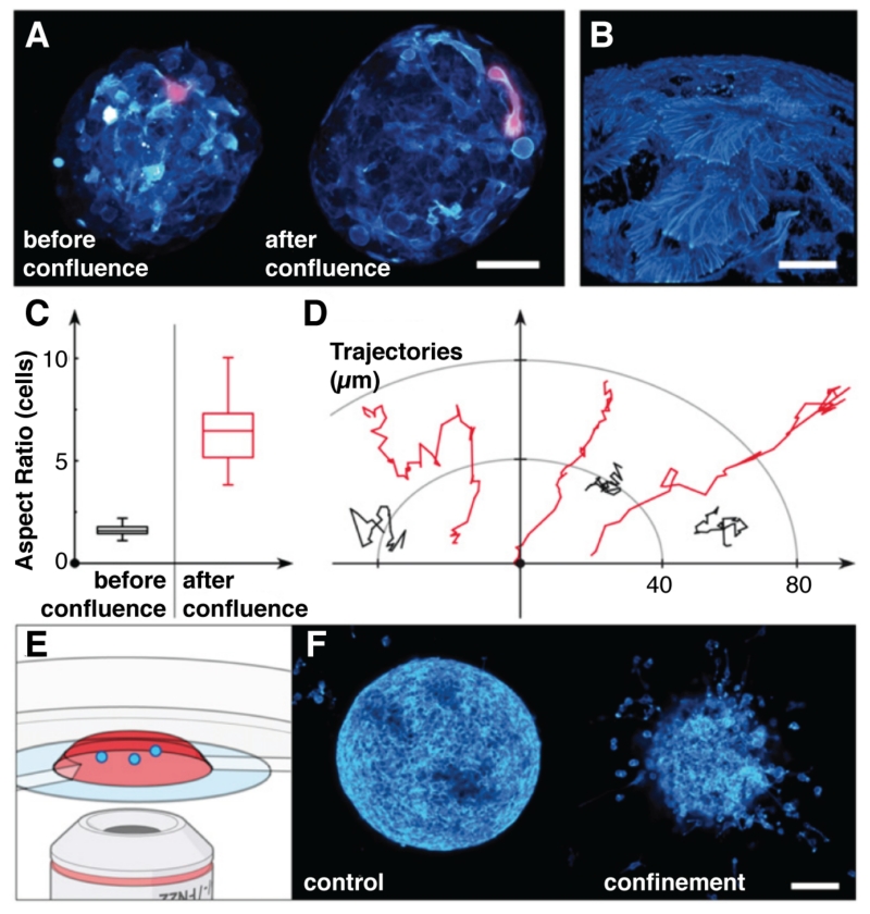
Confinement and confluence in multicellular spheroid cultures (MCSs). (A) before confluence, cells proliferate in the interior of the gel-shell microcapsule and upon confluence, cover the gel surface with phenotypically different cells, magnified in (B). Quantitation of (C) cell morphology and (D) cell motility before and after confluence reveal the emergence of hypermotile and elongated cells. (E) Confocal fluorescence microscopy-based analysis of (F) invasion assays using either spheroids grown freely (control) or confined in gel microcapsules (confinement) support the hypothesis that the confined, postconfluent phenotype is different and invasive, readily escaping the collagen matrix where free-range MCSs display benign behavior. Adapted from Alessandri et al. (ref 36) with permission from the National Academy of Sciences.

Aqueous polyetherdiamine Jeffamine associates with the carboxylate headgroups of Krytox157 solubilized in perfluorous oil generating a dynamic surfactant from commercially available starting materials. Microfluidic emulsions are compatible with PCR and protein binding assays. Adapted with permission from DeJournette, C. J.; Kim, J.; Medlen, H.; Li, X.; Vincent, L. J.; Easley, C. J. Anal. Chem.
2013, 85, 10556–10564 (ref 50). Copyright 2013 American Chemical Society.

Compound library sampling and on-line MS analysis in droplets. Microplate reformatting into indexed droplet flows occurs through a high-throughput sipping system that features sample plates submerged below an insulating layer of oil. (B) Sampled compound droplets proceed toward an electrospray ionization MS front end. Adapted with permission from Sun, S.; Kennedy, R. T. Anal. Chem.
2014, 86, 9309–9314 (ref 53). Copyright 2014 American Chemical Society.
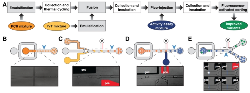
Modular microfluidic droplet-based discovery workflow. (A) Multiple devices accomplish in serial the workflow tasks, including (B) emulsification, (C) reagent addition via electrocoalescence or (D) pico-injection, and (E) fluorescence-activated sorting. Incubation occurs off-line. Figure adapted from Ryckelynck, M.; Baudrey, S.; Rick, C.; Marin, A.; Coldren, F.; Westhof, E.; Griffiths, A. D. RNA
2015, 21, 458–469 (ref 56) under a Creative Commons License.

Compartmentalized cellulase screening platform. Yeast cells expressing the Cel5a cellulase gene secrete the enzyme product into a fluorogenic activity assay reagent. Carboxymethylcellulase (CMC) digestion yields smaller sugar products, such as glucose, which can participate in a redox cascade through hexose oxidase (HOx) and a vanadium bromoperoxidase (VBrPO), yielding conversion of quenched aminophenyl fluorescein substrate into fluorescent product. Adapted with permission from Ostafe, R.; Prodanovic, R.; Lloyd Ung, W.; Weitz, D. A.; Fischer, R. Biomicrofluidics
2014, 8, 041102 (ref 57). Copyright 2014, AIP Publishing LLC.
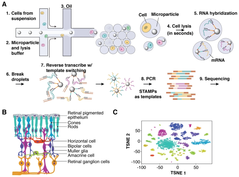
Droplet-parallelized single-cell quantitative mRNA analysis. (A) The workflow begins with encapsulation of individually barcoded poly-dT-functionalized mRNA capture beads and cell suspension into droplets. In-droplet cell lysis and subsequent mRNA hybridization yields templates for reverse transcriptase to extend bead-bound DNA oligonucleotides. PCR amplification of the STAMPs (single-cell transcriptomes attached to microparticles) generates libraries for deep sequencing. (B) Retina tissue samples contain a variety of epithelial, sensor and neuronal cells types, which (C) cluster tightly according to transcriptional profile in a t-distributed stochastic neighbor embedding analysis. Some cell types exhibit multiple distinct transcriptional states. Reprinted from Cell, 161, Macosko, E. Z.; Basu, A.; Satija, R.; Nemesh, J.; Shekhar, K.; Goldman, M.; Tirosh, I.; Bialas, A. R.; Kamitaki, N.; Martersteck, E. M.; Trombetta, J. J.; Weitz, D. A.; Sanes, J. R.; Shalek, A. K.; Regev, A.; McCarroll, S. A., “Highly Parallel Genome-wide Expression Profiling of Individual Cells Using Nanoliter Droplets,” 1202–1214, Copyright 2015, with permission from Elsevier. (ref 46)
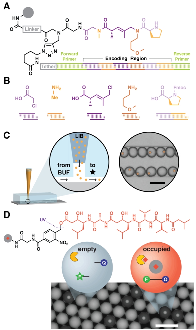
A distributable droplet-scale small molecule discovery platform. (A) DNA-encoded solid-phase synthesis can generate large and arbitrarily complex libraries, encoding each molecule’s structure in a step-wise synthesized DNA tag. All tags feature common PCR primer binding sites for high-sensitivity detection. (B) Each sequence block in the tag correlates with a monomer added during synthesis. (C) Use of a suspension hopper enables large-format (~10 μm) bead loading. Particles settle out of a capped pipette tip into a microfluidic carrier flow and subsequent compartmentalization in droplets. (D) Pepstatin A, an aspartyl protease inhibitor, is attached to resin via a photolabile linker, loaded into droplets of a fluorogenic HIV-1 protease activity assay, and photolytically liberated into the assay. Liberated peptstatin inhibits proteolytic substrate cleavage and concomitant increase in fluorescence. Scale = 100 μm Adapted with permission from MacConnell, A. B.; McEnaney, P. J.; Cavett, V. J.; Paegel, B. M. ACS Comb. Sci.
2015, 17, 518–534 (ref 81). Copyright 2015 American Chemical Society. Adapted with permission from Price, A. K.; MacConnell, A. B.; Paegel, B. M. Anal. Chem.
2014, 86, 5039–5044 (ref 29). Copyright 2014 American Chemical Society.
References
-
- Shim J-U, Ranasinghe RT, Smith CA, Ibrahim SM, Hollfelder F, Huck WTS, Klenerman D, Abell C. ACS Nano. 2013;7:5955–5964. - PubMed
-
- Li Z, Leshansky AM, Pismen LM, Tabeling P. Lab Chip. 2015;15:1023–1031. - PubMed
-
- Leman M, Abouakil F, Griffiths AD, Tabeling P. Lab Chip. 2015;15:753–765. - PubMed
-
- Abate AR, Weitz DA. Lab Chip. 2011;11:1911–1915. - PubMed
Publication types
MeSH terms
Substances
Grants and funding
LinkOut - more resources
Full Text Sources
Other Literature Sources
