Review
. 2016 Jun 1;138(11):2570-8.
doi: 10.1002/ijc.29923. Epub 2015 Nov 28.

Constitutive activation of STAT3 in breast cancer cells: A review

Affiliations
Review

Constitutive activation of STAT3 in breast cancer cells: A review

Kasturi Banerjee et al. Int J Cancer. .

Abstract

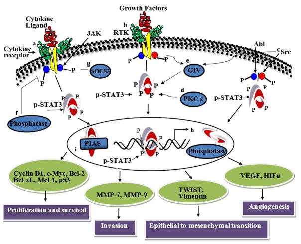
Signal transducer and activator of transcription 3 (STAT3) is constitutively activated in numerous cancer types, including more than 40% of breast cancers. In contrast to tight regulation of STAT3 as a latent transcription factor in normal cells, its signaling in breast cancer oncogenesis is multifaceted. Signaling through the IL-6/JAK/STAT3 pathway initiated by the binding of IL-6 family of cytokines (i.e., IL-6 and IL-11) to their receptors have been implicated in breast cancer development. Receptors with intrinsic kinase activity such as EGFR and VEGFR directly or indirectly induce STAT3 activation in various breast cancer types. Aberrant STAT3 signaling promotes breast tumor progression through deregulation of the expression of downstream target genes which control proliferation (Bcl-2, Bcl-xL, Survivin, Cyclin D1, c-Myc and Mcl-1), angiogenesis (Hif1α and VEGF) and epithelial-mesenchymal transition (Vimentin, TWIST, MMP-9 and MMP-7). These multiple modes of STAT3 regulation therefore make it a central linking point for a multitude of signaling processes. Extensive efforts to target STAT3 activation in breast cancer had no remarkable success in the past because the highly interconnected nature of STAT3 signaling introduces lack of selectivity in pathway identification for STAT3 targeted molecular therapies or because its role in tumorigenesis may not be as critical as it was thought. This review provides a full spectrum of STAT3's involvement in breast cancer by consolidating the knowledge about its role in breast cancer development at multiple levels: its differential regulation by different receptor signaling pathways, its downstream target genes, and modification of its transcriptional activity by its coregulatory transcription factors.

Keywords: STAT3; breast cancer; regulation; target genes; transcription factor; tumor growth.


Figures

Figure 1
Figure 1
Schematic representation of STAT3 activation and its pathways. STAT3 is activated by (a) cytokines (e.g., IL6 and non-IL6 family members), (b) growth factors for receptor tyrosine kinases (e.g., EGF, PDGF, VEGF), (c) non receptor tyrosine kinases (e.g., Src), (d) serine kinases (e.g., PKCε), and (e) Gα-interacting vesicle-associated protein (GIV/Girdin)/guanine nucleotide exchange factor (GEF). Binding of extracellular ligands to their cognate cell surface receptors trigger the signaling events that can lead to the phosphorylation of STAT3 molecules. Tyrosine or serine residue phosphorylation activates STAT3, which then forms homodimers and translocates to the nucleus to bind to the consensus sequence of its downstream target genes. The negative regulators of STAT3 (f) protein phosphatases and (g) suppressor of cytokine signaling (SOCS3) block STAT3 activation in the cytoplasm. In the nucleus, (h) nuclear phosphatases can mediate STAT3 dephosphorylation and block phosphorylation. Interactions with proteins such as (i) PIAS also inhibit STAT3 phosphorylation.
Figure 2
Figure 2
Major mechanism of STAT3 activation in tumor cells occur by autocrine and paracrine secretion of soluble factors. This figure is a diagrammatic illustration of how autocrine and paracrine signaling activates STAT3 in breast cancer cells. (A) Schematic diagram showing the secretion and uptake of autocrine cytokines, chemokines and growth factors by the same cells. Breast cancer cells themselves secrete soluble factors like cytokines, chemokines and growth factors in the tumor microenvironment. These soluble factors behave as ligands and bind to their receptors present on the surface of the same cell which secreted them. Existence of autocrine loops eliminates the dependence on the availability of external secreted factors for growth and survival. (B) Diagrammatic representation of paracrine mechanism by which secreted factors from the nearby neighboring cells are taken up by breast cancer cells. Paracrine factors are produced by the stroma, fibroblast, leukocyte and infiltrating inflammatory cells. In the paracrine mechanism, cancer cells depend on the availability of secreted factors from the extracellular milieu for their survival. Both autocrine and paracrine signaling can lead to the aggressive growth of cancer cells. (C) Diagrams depict different cell types and components that are typically present in breast tumor microenvironment.

References

    1. Levy DE, Darnell JE., Jr Stats: transcriptional control and biological impact. Nature reviews Molecular cell biology. 2002;3(9):651–62. - PubMed
    1. Walker SR, Xiang M, Frank DA. Distinct roles of STAT3 and STAT5 in the pathogenesis and targeted therapy of breast cancer. Molecular and cellular endocrinology. 2014;382(1):616–21. - PMC - PubMed
    1. Yue P, Turkson J. Targeting STAT3 in cancer: how successful are we? Expert opinion on investigational drugs. 2009;18(1):45–56. - PMC - PubMed
    1. Klemm JD, Schreiber SL, Crabtree GR. Dimerization as a regulatory mechanism in signal transduction. Annual review of immunology. 1998;16:569–92. - PubMed
    1. Rane SG, Reddy EP. Janus kinases: components of multiple signaling pathways. Oncogene. 2000;19(49):5662–79. - PubMed

Publication types

MeSH terms