. 2015 Nov;56(12):7338-44.
doi: 10.1167/iovs.15-18038.

Immunofluorescence Tomography of Mouse Ocular Surface Epithelial Stem Cells and Their Niche Microenvironment

Immunofluorescence Tomography of Mouse Ocular Surface Epithelial Stem Cells and Their Niche Microenvironment

Geraint J Parfitt et al. Invest Ophthalmol Vis Sci. 2015 Nov.

Abstract

Purpose: Currently, there are no definitive immunomarkers for epithelial stem cells (corneal and conjunctival) or their poorly understood niche microenvironment. The H2B-GFP/K5tTA mouse enables visualization of label-retaining cells (LRCs), which exhibit the functional marker of stem cell quiescence. We used immunofluorescence tomography to evaluate putative stem cell markers and LRCs of the mouse ocular surface.

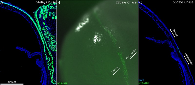
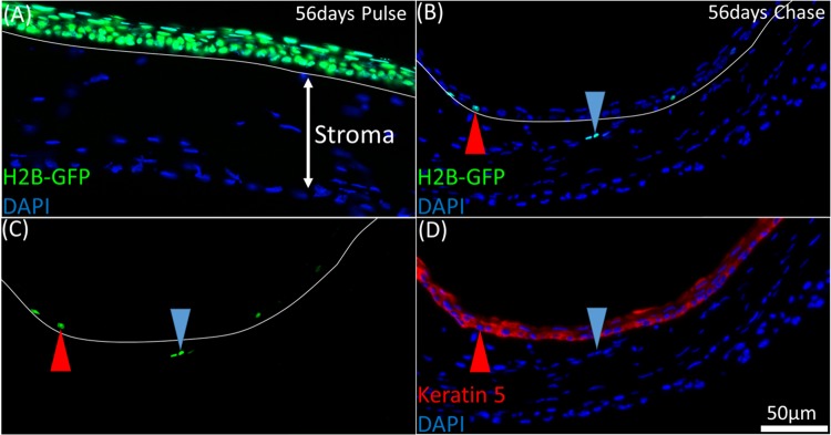
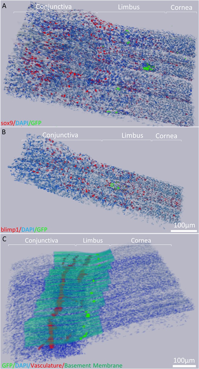
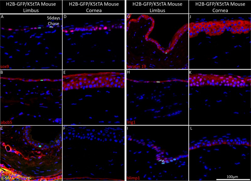
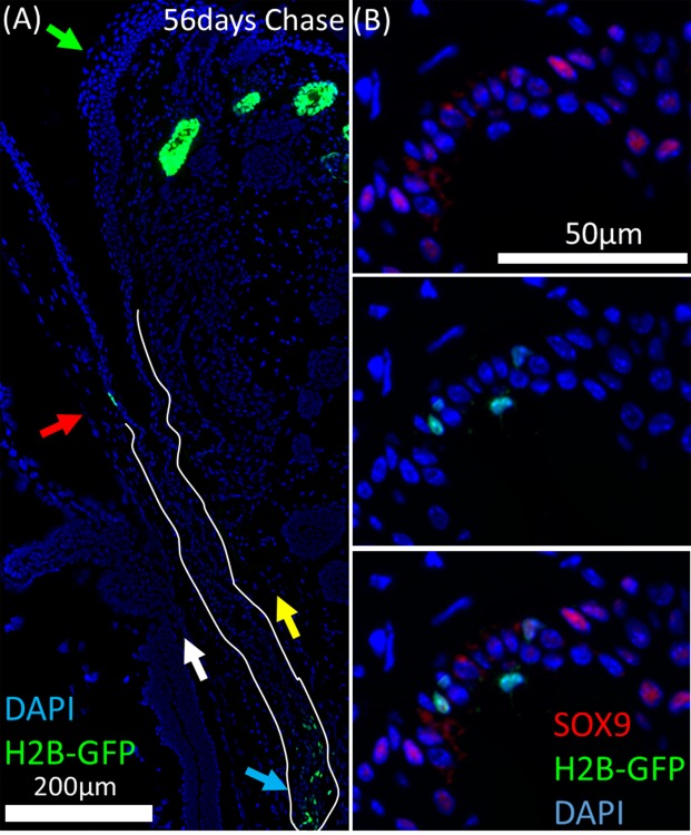
Methods: H2B-GFP/K5tTA mice were pulsed for 56 days and then chased with doxycycline to label LRCs. Limbus and eyelid tissue was 3-dimensionally (3-D) reconstructed using immunofluorescence tomography to identify and characterize LRCs using the putative stem cell markers sox9, keratin 19, lrig1, blimp1, and abcb5.

Results: After 28 days of chase, LRCs were localized to the entire limbus epithelium and, infrequently, the anterior limbal stroma. Label-retaining cells comprised 3% of limbal epithelial cells after 56 days of chase. Conjunctival LRCs were localized to the fornix and comprised 4% of the total fornix epithelial cells. No stem cell immunomarker was specific for ocular surface LRCs; however, blimp1 enriched for limbal basal epithelial cells and 100% of green fluorescent protein-positive (GFP+) cells at the limbus and fornix were found to be lrig1-positive.

Conclusions: Label-retaining cells represent a larger population of the mouse limbus than previously thought. They decrease in number with increased doxycycline chase, suggesting that LRC populations with different cell cycle lengths exist at the limbus. We conclude that current immunomarkers are unable to colocalize with the functional marker of epithelial stem cell quiescence; however, blimp1 may enrich for limbal epithelial basal cells.


Figures

Figure 1
Figure 1
Slow-cycling limbal stem cells responsible for corneal epithelial cell renewal were localized to the limbus in the H2B-GFP/K5tTA mouse after >28 days doxycycline chase. (A) At 56 days pulse (no doxycycline), keratin 5+ cells express nuclear GFP, which includes all the basal keratinocytes of the ocular surface: the cornea, conjunctiva, and meibomian gland. (B) Low-magnification image of H2B-GFP/K5tTA mouse whole eye after 28 days doxycycline chase. At 56 days pulse – 28 days chase, LRCs were seen throughout the circumference of the corneal limbus, although regions devoid of LRCs were also observed (*). (C) After 56 days doxycycline chase, LRCs at the limbus are GFP+ because they divide infrequently compared to other epithelial cells which lose half of their GFP signal with every round of cell division.
Figure 2
Figure 2
LRCs in the H2B-GFP/K5tTA mouse were localized to the limbus epithelium and infrequently, the anterior limbal stroma. (A) At 56 days pulse, all keratin 5+ corneal epithelial cells express nuclear GFP tagged to histone H2B. (B, C) A 2 μm BMMA cross-section of the H2B-GFP/K5tTA mouse limbus after 56 days doxycycline chase. GFP+ cells were primarily localized to the limbus epithelium (red arrow); however, on rare occasion, they were observed in the limbus stroma with varying fluorescence intensities (blue arrow). (D) Keratin 5 staining (B, C) shows that the GFP+ stromal cells are Keratin 5. This suggests that migration of limbal epithelial cells may occur or there is ectopic expression of keratin 5.
Figure 3
Figure 3
Immunofluorescence tomography 3-D reconstruction of the H2B-GFP/K5tTA mouse limbus. (A) Reconstruction of sox9 immunostaining and LRCs after 56 days doxycycline chase using 186, 2-μm BMMA serial sections (372 μm). (B) 3-D Reconstruction of blimp1 immunostaining and LRCs in the mouse limbus using 87 serial sections (174 μm). (C) 3-D reconstruction of the basement membrane and vasculature network beneath the limbus epithelium which harbors LRCs.
Figure 4
Figure 4
Immunohistochemistry staining of the H2B-GFP/K5tTA mouse limbus and central cornea with putative stem cell markers at 56 days doxycycline chase. The 2 μm BMMA sections of the H2B-GFP/K5tTA mouse limbus (AC, GI) and central cornea (DF, JL) after 56 days doxycycline chase were immunostained for (A, D) sox9, (B, E), abcb5, (C, F) α-smooth muscle actin, and collagen IV, (G, I) keratin 19, (H, K) lrig1, (I, L) blimp1. Sox9 is localized to LRCs and dividing basal cells of the ocular surface while collagen IV is expressed in the basement membrane and the vasculature network, which terminates at the limbus. Keratin 19 is found in the basal and suprabasal layers of the ocular surface, while the putative stem cell markers lrig1 and blimp1 are found expressed in LRCs and other basal cells of the limbus and the cornea.
Figure 5
Figure 5
Label-retaining cells in the H2B-GFP/K5tTA mouse conjunctiva were localized to the fornix region and are most likely responsible for the renewal of palpebral and bulbar conjunctiva epithelium. (A) After 56 days doxycycline chase, GFP+ LRCs in the cornea were localized to the limbus (red arrow) whereas conjunctival LRCs were localized to the fornix (blue arrow). The daughter cells from LRCs in the fornix conjunctiva are likely to repopulate the bulbar (white arrow) and palpebral (yellow arrow) conjunctiva with epithelial cells. No LRCs were observed at the eyelid margin (green arrow). (B) Sox9 immunostaining shows LRCs and dividing cells of the fornix conjunctiva are sox9+ or sox9.
Figure 6
Figure 6
Three-dimensional quantification of sox9+, lrig1+, and blimp1+ epithelial cells and LRCs across the ocular surface epithelium. Using the 3-D objects counter in ImageJ, the co-localization of sox9, lrig1, and blimp1 with epithelial cells and LRCs was quantified in the (A) limbus (n = 1987 average nuclei), (B) cornea (n = 3407 average nuclei), and (C) fornix conjunctiva epithelium (n = 2386 average nuclei) of the H2B-GFP/K5tTA mouse by immunofluorescence tomography. The limbus and fornix conjunctiva exhibited lrig1+ LRCs and blimp1± or sox9± LRCs. No LRCs were observed in the central corneal epithelium.

References

    1. Stepp MA,, Zieske JD. The corneal epithelial stem cell niche. Ocul Surf. 2005; 3: 15–26. - PubMed
    1. Alonso L,, Fuchs E. Stem cells of the skin epithelium. Proc Natl Acad Sci U S A. 2003; 100 (suppl 1): 11830–11835. - PMC - PubMed
    1. Slack JM. Stem cells in epithelial tissues. Science. 2000. ; 287: 1431–1433. - PubMed
    1. Thoft RA,, Friend J. The X, Y, Z hypothesis of corneal epithelial maintenance. Invest Ophthalmol Vis Sci. 1983; 24: 1442–1443. - PubMed
    1. Li W,, Hayashida Y,, Chen YT,, Tseng SC. Niche regulation of corneal epithelial stem cells at the limbus. Cell Res. 2007; 17: 26–36. - PMC - PubMed

Publication types