. 2015 Nov 11;90(3):1278-89.
doi: 10.1128/JVI.02750-15. Print 2016 Feb 1.

A Comprehensive RNA Sequencing Analysis of the Adeno-Associated Virus (AAV) Type 2 Transcriptome Reveals Novel AAV Transcripts, Splice Variants, and Derived Proteins

Affiliations

A Comprehensive RNA Sequencing Analysis of the Adeno-Associated Virus (AAV) Type 2 Transcriptome Reveals Novel AAV Transcripts, Splice Variants, and Derived Proteins

Catrin Stutika et al. J Virol. .

Abstract

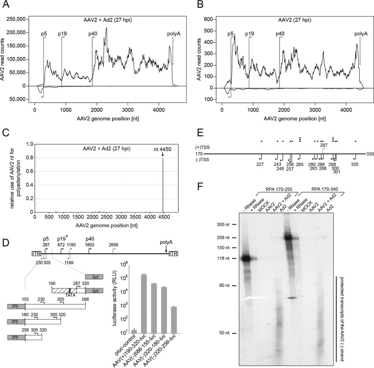
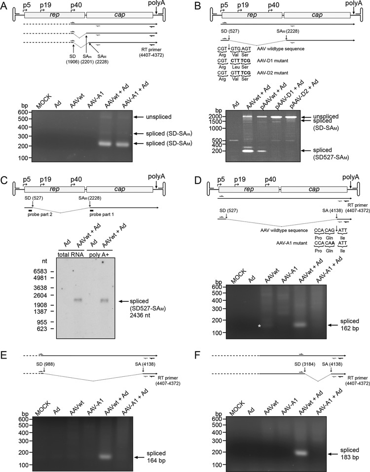
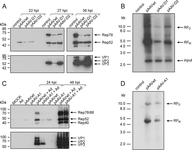
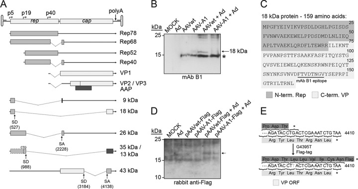
Adeno-associated virus (AAV) is recognized for its bipartite life cycle with productive replication dependent on coinfection with adenovirus (Ad) and AAV latency being established in the absence of a helper virus. The shift from latent to Ad-dependent AAV replication is mostly regulated at the transcriptional level. The current AAV transcription map displays highly expressed transcripts as found upon coinfection with Ad. So far, AAV transcripts have only been characterized on the plus strand of the AAV single-stranded DNA genome. The AAV minus strand is assumed not to be transcribed. Here, we apply Illumina-based RNA sequencing (RNA-Seq) to characterize the entire AAV2 transcriptome in the absence or presence of Ad. We find known and identify novel AAV transcripts, including additional splice variants, the most abundant of which leads to expression of a novel 18-kDa Rep/VP fusion protein. Furthermore, we identify for the first time transcription on the AAV minus strand with clustered reads upstream of the p5 promoter, confirmed by 5' rapid amplification of cDNA ends and RNase protection assays. The p5 promoter displays considerable activity in both directions, a finding indicative of divergent transcription. Upon infection with AAV alone, low-level transcription of both AAV strands is detectable and is strongly stimulated upon coinfection with Ad.

Importance: Next-generation sequencing (NGS) allows unbiased genome-wide analyses of transcription profiles, used here for an in depth analysis of the AAV2 transcriptome during latency and productive infection. RNA-Seq analysis led to the discovery of novel AAV transcripts and splice variants, including a derived, novel 18-kDa Rep/VP fusion protein. Unexpectedly, transcription from the AAV minus strand was discovered, indicative of divergent transcription from the p5 promoter. This finding opens the door for novel concepts of the switch between AAV latency and productive replication. In the absence of a suitable animal model to study AAV in vivo, combined in cellulae and in silico studies will help to forward the understanding of the unique, bipartite AAV life cycle.


Figures

FIG 1
FIG 1
RNA-Seq: mapping and transcription analysis on both strands of the AAV2 genome. (A) Coverage map of AAV2-specific RNA-Seq reads to the AAV2 genome of AAV2/Ad2-coinfected cells at 27 hpi. The AAV2 genome scale is displayed in the center. Reads mapped to the plus strand are presented above, and reads mapped to the minus strand are presented below. (B) Display as in panel A. RNA-Seq reads of AAV2-infected cells at 27 hpi are presented. Note the different scales of the read counts in panels A and B. (C) Mapping of RNA-Seq reads ending with a poly(A) tail. The genome position of poly(A) addition is displayed relative to the totality of polyadenylated AAV reads in the data set of AAV2/Ad2-coinfected cells. (D) Promoter activity of the AAV2 p5 region in reverse direction. The upper part shows the AAV2 genome with the nucleotide positions of the transcription start sites of the p5, p19, and p40 promoters (black arrows). In addition, putative promoters (gray arrows) originating from a promoter prediction program with a cutoff score above 0.9 are indicated on the plus or minus strand of the AAV genome with the respective nucleotide positions of the TSS. An asterisk (*) highlights the established p19 promoter, detected with a cutoff score below 0.9. The lower part shows the relative luciferase activities of the indicated candidate reverse p5 promoter sequences on the AAV2 minus strand. AAV(+)190-320-luc represents the AAV2 p5 promoter in the forward orientation serving as a positive control and empty pluc serving as a negative control. The relative luciferase activities of three different experiments were determined in the presence of Ad2 and are depicted as means ± standard deviations (SD). (E) 5ˈRACE analysis of AAV minus strand transcription. Displayed are the TSSs (arrows) derived from DNA sequences of cloned 5ˈRACE products from the AAV minus strand (nt 330 to 170). Single TSSs are displayed as black squares above the genome. The p5 promoter TSS at nt 287 (gray arrow) on the AAV plus strand is shown for orientation. (F) An RNase protection assay was performed with in vitro-transcribed hybridization probes covering AAV2 nt 170 to 250 (left part) or AAV2 nt 170 to 340 (right part) in a sense orientation for the detection of AAV minus strand transcripts in total RNA from HeLa cells either mock infected, AAV2 infected, Ad2 infected, or AAV2/Ad2 coinfected for 27 h. The undigested probes (–RNase) and the probes digested in the presence of yeast t-RNA (+RNase) are shown as controls. Both probes contain additional non-AAV2 sequences from the pBluescript vector used for T7 polymerase-dependent in vitro transcription. The maximal protected AAV2 regions are 81 nt for probe RPA 170-250, and 171 nt for probe RPA 170-340, respectively.
FIG 2
FIG 2
Validation of RNA-Seq-derived AAV2 splice events by RT-PCR and Northern blot analysis. (A) The AAV2 genome with unspliced/spliced transcripts is displayed at the top. The primer for reverse transcription is shown with its binding position. The forward primer (nt 1862 to 1889) and reverse primer (nt 2440 to 2421) are displayed for PCR analysis of 1906:2201/2228 splicing. Below, the results of RT-PCR for AAV wild-type- and mutant A1-infected cells are displayed, separated on an agarose gel. PCR products are indicated by arrows. (B) RT-PCR of splice event 527:2228, performed as in panel A with cells transfected with plasmids for AAV wild-type or mutants D1 or D2. Recoded nucleotide sequences of AAV-D1 and AAV-D2 are displayed below the AAV genome. (C) Northern blot analysis of total and poly(A)+ selected RNA of cells coinfected with AAV2 and Ad2 using a 24-nt oligomer as a probe that spans the AAV2 splice junction 527:2228, as depicted below the AAV genome. The arrow points to the spliced transcripts. (D to F) RT-PCR analysis of additional AAV RNA splice events, performed as in panel A. (D) Forward primer (nt 415 to 434) and reverse primer (nt 4186 to 4161) used for the analysis of 527:4138 splicing. The corresponding mutation of AAV-A1 is displayed below the AAV genome. The asterisk (*) marks a faint band of the splice product in the absence of Ad. (E) Forward primer (nt 874 to 893) and reverse primer (nt 4186 to 4161) for the analysis of 988:4138 splicing. (F) Forward primer (nt 3051 to 3070) and reverse primer (nt 4186 to 4161) for the analysis of 3184:4138 splicing.
FIG 3
FIG 3
Influence of alternative splicing on AAV replication. (A) Analysis of AAV2 Rep and VP expression in AAV wild type-, splice mutant AAV-D1-, or AAV-D2 (see Fig. 2B)-transfected HeLa cells at the indicated time points after Ad infection. AAV Rep and VP proteins are detected by MAb 303.9 or MAb B1, respectively. (B) Southern blot analysis of Hirt extracts of AAV wild type-, AAV-D1-, or AAV-D2-transfected and Ad-infected HeLa cells. AAV-specific monomeric (RFM) and dimeric (RFD) replicative forms, detected by probing with a labeled 1.6-kb cap-derived DNA sequence, are indicated by arrows. Input refers to undigested input DNA. (C) Western blot analysis as described in panel A with mutant AAV-A1 (see Fig. 2D). Mock- and Ad-infected cells served as controls. (D) Southern blot analysis as performed for panel B with mutant AAV-A1.
FIG 4
FIG 4
Detection of novel AAV proteins derived from alternative splicing. (A) The AAV2 genome and current transcription/translation map is represented at the top. The different shading patterns indicate alternative reading frames. Below, the newly identified spliced transcripts are displayed with the deduced novel fusion ORFs. The molecular mass of the derived protein candidates is indicated to the right in kilodaltons. (B) Analysis of the predicted protein (18 kDa) expressed from splice variant 527:4138 on a Western blot reacted with MAb B1. AAV-A1 represents the AAV2 mutant at nt 4138 (see Fig. 2D). An asterisk (*) marks the previously described 15- to 17-kDa band, which is indicative of degraded input capsids. (C) Amino acid sequence of the novel 18-kDa protein. The first 69 amino acids correspond to the N-terminal part of Rep78; the last 90 amino acids correspond to the C terminus of the VP proteins, including the epitope for MAb B1. (D) Detection of AAVwt-Flag and AAV-A1-Flag constructs on a Western blot reacted with rabbit anti-Flag upon extensive exposure. The arrow marks a faint band of ∼18 kDa consistent with the previously proposed X protein (38). (E) Displayed is the proposed X-gene-derived C terminus of the resulting protein (dark gray) before and after insertion of the Flag tag in comparison to the confirmed capsid (VP) protein sequence (light gray). The asterisk (*) denotes the stop codon in the wild-type sequence.
FIG 5
FIG 5
Alignment of splice donor and acceptor sites for AAV serotypes 1 to 9. (A) Splice donor sites at nt positions 1906, −527, −988, and −3184 of AAV2 were aligned to the corresponding sites of AAV serotypes 1 to 9 and compared to the mammalian consensus sequence of splice donor sites (34). Exon-intron boundaries are indicated by dashed lines. Below the consensus sequence the frequency (%) of a particular nucleotide in average splice donor sites is displayed. White areas indicate aberrations to the consensus sequence in the given AAV serotype. (B) Splice acceptor sites at nt positions 2228, −2201, −807, and −4138 of AAV2 were aligned to the corresponding sites of AAV serotypes 1 to 9 and compared to the mammalian consensus sequence of splice acceptor sites. The display is as described for panel A. Py, pyrimidine.

Similar articles

Cited by

References

    1. Blacklow NR, Hoggan MD, Rowe WP. 1967. Isolation of adenovirus-associated viruses from man. Proc Natl Acad Sci U S A 58:1410–1415. doi:10.1073/pnas.58.4.1410. - DOI - PMC - PubMed
    1. Srivastava A, Lusby EW, Berns KI. 1983. Nucleotide sequence and organization of the adeno-associated virus 2 genome. J Virol 45:555–564. - PMC - PubMed
    1. Sonntag F, Schmidt K, Kleinschmidt JA. 2010. A viral assembly factor promotes AAV2 capsid formation in the nucleolus. Proc Natl Acad Sci U S A 107:10220–10225. doi:10.1073/pnas.1001673107. - DOI - PMC - PubMed
    1. Green MR, Roeder RG. 1980. Definition of a novel promoter for the major adenovirus-associated virus mRNA. Cell 22:231–242. doi:10.1016/0092-8674(80)90171-3. - DOI - PubMed
    1. Lusby EW, Berns KI. 1982. Mapping of the 5ˈ termini of two adeno-associated virus 2 RNAs in the left half of the genome. J Virol 41:518–526. - PMC - PubMed

Publication types

LinkOut - more resources