. 2015 Nov 11;90(3):1259-77.
doi: 10.1128/JVI.02651-15. Print 2016 Feb 1.

Trehalose, an mTOR-Independent Inducer of Autophagy, Inhibits Human Cytomegalovirus Infection in Multiple Cell Types

Affiliations

Trehalose, an mTOR-Independent Inducer of Autophagy, Inhibits Human Cytomegalovirus Infection in Multiple Cell Types

Jean-Philippe Belzile et al. J Virol. .

Abstract

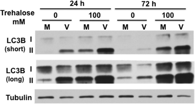
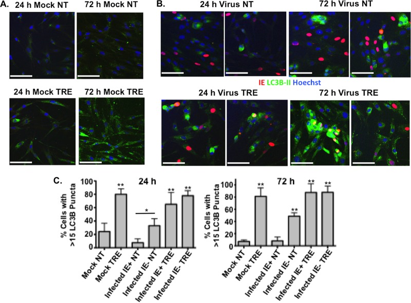
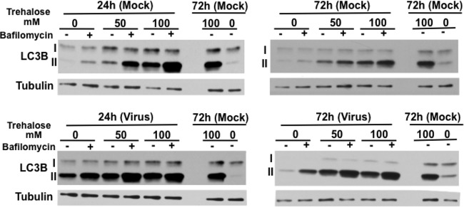
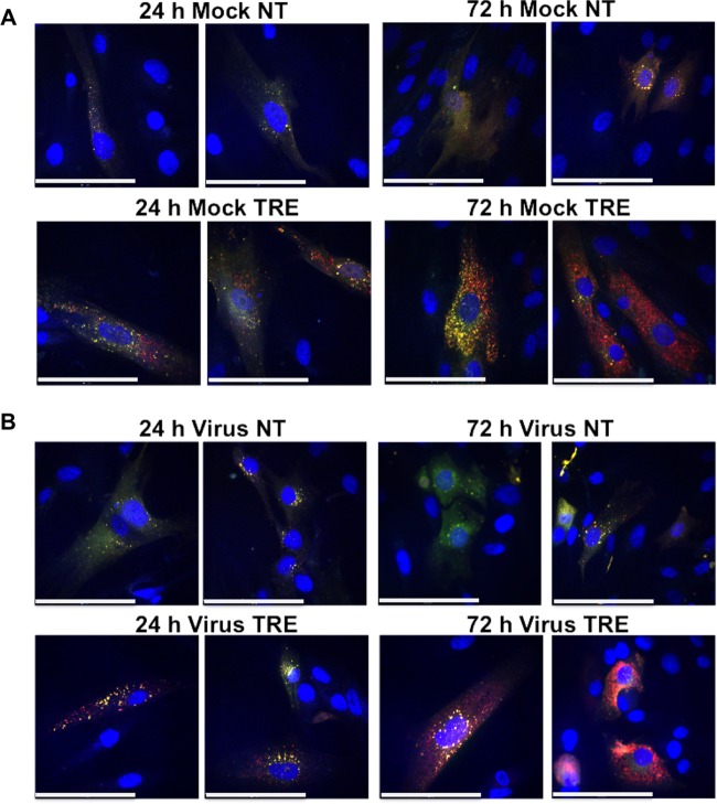
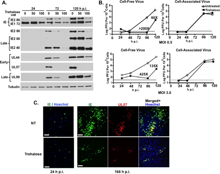
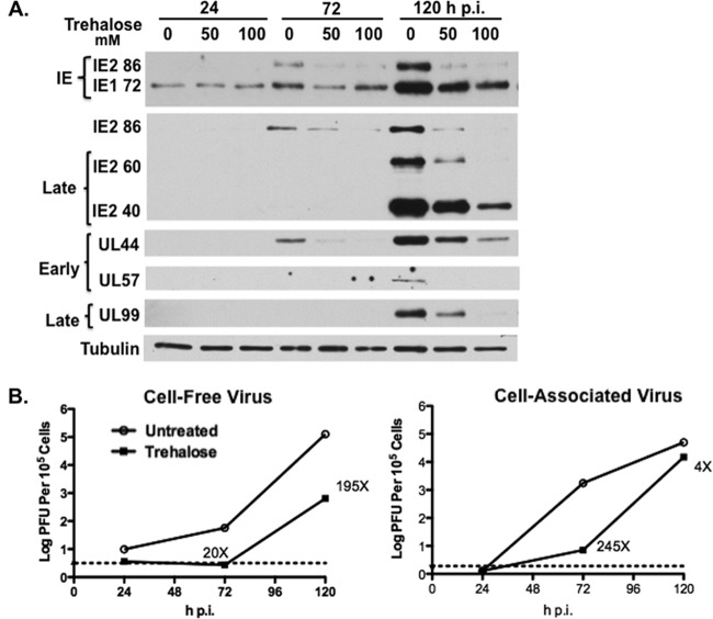
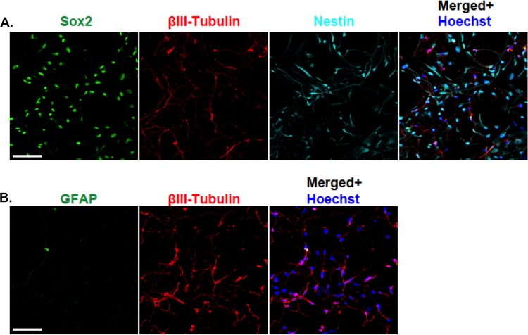
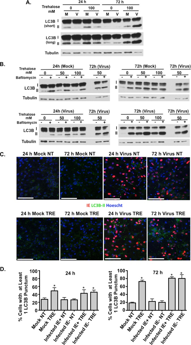
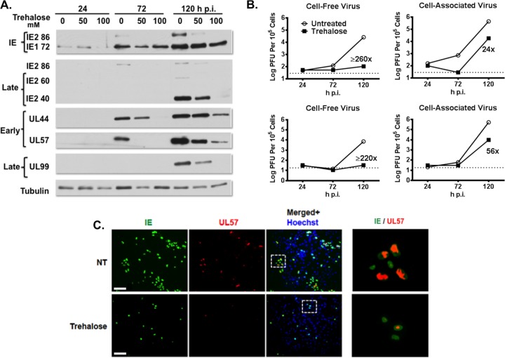
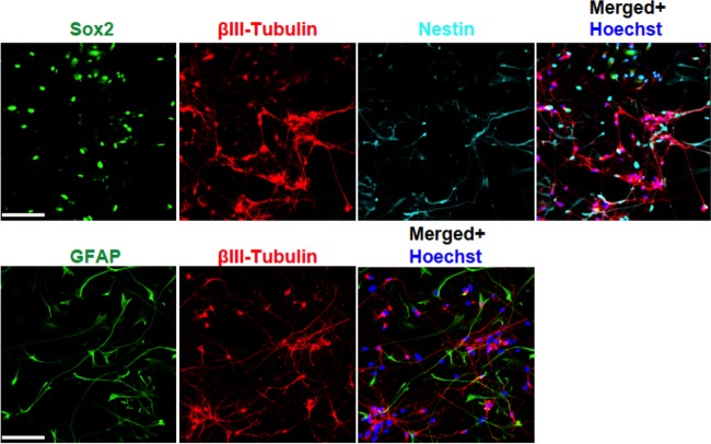
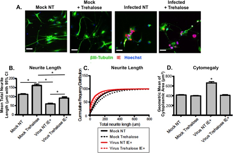
Human cytomegalovirus (HCMV) is the major viral cause of birth defects and a serious problem in immunocompromised individuals and has been associated with atherosclerosis. Previous studies have shown that the induction of autophagy can inhibit the replication of several different types of DNA and RNA viruses. The goal of the work presented here was to determine whether constitutive activation of autophagy would also block replication of HCMV. Most prior studies have used agents that induce autophagy via inhibition of the mTOR pathway. However, since HCMV infection alters the sensitivity of mTOR kinase-containing complexes to inhibitors, we sought an alternative method of inducing autophagy. We chose to use trehalose, a nontoxic naturally occurring disaccharide that is found in plants, insects, microorganisms, and invertebrates but not in mammals and that induces autophagy by an mTOR-independent mechanism. Given the many different cell targets of HCMV, we proceeded to determine whether trehalose would inhibit HCMV infection in human fibroblasts, aortic artery endothelial cells, and neural cells derived from human embryonic stem cells. We found that in all of these cell types, trehalose induces autophagy and inhibits HCMV gene expression and production of cell-free virus. Treatment of HCMV-infected neural cells with trehalose also inhibited production of cell-associated virus and partially blocked the reduction in neurite growth and cytomegaly. These results suggest that activation of autophagy by the natural sugar trehalose or other safe mTOR-independent agents might provide a novel therapeutic approach for treating HCMV disease.

Importance: HCMV infects multiple cell types in vivo, establishes lifelong persistence in the host, and can cause serious health problems for fetuses and immunocompromised individuals. HCMV, like all other persistent pathogens, has to finely tune its interplay with the host cellular machinery to replicate efficiently and evade detection by the immune system. In this study, we investigated whether modulation of autophagy, a host pathway necessary for the recycling of nutrients and removal of protein aggregates, misfolded proteins, and pathogens, could be used to target HCMV. We found that autophagy could be significantly increased by treatment with the nontoxic, natural disaccharide trehalose. Importantly, trehalose had a profound inhibitory effect on viral gene expression and strongly impaired viral spread. These data constitute a proof-of-concept for the use of natural products targeting host pathways rather than the virus itself, thus reducing the risk of the development of resistance to treatment.


Figures

FIG 1
FIG 1
Trehalose increases LC3B-II levels in uninfected and infected HFFs. Synchronized HFFs were infected with TB40E at an MOI of 3 (V) or mock infected (M) upon seeding in the presence or absence of 100 mM trehalose. Cells were harvested at the indicated time points, and extracts were analyzed by Western blotting using a rabbit polyclonal antibody against LC3B. Both native 18-kDa (I) and lipid-associated 16-kDa (II) forms of LC3B are detectable using this antibody. An antibody against α-tubulin was used to control for loading. Short and long exposures are shown. This experiment was repeated at least twice.
FIG 2
FIG 2
Autophagosome formation in HCMV-infected cells is induced by trehalose. Synchronized HFFs were mock infected (A) or infected with TB40E at an MOI of 0.5 (B) upon seeding on coverslips in the absence (not treated, NT) or presence (TRE) of 100 mM trehalose. Cells were fixed at the indicated time points and permeabilized with saponin as described in Materials and Methods. Cells were stained with mouse monoclonal antibodies against LC3B (green) and IE1 (red). Nuclei were counterstained with Hoechst 33342 (blue). Pictures were acquired by confocal microscopy. Representative images are shown for each condition. Scale bar, 100 μm. (C) Quantification of the percentage of cells represented in panels A and B with more than 15 LC3B puncta. At least five fields (with approximately 25 cells in each) were counted for each condition. Histograms represent the means (bars), and the standard deviations (error bars) of the five fields are shown. Statistical significance was determined by one-way ANOVA combined with Tukey's multiple-comparison test (**, P < 0.01 versus results for mock uninfected and infected IE+ untreated cells; *, P < 0.05 versus results for infected IE+ untreated cells only). This experiment was repeated at least twice.
FIG 3
FIG 3
Trehalose increases the basal levels of autophagy and maintains autophagic flux in infected HFFs. Synchronized HFFs were infected with TB40E at an MOI of 3 (or mock infected) upon seeding in the absence or presence of trehalose (50 mM or 100 mM). At the indicated time points, cells were pulsed with bafilomycin A1 (or with DMSO as a control) for 2 h prior to harvest. Cell extracts were analyzed by Western blotting using antibodies against LC3B. An antibody against α-tubulin was used to control for loading. Repeated samples on each gel were used to calibrate blotting and exposures between the different gels. This experiment was repeated at least twice.
FIG 4
FIG 4
Trehalose promotes acidification of autophagosomes in infected HFFs. HFFs were transduced with a baculovirus expressing LC3B fused to GFP (green) and RFP (red). At 1 day postransduction, cells were mock infected (A) or infected with TB40E at an MOI of 3 (B) upon seeding on coverslips in the absence (not treated, NT) or presence (TRE) of 100 mM trehalose. At the indicated time points, cells were fixed, and nuclei were counterstained with Hoechst 33342. Z-stacks composed of individual 0.4-μm-thick slices were acquired at high magnification using a spinning-disk microscope. Between 8 and 10 fields were acquired for each condition. Representative phenotypes are shown. Colocalization (yellow) between the direct fluorescence of GFP and RFP results from a neutral pH, whereas red puncta correspond to a more acidic environment in which the GFP signal was quenched. Scale bar, 100 μm. This experiment was repeated at least twice.
FIG 5
FIG 5
Trehalose inhibits HCMV gene expression, virus production, and viral spread in HFFs. (A) Synchronized HFFs were infected with TB40E at an MOI of 0.5 upon seeding in the absence or presence of 50 mM or 100 mM trehalose. Cell extracts prepared at 24, 72, and 120 hpi were analyzed by Western blotting using antibodies against HCMV IE1/IE2 (CH160), IE2-86, late IE2 proteins IE2-60 and IE2-40, UL44, UL57, and UL99. An antibody against α-tubulin was used to control for loading. The experiment was repeated three times, and a representative Western blot is shown. (B) Synchronized HFFs were infected with TB40E at an MOI of 0.5 or 3 upon seeding in the absence or presence of 100 mM trehalose. At the indicated time points, cell-associated and cell-free virus were prepared as described in Materials and Methods. Titers of viral preparations were determined by plaque assays. Graphs represent titer results obtained from a representative experiment. Relevant fold decreases in titers in trehalose-treated cells, relative to those nontreated cells, are indicated. Dotted lines represent assay limits of detection. (C) Synchronized HFFs were infected with TB40E at an MOI of 0.025 upon seeding on coverslips in the absence (not treated, NT) or presence of 100 mM trehalose. Cells were fixed at 24 and 168 hpi and stained with mouse monoclonal antibodies against HCMV IE (green) and UL57 (red) proteins. Nuclei were counterstained with Hoechst 33342 (blue). Images were acquired by epifluorescence microscopy. Representative images of phenotypes obtained at 24 and 168 hpi are shown. Scale bar, 100 μm. Each experiment was repeated three times.
FIG 6
FIG 6
Trehalose increases the basal levels of autophagy and maintains autophagic flux in infected aortic endothelial cells. Human aortic endothelial cells (HAECs) were infected with TB40E at an MOI of 3 (or mock infected) 1 day after seeding in the absence or presence of 50 or 100 mM trehalose. (A) At the indicated time points, cell extracts were prepared and analyzed by Western blotting using antibodies against LC3B. Short and long exposures are shown. (B) At the indicated time points, cells were pulsed with bafilomycin A1 (or with DMSO as a control) for 2 h prior to harvest. Cell extracts were analyzed by Western blotting using antibodies against LC3B. An antibody against α-tubulin was used to control for loading. Repeated samples on each gel were used to calibrate blotting and exposures between the different gels. Each experiment was repeated at least twice.
FIG 7
FIG 7
Trehalose inhibits HCMV gene expression and virus production in endothelial cells. HAECs were infected with TB40E at an MOI of 0.5 in the absence or presence of trehalose (50 mM or 100 mM). (A) Cell extracts prepared at 24, 72, and 120 hpi were analyzed by Western blotting using antibodies against HCMV IE1/IE2 (CH160), IE2-86, late IE2 proteins IE2-60 and IE2-40, UL44, UL57, and UL99. An antibody against α-tubulin was used to control for loading. (B) At the indicated time points, cell-free and cell-associated virus was prepared as described in Materials and Methods, and virus titers were determined by plaque assays. Graphs represent titer results obtained from a representative experiment. Dotted lines represent assay limits of detection. Relevant fold decreases in titers from trehalose-treated cells, relative to those of nontreated cells, are indicated. Each experiment was repeated at least twice.
FIG 8
FIG 8
Differentiation of H9-derived human primitive neural stem cells for 8 days yields a mixed population of stem cells and neurons. H9-derived pNSCs were patterned for 10 days and differentiated on coverslips as described in Materials and Methods. At 8 days postdifferentiation, cells were fixed, permeabilized, and stained with antibodies against βIII-tubulin (red), Sox2 (green), and nestin (cyan) (A) or with antibodies against βIII-tubulin (red) and GFAP (green) (B). Nuclei were counterstained with Hoechst 33342 (blue). Between six and eight fields were acquired for each condition. Representative phenotypes are shown. Scale bar, 100 μm. The experiment was repeated twice.
FIG 9
FIG 9
Trehalose increases the basal levels of autophagy and maintains autophagic flux in infected neuronal cultures. H9-derived neurons at day 8 (H9 d8 neurons) postdifferentiation were infected with TB40E at an MOI of 3 (or mock infected) in the absence or presence of trehalose (50 mM or 100 mM). (A) Cells were harvested at the indicated time points, and extracts were analyzed by Western blotting using a rabbit polyclonal antibody against LC3B. An antibody against α-tubulin was used to control for loading. Short and long exposures are shown. (B) At the indicated time points, cells were pulsed with bafilomycin A1 (or with DMSO as a control) for 2 h prior to harvest. Cell extracts were analyzed by Western blotting using antibodies against LC3B. Repeated samples on each gel were used to calibrate blotting and exposures between the different gels. Experiments were repeated at least twice. (C) H9 day 8 neurons were infected with TB40E at an MOI of 0.5 (or mock infected) in the absence (not treated, NT) or presence (TRE) of 100 mM trehalose. Cells were fixed at the indicated time points and permeabilized with saponin as described in Materials and Methods. Cells were stained with mouse monoclonal antibodies against LC3B (green) and IE1 (red). Nuclei were counterstained with Hoechst 33342 (blue). Pictures were acquired by confocal microscopy. Scale bar, 100 μm. Representative images are shown for each condition. (D) Quantification of the percentage of cells in panel C with at least one LC3B punctum. At least five fields (with approximately 150 cells in each) were counted for each condition, and the means (bars) and standard deviations (error bars) of the five fields are shown. Statistical significance was determined with a one-way ANOVA test combined with Tukey's multiple-comparison test (*, P < 0.05 versus results in mock untreated and infected IE+ untreated cells). Each experiment was repeated at least twice.
FIG 10
FIG 10
Trehalose promotes acidification of autophagosomes in infected neuronal cultures. H9 day 8 neurons were transduced with a baculovirus expressing LC3B fused to GFP (green) and RFP (red). At 1 day postransduction, cells were mock infected (A) or infected with TB40E at an MOI of 3 (B) in the absence (not treated, NT) or presence (TRE) of 100 mM trehalose. At the indicated time points, cells were fixed, and nuclei were counterstained with Hoechst 33342. Z-stacks composed of individual 0.4-μm-thick slices were acquired at high magnification using a spinning-disk microscope. Between 8 and 10 fields were acquired for each condition. Representative phenotypes are shown. Colocalization (yellow) between the direct fluorescence of GFP and RFP results from a neutral pH, whereas red puncta correspond to a more acidic environment in which the GFP signal was quenched. Scale bar, 25 μm. Each experiment was repeated twice.
FIG 11
FIG 11
Trehalose inhibits HCMV gene expression, virus production, and viral spread in neuronal cultures. H9 day 8 neurons were infected with TB40E at an MOI of 0.5 in the absence or presence of trehalose (50 mM or 100 mM). (A) Cell extracts prepared at 24, 72, and 120 hpi were analyzed by Western blotting using antibodies against HCMV IE1/IE2 (CH160), IE2-86, late IE2 proteins IE2-60 and IE2-40, UL44, UL57, and UL99. An antibody against α-tubulin was used to control for loading. The experiment was repeated three times. (B) At the indicated time points, cell-associated and cell-free virus was obtained from cells infected in the absence (untreated) or presence of 100 mM trehalose as described in Materials and Methods. Titers of viral preparations were determined by plaque assays. Graphs showing titers from two representative experiments are shown. Triplicate assays were performed for each sample. Relevant fold decreases in titers from trehalose-treated cells, relative to levels in nontreated cells, are indicated. Dotted lines represent assay limits of detection. (C) H9 day 8 neurons were infected with TB40E at an MOI of 0.025 in the absence (not treated, NT) or presence of 100 mM trehalose. Cells were fixed at 24, 96, and 168 hpi and stained with mouse monoclonal antibodies against HCMV IE (green) and UL57 (red). Nuclei were counterstained with Hoechst 33342 (blue). Images were acquired by epifluorescence microscopy. Only representative images of phenotypes obtained at 168 hpi are shown. Scale bar, 100 μm. The right panels show enlarged images of the boxed regions.
FIG 12
FIG 12
Differentiation of H9-derived human primitive neural stem cells for 21 days yields a mixed population of stem cells, astroglial cells, and mature neurons with long neurites. H9-derived pNSCs were patterned for 10 days and differentiated on coverslips as described in Materials and Methods. At 21 days postdifferentiation, cells were fixed, permeabilized, and stained with antibodies against βIII-tubulin (red), Sox2 (green), and nestin (cyan) (A) or with antibodies against βIII-tubulin (red) and GFAP (green) (B). Nuclei were counterstained with Hoechst 33342 (blue). Between 6 and 8 fields were acquired for each condition. Representative phenotypes are shown. Scale bar, 100 μm. The experiment was repeated twice.
FIG 13
FIG 13
Trehalose significantly protects against defects in neurite growth and cytomegaly in infected neurons. (A) H9 neurons differentiated for 21 days were harvested and reseeded in 96-well plates. One day after seeding, neurons were mock infected or infected with TB40E at an MOI of 0.1 in the absence (not treated, NT) or presence of 100 mM trehalose. Cells were fixed at 168 hpi and stained with antibodies against βIII-tubulin (green) and HCMV IE (red). Nuclei were counterstained with Hoechst 33342. For each condition, images (five wells and 25 fields for each well) were acquired using the CellInsight high-content microscopy platform. Representative images are shown. Scale bar, 25 μm. (B and C) Quantification of neurite length was performed on approximately 1,000 neurons for each condition using the Cellomics neuronal profiling algorithm. Histograms represent the mean total neurite length per neuron (bar) with 95% confidence intervals for a representative experiment (B). Statistical significance was determined by one-way ANOVA combined with Bonferroni's multiple-comparison test (*, P < 0.001 versus all other conditions). Cumulative frequency distribution (y axis, cumulative percentage of neurons) of total neurite length per neuron (x axis) highlights the defects in neurite length in infected neurons (shorter neurites are observed at the median of the distribution as well as for the highest quartile) (C). Treatment of infected neurons with 100 mm trehalose partially restored neurite length at 50% of the distribution (median) and completely restored neurons with very long neurites (highest quartile). (D) Quantification of cell body area was performed on approximately 1,000 neurons for each condition using the Cellomics neuronal profiling algorithm. Histograms represent the geometric mean of cell body areas (bars) with 95% confidence intervals (error bars). Statistical significance was determined by one-way ANOVA combined with Bonferroni's multiple-comparison test (*, P < 0.001 versus all other conditions). For all panels, each experiment was repeated three times.

Similar articles

Cited by

References

    1. Mocarski ES, Shenk T, Pass RF. 2007. Cytomegaloviruses, p 2701–2772. In Knipe DM, Howley PM, Griffin DE, Lamb RA, Martin MA, Roizman B, Straus SE (ed), Fields virology, 5th ed, vol 2 Lippincott Williams & Wilkins, Philadelphia, PA.
    1. Bentz GL, Yurochko AD. 2008. Human CMV infection of endothelial cells induces an angiogenic response through viral binding to EGF receptor and β1 and β3 integrins. Proc Natl Acad Sci U S A 105:5531–5536. doi:10.1073/pnas.0800037105. - DOI - PMC - PubMed
    1. Berencsi K, Endresz V, Klurfeld D, Kari L, Kritchevsky D, Gönczöl E. 1998. Early atherosclerotic plaques in the aorta following cytomegalovirus infection of mice. Cell Adhes Commun 5:39–47. doi:10.3109/15419069809005597. - DOI - PubMed
    1. Span AH, Grauls G, Bosman F, Boven F, van Boven CP, Bruggeman CA. 1992. Cytomegalovirus infection induces vascular injury in the rat. Atherosclerosis 93:41–52. doi:10.1016/0021-9150(92)90198-P. - DOI - PubMed
    1. Roberts RT, Haan MN, Dowd JB, Aiello AE. 2010. Cytomegalovirus antibody levels, inflammation, and mortality among elderly Latinos over 9 years of follow-up. Am J Epidemiol 172:363–371. doi:10.1093/aje/kwq177. - DOI - PMC - PubMed

Publication types

LinkOut - more resources