. 2015 Dec 25;290(52):30713-25.
doi: 10.1074/jbc.M115.679407. Epub 2015 Nov 11.

The Structure of the Cyprinid herpesvirus 3 ORF112-Zα·Z-DNA Complex Reveals a Mechanism of Nucleic Acids Recognition Conserved with E3L, a Poxvirus Inhibitor of Interferon Response

Affiliations

The Structure of the Cyprinid herpesvirus 3 ORF112-Zα·Z-DNA Complex Reveals a Mechanism of Nucleic Acids Recognition Conserved with E3L, a Poxvirus Inhibitor of Interferon Response

Krzysztof Kuś et al. J Biol Chem. .

Abstract

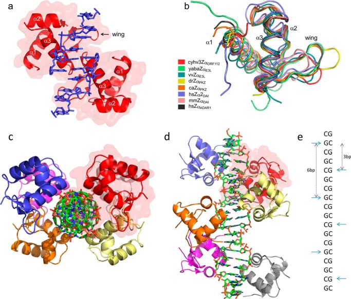
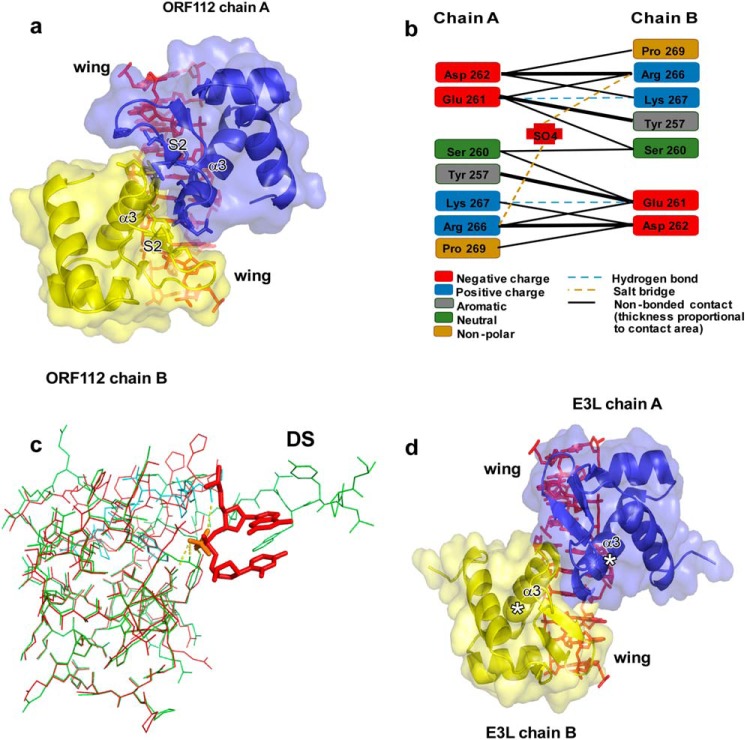
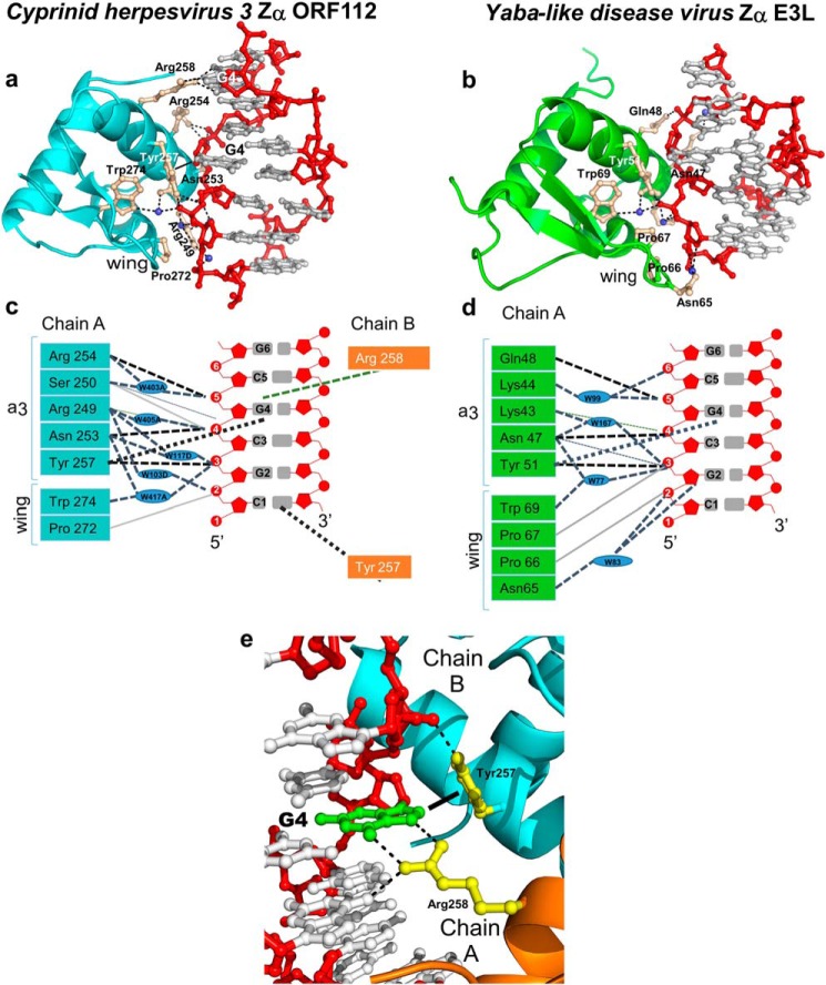
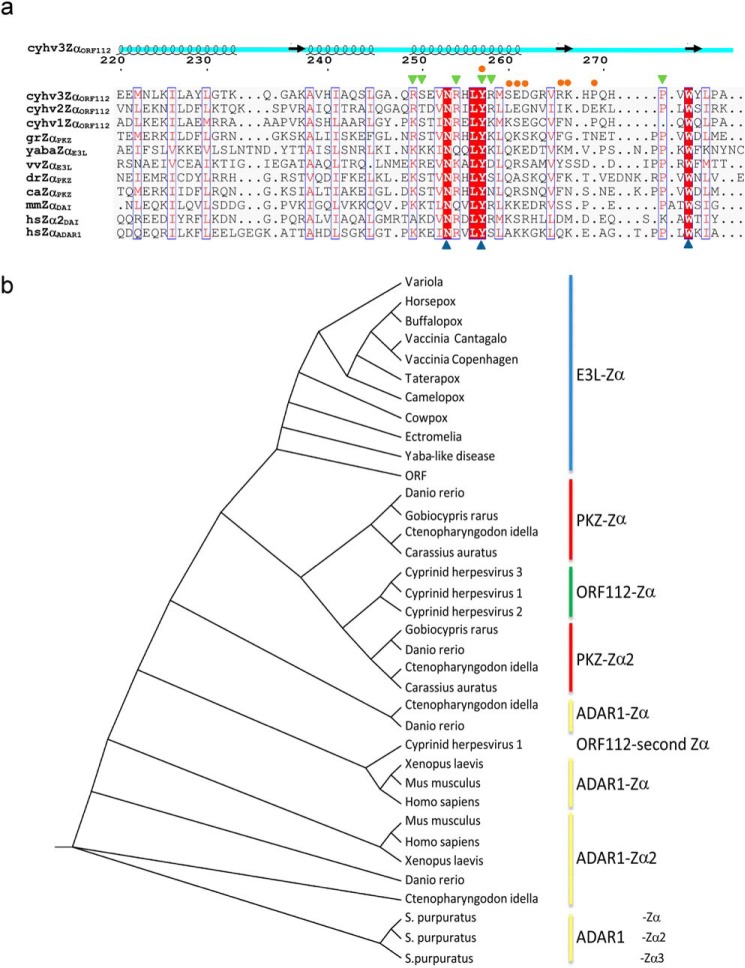
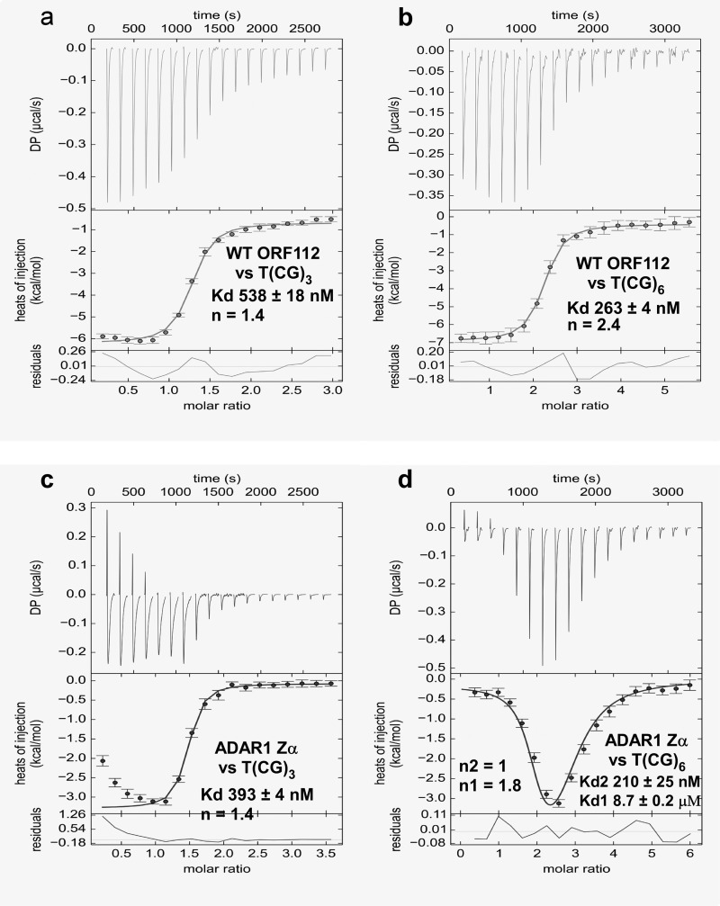
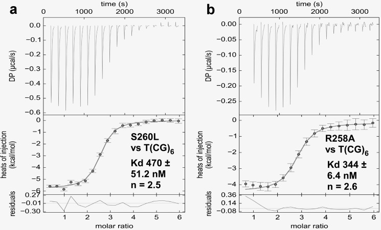
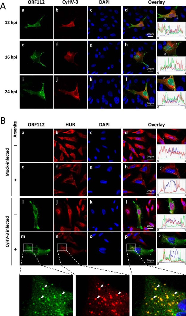
In vertebrate species, the innate immune system down-regulates protein translation in response to viral infection through the action of the double-stranded RNA (dsRNA)-activated protein kinase (PKR). In some teleost species another protein kinase, Z-DNA-dependent protein kinase (PKZ), plays a similar role but instead of dsRNA binding domains, PKZ has Zα domains. These domains recognize the left-handed conformer of dsDNA and dsRNA known as Z-DNA/Z-RNA. Cyprinid herpesvirus 3 infects common and koi carp, which have PKZ, and encodes the ORF112 protein that itself bears a Zα domain, a putative competitive inhibitor of PKZ. Here we present the crystal structure of ORF112-Zα in complex with an 18-bp CpG DNA repeat, at 1.5 Å. We demonstrate that the bound DNA is in the left-handed conformation and identify key interactions for the specificity of ORF112. Localization of ORF112 protein in stress granules induced in Cyprinid herpesvirus 3-infected fish cells suggests a functional behavior similar to that of Zα domains of the interferon-regulated, nucleic acid surveillance proteins ADAR1 and DAI.

Keywords: DNA viruses; X-ray crystallography; Zalpha domain; interferon; protein-nucleic acid interaction; stress granule.


Figures

FIGURE 1.
FIGURE 1.
Overall structure of Cyprinid herpesvirus 3 Zα domain in complex with Z-DNA and its fold similarity to other members of Zα family. a, representation of the asymmetric unit of CyHV-3 ORF112-Zα: two protein domains (red with semi-transparent surface) and two Z-DNA strands (in blue). b, structural alignments (Cα trace) of Zα family members: Cyprinid herpesvirus 3 ORF112-Zα (cyhv3ZαORF112, PDB code 4WCG), Yaba-like E3L-Zα (yabaZαE3L, PDB code 1SFU), vaccinia virus E3L-Zα (vvZαE3L, PDB code 1OYI), D. rerio PKZ-Zα (drZαPKZ, PDB code 4LB5), C. auratus PKZ-Zα (caZαPKZ, PDB code 4KMF), Homo sapiens DAI-Zα2 (hsZα2DAI, PDB code 3EYI), Mus musculus DAI-Zα (mmZαDAI, PDB code 1J75), and Homo sapiens ADAR1-Zα (hsZαADAR1, PDB code 1QBJ). c and d, representation of reconstructed biological assembly of T(CG)9 oligonucleotide. The view is perpendicular to (c) or along (d) the DNA axis. DNA duplex is represented as a stick model. Proteins are depicted as the ribbons; six monomers decorate T(CG)9 oligonucleotide. Red transparent surface marking one of the monomers serves as a reference. e, schematic representation of the arrangement of the ORF112-Zα domains along the Z-DNA helix. The CH-π interaction between the guanosines in the syn conformation and Tyr-257 is used as the reference and is indicated by horizontal cyan arrows. Vertical arrows indicate binding site spacing (number of base pairs) between adjacent monomers.
FIGURE 2.
FIGURE 2.
DNA-mediated protein-protein interaction. a, representation of the protein-protein interface. CyHV-3 ORF112-Zα domain is shown as a blue and yellow cartoon with transparent surface. A 10-bp DNA was reconstructed from the asymmetric unit and is shown as a stick model. b, schematic representation of interactions between ORF112-Zα monomers. Hydrogen bonding involves the protein backbone. c, the structure of the free ORF112-Zα (green) (PDB code 4HOB) superimposed to the DNA bound form (red) with 0.33 Å RMSD. The C-terminal part of a second monomer (cyan) that is exchanged and completes the free protein structure is also shown. For clarity, only two DNA bases and their phosphate linkage from the complex structure are shown along with the sulfate ion S302 (orange) that occupies the same position as the phosphate in the free protein. In the free protein, the sulfate ion interacts with Tyr-257, Asn-253, and Arg-249, as well as with Lys-267 and the backbone of Gln-270 of the second monomer that are being exchanged. The C-terminal part of the domain that is involved in domain swapping is indicated (DS). d, putative complex formed by Yaba-like disease virus E3L-Zα monomers (modeled based on ORF112-Zα) with long Z-DNA. No clashes between backbone atoms of E3L-Zα monomers are observed.
FIGURE 3.
FIGURE 3.
Z-DNA binding modes of viral Zα domains from ORF112 (Cyprinid herpesvirus 3) and E3L (Yaba-like disease virus). a, binding interface between CyHV-3 ORF112-Zα and a reconstructed Z-DNA 7-bp duplex. The protein is represented as a light blue cartoon and with ball and stick key residues (wheat color) involved in the interaction with Z-DNA. Phosphate backbone and sugars are colored in red, and bases are in gray. Waters are depicted as the blue spheres. b, Z-DNA recognition by Yaba-like disease virus E3L-Zα (6 bp, PDB code 1SFU). The protein is shown as green cartoon; all other depictions are represented as in a. c and d, schematic representation of the interactions between ORF112-Zα (c) and Yaba-like disease virus E3L-Zα (d) with Z-DNA. The phosphate backbone and sugars are depicted in red, and bases are in gray. Direct hydrogen bonds are represented as black dashed lines, and water-mediated bonds shown as light blue dashed lines. Nonbonded contacts are drawn as solid light gray lines. The characteristic CH-π interaction between tyrosine and guanosine in syn conformation is shown as dotted black line. Blue ovals represent water molecules. e, close view of Arg-258A and Tyr-257B interactions with G4 (green) in syn conformation. The dotted lines indicate hydrogen bonds, whereas the filled line distinguishes the CH-π-bond of Tyr-257 to the G4 ring.
FIGURE 4.
FIGURE 4.
ORF112-Zα phylogenetic analysis. a, structure-aided alignment of CyHV-3 ORF112-Zα with other Zα domains (for PDB codes see Fig. 1). Three sequences without structural information are included: Cyprinid herpesvirus 1 (cyhv1ZαORF112), Cyprinid herpesvirus 2 (cyhv2ZαORF112), and G. rarus PKZ-Zα (grZαPKZ). On the top of the alignment, the schematic representation of ORF112-Zα (cyhv3ZαORF112) secondary structure is drawn. Blue triangles below the alignment mark the triad of critical Z-DNA/Z-RNA binding residues: Asn, Tyr, and Trp. Blue boxes mark positions with conservation higher than 50%, and red shading highlights absolute conservation in this alignment. Residues involved in protein·DNA (green triangles) and protein-protein interactions (orange circles) are indicated above the alignment. b, cladogram generated based on curated (gaps removed) Muscle alignment with PhyML using Jones-Taylor-Thornton substitution model with the Shimodaira-Hasegawa approximate likelihood ratio test.
FIGURE 5.
FIGURE 5.
DNA binding affinities of ORF112 and ADAR1 Zα. a and b, isothermal titration calorimetry was used to determine the binding affinity of ORF112 Zα against a T(CG)3 (675 μm in the syringe versus 45 μm in the cell) (a) and a T(CG)6 (600 μm versus 20 μm) (b) double-stranded oligonucleotide. c and d, the same experiment but for ADAR1 Zα is shown in c (1200 μm versus 70 μm) and d (1200 μm versus 40 μm). A two-binding site model was used to fit the ADAR1 Zα against T(GC)6 data; for all other experiments, a one-site model was used.
FIGURE 6.
FIGURE 6.
DNA binding affinities of ORF112 Zα mutants. a and b, isothermal titration calorimetry of S260L (1200 μm versus 40 μm) (a) and R258A (600 μm versus 20 μm) (b) mutants against T(CG)6. The results were interpreted using a one-site model.
FIGURE 7.
FIGURE 7.
ORF112 expression and subcellular localization during CyHV-3 infection and oxidative stress. A, CCB cells were infected with CyHV-3 at the multiplicity of infection of 0.01 plaque forming units per cell. At the indicated time postinfection, cells were treated for indirect immunofluorescent labeling of ORF112 (green signal; panels a, e, and i), CyHV-3 structural proteins (red signal; panels b, f, and j), and DNA staining (panels c, g, and k). Overlays of the three staining are shown (panels d, h, and l). The right column of panels illustrates the relative quantification of the intensities of the three fluorochromes assessed along the line indicated on the magnification of the overlay. B, CCB cells were mock infected or infected with CyHV-3 at the multiplicity of infection of 0.01 plaque forming units per cell. After an incubation of 16 h, cells were mock treated or treated with sodium arsenite (1 mm). Cells were then subjected to immunofluorescent staining of CyHV-3 ORF112 (green signal; panels a, e, i, and m), carp HuR/ELAVL1 (red signal; panels b, f, j, and n), and DNA staining (panels c, g, k, and o). Overlays of the three staining are shown (panels d, h, l, and p). The right column of panels illustrates the relative quantification of the intensities of the three fluorochromes assessed along the line indicated on the magnification of the overlay. The bottom row of panels represent magnification of a defined area of panels m, n, and p. Arrows indicate co-localizing signals. hpi, hours postinfection.

References

    1. Wang A. H., Quigley G. J., Kolpak F. J., Crawford J. L., van Boom J. H., van der Marel G., and Rich A. (1979) Molecular structure of a left-handed double helical DNA fragment at atomic resolution. Nature 282, 680–686 - PubMed
    1. Rich A., and Zhang S. (2003) Timeline: Z-DNA: the long road to biological function. Nat. Rev. Genet. 4, 566–572 - PubMed
    1. Athanasiadis A. (2012) Zalpha-domains: at the intersection between RNA editing and innate immunity. Semin. Cell Dev. Biol. 23, 275–280 - PubMed
    1. Schwartz T., Rould M. A., Lowenhaupt K., Herbert A., and Rich A. (1999) Crystal structure of the Zalpha domain of the human editing enzyme ADAR1 bound to left-handed Z-DNA. Science 284, 1841–1845 - PubMed
    1. Schwartz T., Behlke J., Lowenhaupt K., Heinemann U., and Rich A. (2001) Structure of the DLM-1-Z-DNA complex reveals a conserved family of Z-DNA-binding proteins. Nat. Struct. Biol. 8, 761–765 - PubMed

Publication types

MeSH terms