

Coccidioidomycosis and the skin: a comprehensive review
- PMID: 26560205
- PMCID: PMC4631225
- DOI: 10.1590/abd1806-4841.20153805
Coccidioidomycosis and the skin: a comprehensive review
Abstract
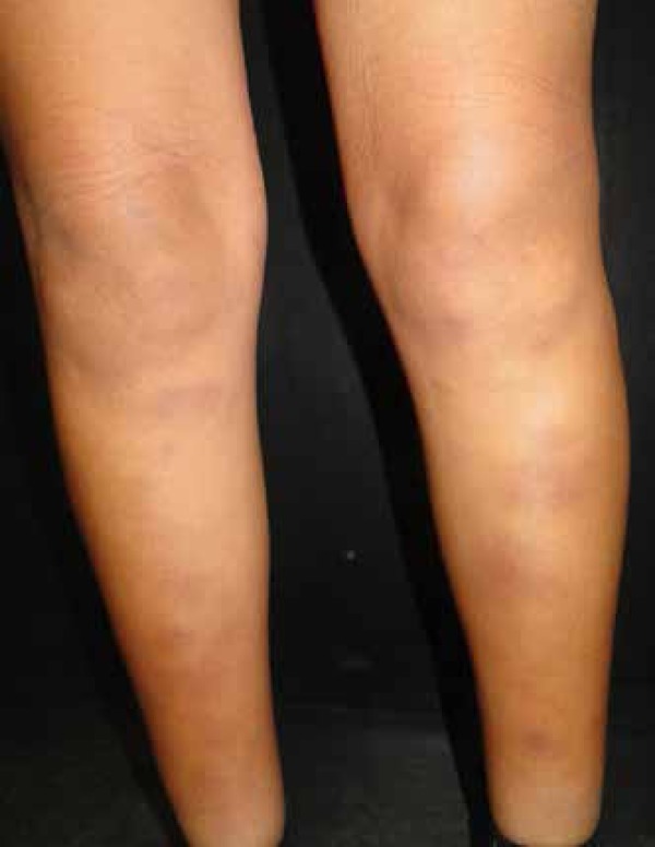
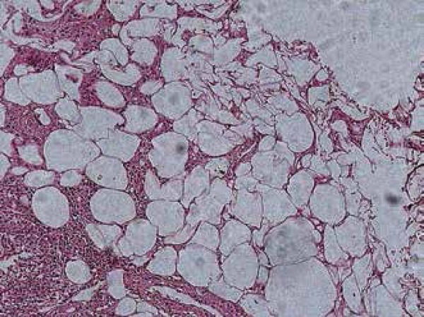
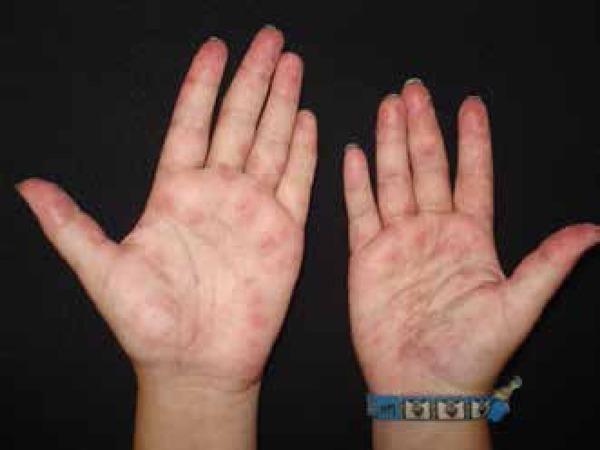
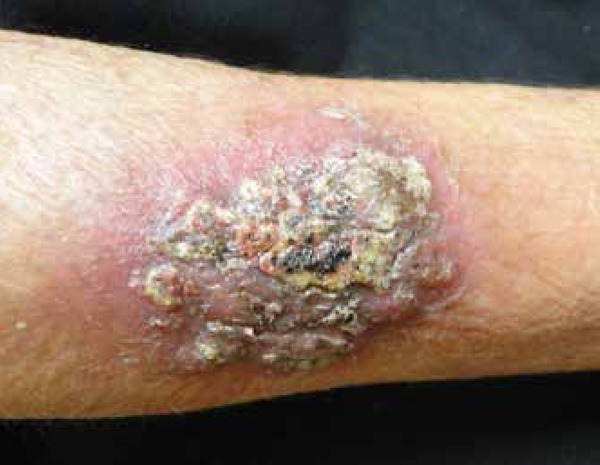
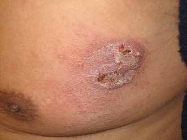
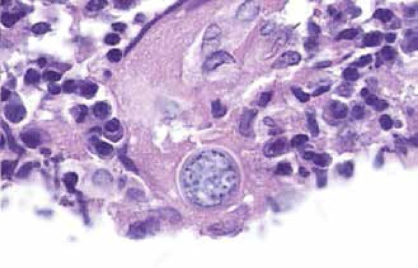
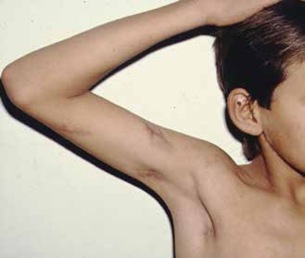
Coccidioidomycosis is a highly prevalent disease in the Western hemisphere. It is considered one of the most virulent primary fungal infections. Coccidioides species live in arid and semi-arid regions, causing mainly pulmonary infection through inhalation of arthroconidia although many other organs can be affected. Primary inoculation is rare. Since the first case of coccidioidomycosis was reported in 1892, the skin has been identified as an important target of this disease. Knowledge of cutaneous clinical forms of this infection is important and very useful for establishing prompt diagnosis and treatment. The purpose of this article is to provide a review of this infection, emphasizing its cutaneous manifestations, diagnostic methods and current treatment.
Conflict of interest statement
Conflicts of Interest: None
Figures
References
-
- Laniado-Laborín R, Alcandar-Schramm JM, Cazares-Adame R. Coccidioidomycosis: An Update. Curr Fungal Infect Rep. 2012;6:113–120.
-
- Dixon DM. Coccidioides immitis as a Select Agent of bioterrorism. J Appl Microbiol. 2001;91:602–605. - PubMed
-
- Sharma S, Thompson III GR. How I Treat Coccidioidomycosis. Curr Fungal Infect Rep. 2012;7:29–35.
-
- Welsh O, Vera-Cabrera L, Rendon A, Gonzalez G, Bonifaz A. Coccidioidomycosis. Clin Dermatol. 2012;30:573–591. - PubMed
Publication types
MeSH terms
LinkOut - more resources
Full Text Sources
Other Literature Sources
Medical