. 2015 Nov 11:10:145.
doi: 10.1186/s13023-015-0360-4.

Bruch's membrane abnormalities in PRDM5-related brittle cornea syndrome

Affiliations

Bruch's membrane abnormalities in PRDM5-related brittle cornea syndrome

Louise F Porter et al. Orphanet J Rare Dis. .

Abstract

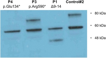
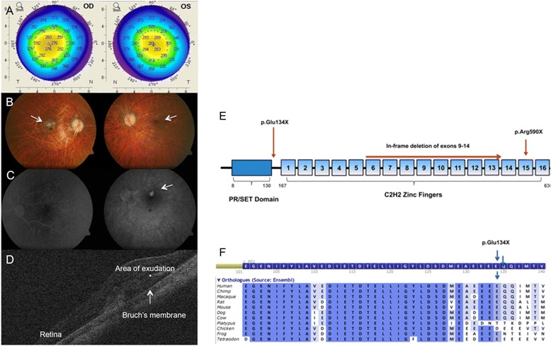
Background: Brittle cornea syndrome (BCS) is a rare, generalized connective tissue disorder associated with extreme corneal thinning and a high risk of corneal rupture. Recessive mutations in transcription factors ZNF469 and PRDM5 cause BCS. Both transcription factors are suggested to act on a common pathway regulating extracellular matrix genes, particularly fibrillar collagens. We identified bilateral myopic choroidal neovascularization as the presenting feature of BCS in a 26-year-old-woman carrying a novel PRDM5 mutation (p.Glu134*). We performed immunohistochemistry of anterior and posterior segment ocular tissues, as expression of PRDM5 in the eye has not been described, or the effects of PRDM5-associated disease on the retina, particularly the extracellular matrix composition of Bruch's membrane.

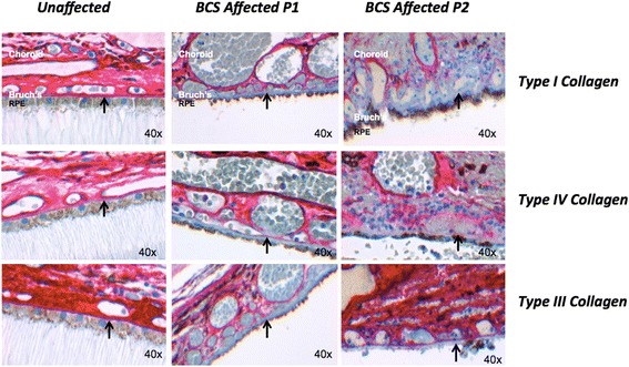
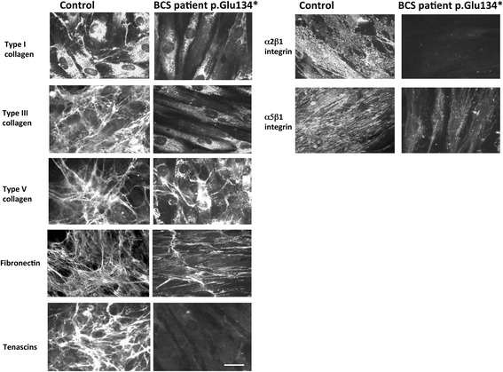
Methods: Immunohistochemistry using antibodies against PRDM5, collagens type I, III, and IV was performed on the eyes of two unaffected controls and two patients (both with Δ9-14 PRDM5). Expression of collagens, integrins, tenascin and fibronectin in skin fibroblasts of a BCS patient with a novel p.Glu134* PRDM5 mutation was assessed using immunofluorescence.

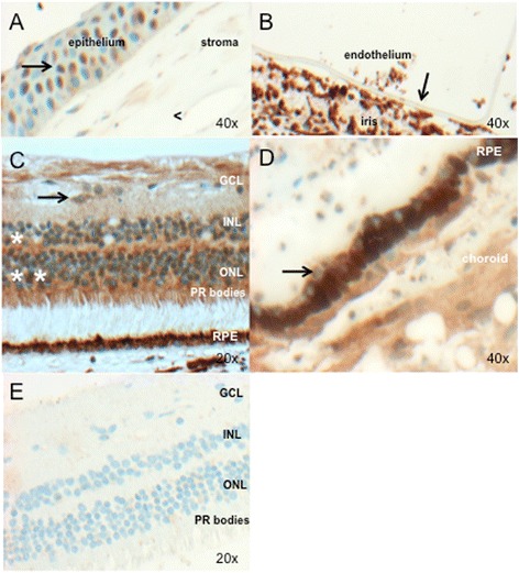
Results: PRDM5 is expressed in the corneal epithelium and retina. We observe reduced expression of major components of Bruch's membrane in the eyes of two BCS patients with a PRDM5 Δ9-14 mutation. Immunofluorescence performed on skin fibroblasts from a patient with p.Glu134* confirms the generalized nature of extracellular matrix abnormalities in BCS.

Conclusions: PDRM5-related disease is known to affect the cornea, skin and joints. Here we demonstrate, to the best of our knowledge for the first time, that PRDM5 localizes not only in the human cornea, but is also widely expressed in the retina. Our findings suggest that ECM abnormalities in PRDM5-associated disease are more widespread than previously reported.


Figures

Fig. 1
Fig. 1
Functional effects of PRDM5 mutations. Western blot using antibody PRDM5 Ab2 in skin fibroblasts from patients P1 with deletion of exons 9–14 and P3 with p.Arg590* (described by Burkitt-Wright et al. [4]), and an unaffected age-matched control (WT). The in-frame deletion of exons 9–14 (P1) produces a smaller protein of approximately 45 kDa that is consistent with the predicted mass. The p.Arg590* nonsense mutation is in the last exon of PRDM5 and results in a protein of approximately 70 kDa, consistent with the predicted mass of the PRDM5 protein resulting from the truncated transcript. The WT protein from the unaffected control gives a band of the expected mass (78 kDa). A non-specific band at 60 kDa is present in all samples. P4 protein from a patient with the presumed null mutation p.Glu134* does not produce any bands other than the non-specific band at 60 kDa
Fig. 2
Fig. 2
A case of PRDM5-associated disease presenting with myopic choroidal neovascularization. a. Corneal topography of right eye (OD) and left eye (OS) in patient P4 demonstrating marked corneal thinning, with central corneal thickness measuring 276 μm right eye and 281 μm left eye. b. Retinal photograph of RE (upper left panel) and LE (upper right panel) demonstrating evidence of disciform-like scarring in the macular region of the RE (arrow), and active choroidal neovascularization in the LE (arrow) leading to retinal exudation. Disc pallor and choroidal thinning are also present, related to the high refractive error of P4. c. Two fluorescein angiogram images of the LE taken at the time of presentation. Early phase (left panel) and late phase (right panel). An area of exudation indicative of vascular leakage into the retina is evident, suggestive of predominantly classic choroidal neovascularization. d. Optical coherence tomography scan of the LE at baseline demonstrating the presence of vascular exudation into the retina (*). e. Schematic of PRDM5 protein showing the novel mutation p.Glu134*; PRDM5 deletion exons 9–14; and p.Arg590*. f. Amino acid conservation in 11 species upstream and flanking the mutation p.Glu134*. The alignment was generated using Alamut v2.3 (Interactive Biosoftware, 2013)
Fig. 3
Fig. 3
PRDM5 expression in the adult human eye (ad) (control #2, Table 1). Image objective magnifications (OM) are shown. PRDM5 staining is brown (obtained using DAB as the chromogen). a. Nuclei of the corneal epithelium are positive for PRDM5 (arrow). The corneal stroma shows mild cytoplasmic staining, but nuclei are negative (<). b. PRDM5 is not expressed in the corneal endothelium (arrow) (in this image the endothelium has become detached from the overlying stroma). c. Positive PRDM5 nuclear and cytoplasmic staining in the inner (*) and outer nuclear layers (**), with cones staining more strongly than rods. Ganglion cell cytoplasm are also positive (arrow). Ocular tissue from patient P1 with deletion exons 9–14 is shown in (e). GCL, ganglion cell layer; INL, inner nuclear layer; ONL, outer nuclear layer; PR, photoreceptors, RPE; retinal pigment epithelium. d. Retinal pigment epithelium cell nuclei (arrow) appear negative. e. PRDM5 staining in P1 with PRDM5 deletion exons 9–14, demonstrating loss of PRDM5 nuclear and most cytoplasmic staining in the retina associated with loss of antibody epitope
Fig. 4
Fig. 4
Changes in extracellular matrix collagens in Bruch’s membrane in PRDM5-associated disease. Immunohistochemistry performed for collagens I, III and IV (red stain, obtained using the XT ultraView Universal Red Alkaline Phosphatase detection system) in retinas of a control individual, and BCS patients P1 and P2 (OM 40×). Absence of staining for collagens I, III and IV in Bruch’s membrane (arrow) is present in sample P1, and absent staining for collagen I with reduced staining for collagens III and IV in P2. Images were recorded and processed identically to allow direct comparisons to be made between them
Fig. 5
Fig. 5
Expression of ECM components in dermal fibroblasts expressing or lacking PRDM5. Indirect immunofluorescence microscopy performed on dermal fibroblasts from a control (#2) and BCS patient P4 carrying the PRDM5 p.Glu134* mutation for collagens I, III and V, fibronectin, tenascins and integrin receptors α2β1 and α5β1. In control cells, collagen I is primarily expressed in the cytoplasm, with only limited expression extracellularly. Collagen I labelling is substantially reduced in PRDM5 mutant cells. Collagen III appears well organized in the ECM of control cells but was absent in the mutant fibroblasts where only diffuse cytoplasmic staining was visible. Furthermore, disarray of collagen V was evident with cytoplasmic accumulation and reduced extracellular matrix in the PRDM5 mutant cells. Expression of the collagen integrin receptor α2β1 was essentially abolished, with marked reduction in the fibronectin integrin receptor α5β1 and disorganization of fibronectin matrix, in PRDM5 mutant cells compared to control cells. Tenascins were organized in an abundant extracellular matrix in control cells whereas they were not detectable in the PRDM5 mutant cells. 1 cm on the image scale corresponds to 16 μm. Images were all recorded under identical parameters to allow for direct comparison

References

    1. Stein R, Lazar M, Adam A. Brittle cornea. A familial trait associated with blue sclera. Am J Ophthalmol. 1968;66:67–69. doi: 10.1016/0002-9394(68)91789-3. - DOI - PubMed
    1. Burkitt Wright EM, Porter LF, Spencer HL, Clayton-Smith J, Au L, Munier FL, et al. Brittle cornea syndrome: recognition, molecular diagnosis and management. Orphanet J Rare Dis. 2013;8:68. doi: 10.1186/1750-1172-8-68. - DOI - PMC - PubMed
    1. Abu A, Frydman M, Marek D, Pras E, Nir U, Reznik-Wolf H, et al. Deleterious mutations in the Zinc-Finger 469 gene cause brittle cornea syndrome. Am J Hum Genet. 2008;82(5):1217–22. doi: 10.1016/j.ajhg.2008.04.001. - DOI - PMC - PubMed
    1. Burkitt Wright EM, Spencer HL, Daly SB, Manson FD, Zeef LA, Urquhart J, et al. Mutations in PRDM5 in brittle cornea syndrome identify a pathway regulating extracellular matrix development and maintenance. Am J Hum Genet. 2011;88:767–77. doi: 10.1016/j.ajhg.2011.05.007. - DOI - PMC - PubMed
    1. Rohrbach M, Spencer H, Porter LF, Burkitt-Wright EM, Bürer C, Janecke A, et al. ZNF469, frequently mutated in the Brittle Cornea Syndrome (BCS), is a single exon gene possibly regulating the expression of several extracellular matrix components. Mol Genet Metab. 2013;109(3):289–95. doi: 10.1016/j.ymgme.2013.04.014. - DOI - PMC - PubMed

Publication types

MeSH terms

Supplementary concepts

LinkOut - more resources