. 2015 Nov 12;11(11):e1005263.
doi: 10.1371/journal.ppat.1005263. eCollection 2015.

Interferon-γ Inhibits Ebola Virus Infection

Affiliations

Interferon-γ Inhibits Ebola Virus Infection

Bethany A Rhein et al. PLoS Pathog. .

Abstract

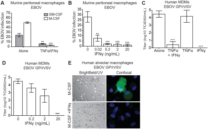
Ebola virus outbreaks, such as the 2014 Makona epidemic in West Africa, are episodic and deadly. Filovirus antivirals are currently not clinically available. Our findings suggest interferon gamma, an FDA-approved drug, may serve as a novel and effective prophylactic or treatment option. Using mouse-adapted Ebola virus, we found that murine interferon gamma administered 24 hours before or after infection robustly protects lethally-challenged mice and reduces morbidity and serum viral titers. Furthermore, we demonstrated that interferon gamma profoundly inhibits Ebola virus infection of macrophages, an early cellular target of infection. As early as six hours following in vitro infection, Ebola virus RNA levels in interferon gamma-treated macrophages were lower than in infected, untreated cells. Addition of the protein synthesis inhibitor, cycloheximide, to interferon gamma-treated macrophages did not further reduce viral RNA levels, suggesting that interferon gamma blocks life cycle events that require protein synthesis such as virus replication. Microarray studies with interferon gamma-treated human macrophages identified more than 160 interferon-stimulated genes. Ectopic expression of a select group of these genes inhibited Ebola virus infection. These studies provide new potential avenues for antiviral targeting as these genes that have not previously appreciated to inhibit negative strand RNA viruses and specifically Ebola virus infection. As treatment of interferon gamma robustly protects mice from lethal Ebola virus infection, we propose that interferon gamma should be further evaluated for its efficacy as a prophylactic and/or therapeutic strategy against filoviruses. Use of this FDA-approved drug could rapidly be deployed during future outbreaks.


Conflict of interest statement

The authors have declared that no competing interests exist.

Figures

Fig 1
Fig 1. IFNγ-treated macrophages are resistant to EBOV infection.
(A) IFNγ/TNFα treatment inhibits EBOV infection of BALB/c peritoneal macrophages. Cultures maintained in media containing GM-CSF or M-CSF were treated with murine IFNγ/TNFα 24 hours prior to EBOV-GFP infection (MOI = 0.05 in Vero E6 cells). Shown are the percent GFP positive cells at 24 hours following infection. (B) IFNγ inhibits EBOV in a dose-dependent manner. BALB/c peritoneal macrophages were infected with EBOV-GFP (MOI = 0.05) 24 hours following IFNγ treatment. (C) IFNγ, but not TNFα, treatment of M-CSF differentiated hMDM cultures inhibits EBOV GP/rVSV infection. Virus titers were determined by end-point dilution on hMDMs from 4 independent donors. (D) Dose response curve of IFNγ inhibition of EBOV GP/rVSV infection of M-CSF-treated hMDMs. (E) IFNγ inhibits EBOV GP/rVSV infection in M-CSF cultured human alveolar macrophages. Left, bright field/UV (magnification: 10X). Right, confocal images (magnification: 100X). All infections were assessed 24 hours following addition of virus. Data are shown as means ± s.e.m. Significance was determined by Student’s t-test compared to no IFNγ control, **p < 0.01, ***p < 0.001.
Fig 2
Fig 2. IFNγ blocks EBOV GP/rVSV and EBOV RNA synthesis.
(A) M-CSF cultured peritoneal macrophages pre-treated for 24 hours with IFNγ have decreased EBOV GP/rVSV RNA synthesis at 6 and 24 hours following infection. Total RNA from IFNγ-treated or untreated peritoneal macrophages at indicated time points was assessed by qRT-PCR for VSV matrix (M) and polymerase (L) RNA. Log2 value of 2 hour M-CSF alone = 1 and data is shown as Log10 of the fold change. Significance was determined by one sample t-test compared to 2 hour M-CSF only control. *p < 0.05, **p < 0.01, ***p < 0.001. (B) IFNγ treatment decreases EBOV RNA levels to the same extent as the protein synthesis inhibitor cycloheximide. Total RNA isolated at 6 or 14 hours following EBOV infection of M-CSF cultured peritoneal macrophages treated with or without IFNγ and/or CHX was quantified by qRT-PCR for EBOV nucleoprotein (NP) or polymerase (L) RNA. Data is shown as log2 values. Significance was determined by ANOVA with a Tukey post-test. *p < 0.05, **p < 0.01 (compared to 6 hours M-CSF alone). ##p < 0.01 (compared to 14 hours M-CSF alone). ns, not significantly different.
Fig 3
Fig 3. Human MDM gene expression is altered by 24 hours of IFNγ treatment.
M-CSF cultured hMDMs were incubated with or without IFNγ for 24 hours and RNA was harvested for gene arrays. (A) Heat map summaries of hMDM gene expression significantly altered by IFNγ. Human MDMs were obtained from four different volunteers and treated with M-CSF or M-CSF plus IFNγ. Genes were clustered into annotated ontology groups and listed in the corresponding GO groups table with their respective GO terms. Significance was determined by paired t-test analysis with cutoff values of at least two-fold change and p < 0.01. NUSE analysis of the array demonstrated that the means were centered at a value of 1 and minimum and maximum values between 0.95 and 1.05. ISGs that were assessed further in this study are bolded in the GO table. (B) Venn diagram representing the statistically significant individual and shared genes altered by IFNγ in hMDMs and human alveolar macrophages.
Fig 4
Fig 4. Identification of IFNγ-stimulated genes that inhibit EBOV infection.
(A) mRNA validation of hMDM profiling results for several of the top IFNγ-stimulated genes identified in our gene arrays. RNA obtained for the microarray analysis was assessed for mRNA levels of the selected genes by qRT-PCR. Results are represented as the log2 values. (B) Identification of IFNγ-stimulated genes that inhibit EBOV GP/rVSV infection. Highly permissive HEK 293T cells stably expressing TIM -1 (H3 cells) were infected with EBOV GP/rVSV 48 hours following transfection of 2 μg of ISG-RFP lentiviral constructs. Cells gated for RFP expression were assessed for EBOV GP/rVSV infection by detection of GFP. Shown is infection of EBOV GP/rVSV in ISG expressing cells relative to infection of cells transfected with a fluc-RFP expressing lentivirus (control). (C) Novel ISGs that inhibit EBOV GP/rVSV also block EBOV infection. HeLa cells were infected with EBOV 48 hours following electroporation of 5 μg of ISG-RFP lentiviral constructs. Infection was assessed by microscopy 24 hours later and percent of cells that were GFP positive were calculated by CellProfiler image analysis software. A lentiviral construct expressing IRF1 served as a positive control in these studies. (D) IRF1 knock down increases EBOV GP/rVSV infection following IFNγ stimulation. IRF1 or scrambled (Scr) siRNA were loaded into HEK 293T derived exosomes. SiRNA loaded exosomes (2.5 μg) were delivered and IFNγ added to BALB/c IFNAR-/- peritoneal macrophages 24 hours prior to EBOV GP/rVSV infection (MOI = 0.1). Twenty-four hours following infection, total RNA was isolated from the macrophages. Amount of IRF1 expression and infection (by detection of VSV polymerase (L)) was quantified by qRT-PCR. Results represent the means ± s.e.m. Significance was determined by Student’s t-test analysis, *p < 0.05, **p < 0.01, ***p < 0.001.
Fig 5
Fig 5. IFNγ reduces EBOV-GP/rVSV morbidity and mortality.
(A) IFNγ enhances survival of EBOV GP/rVSV infected mice. IFNγ (10 μg) or PBS was administered by i.p. injection to BALB/c IFNAR-/- mice 24 hours prior to or 2, 6, 12 or 48 hours following EBOV GP/rVSV infection (n≥8/treatment). (B) IFNγ treatment as a 24 hour pre-treatment or a 2 hour post-treatment reduces serum viremia and organ titers of EBOV GP/rVSV-infected mice. Sera and organs were harvested at 48 hours following infection (n≥ 4/treatment) in mice treated with 3.3μg of IFNγ. Viremia and organ virus titers were determined by endpoint dilution of serum or homogenized organ samples on Vero cells. Significance was calculated by Mann-Whitney test compared to PBS control, *p < 0.05, **p < 0.01. ns, not significant. (C) Intraperitoneal IFNγ treatment of mice significantly inhibits EBOV GP/rVSV infection of peritoneal cells. Peritoneal cells were isolated from EBOV GP/rVSV infected mice treated with 3.3μg of IFNγ at times noted prior to or following challenge. Amount of VSV-L RNA was determined by qRT-PCR. Significance was determined by ANOVA with a Tukey post-test, ***p < 0.001. (D) Intramuscular administration of IFNγ increases survival of IFNAR-/- mice. PBS or IFNγ at the indicated concentration was administered by i.m. injection 24 hours prior to i.p. injection of EBOV GP/rVSV. For A & D, significance was determined by Mantel-Cox Test, **p < 0.01, ***p < 0.001.
Fig 6
Fig 6. IFNγ protects mice from EBOV morbidity and mortality.
(A) IFNγ protects mice from lethal MA-EBOV infection. IFNγ (10 μg) or PBS was administered by i.p. injection to BALB/c mice 24 hours prior to, at the time of infection, 6 or 24 hours following infection (n ≥ 7/treatment). Significance was determined by Mantel-Cox Test, **p < 0.01, ***p < 0.001. ns, not significantly different. (B) IFNγ treatment reduces MA-EBOV morbidity. Results represent mean clinical sickness scores ± s.e.m (n ≥ 7/treatment). Significance was determined by Student’s t-test compared to PBS control. (C) IFNγ treatment significantly reduces MA-EBOV viremia in mice treated 24 hours following infection. Serum was collected 4 days following infection (n = 3–6 mice/treatment) and serial dilution of sera on Vero-E6 cells to assess plaques formed determined viremia titers. Results represent means ± s.e.m. Significance was determined by Mann-Whitney test compared to PBS control, * p < 0.05.

Similar articles

Cited by

References

    1. Leroy EM, Gonzalez JP, Baize S. Ebola and Marburg haemorrhagic fever viruses: major scientific advances, but a relatively minor public health threat for Africa. Clinical microbiology and infection: the official publication of the European Society of Clinical Microbiology and Infectious Diseases. 2011;17(7):964–76. . - PubMed
    1. Gire SK, Goba A, Andersen KG, Sealfon RS, Park DJ, Kanneh L, et al. Genomic surveillance elucidates Ebola virus origin and transmission during the 2014 outbreak. Science (New York, NY). 2014;345(6202):1369–72. 10.1126/science.1259657 . - DOI - PMC - PubMed
    1. Geisbert TW, Young HA, Jahrling PB, Davis KJ, Larsen T, Kagan E, et al. Pathogenesis of Ebola hemorrhagic fever in primate models: evidence that hemorrhage is not a direct effect of virus-induced cytolysis of endothelial cells. The American journal of pathology. 2003;163(6):2371–82. Epub 2003/11/25. - PMC - PubMed
    1. Bray M, Geisbert TW. Ebola virus: the role of macrophages and dendritic cells in the pathogenesis of Ebola hemorrhagic fever. The international journal of biochemistry & cell biology. 2005;37(8):1560–6. Epub 2005/05/18. 10.1016/j.biocel.2005.02.018 . - DOI - PubMed
    1. Gupta M, Mahanty S, Ahmed R, Rollin PE. Monocyte-derived human macrophages and peripheral blood mononuclear cells infected with ebola virus secrete MIP-1alpha and TNF-alpha and inhibit poly-IC-induced IFN-alpha in vitro. Virology. 2001;284(1):20–5. . - PubMed

Publication types