Quantitative Assessment of the Heterogeneity of PD-L1 Expression in Non-Small-Cell Lung Cancer
- PMID: 26562159
- PMCID: PMC4941982
- DOI: 10.1001/jamaoncol.2015.3638
Quantitative Assessment of the Heterogeneity of PD-L1 Expression in Non-Small-Cell Lung Cancer
Erratum in
-
Error in Figure Presentation.JAMA Oncol. 2016 Jan;2(1):146. doi: 10.1001/jamaoncol.2015.5816. JAMA Oncol. 2016. PMID: 26767561 No abstract available.
Abstract
Importance: Early-phase trials with monoclonal antibodies targeting PD-1 (programmed cell death protein 1) and PD-L1 (programmed cell death 1 ligand 1) have demonstrated durable clinical responses in patients with non-small-cell lung cancer (NSCLC). However, current assays for the prognostic and/or predictive role of tumor PD-L1 expression are not standardized with respect to either quantity or distribution of expression.
Objective: To demonstrate PD-L1 protein distribution in NSCLC tumors using both conventional immunohistochemistry (IHC) and quantitative immunofluorescence (QIF) and compare results obtained using 2 different PD-L1 antibodies.
Design, setting, and participants: PD-L1 was measured using E1L3N and SP142, 2 rabbit monoclonal antibodies, in 49 NSCLC whole-tissue sections and a corresponding tissue microarray with the same 49 cases. Non-small-cell lung cancer biopsy specimens from 2011 to 2012 were collected retrospectively from the Yale Thoracic Oncology Program Tissue Bank. Human melanoma Mel 624 cells stably transfected with PD-L1 as well as Mel 624 parental cells, and human term placenta whole tissue sections were used as controls and for antibody validation. PD-L1 protein expression in tumor and stroma was assessed using chromogenic IHC and the AQUA (Automated Quantitative Analysis) method of QIF. Tumor-infiltrating lymphocytes (TILs) were scored in hematoxylin-eosin slides using current consensus guidelines. The association between PD-L1 protein expression, TILs, and clinicopathological features were determined.
Main outcomes and measures: PD-L1 expression discordance or heterogeneity using the diaminobenzidine chromogen and QIF was the main outcome measure selected prior to performing the study.
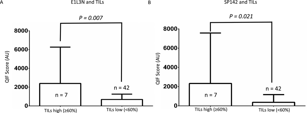
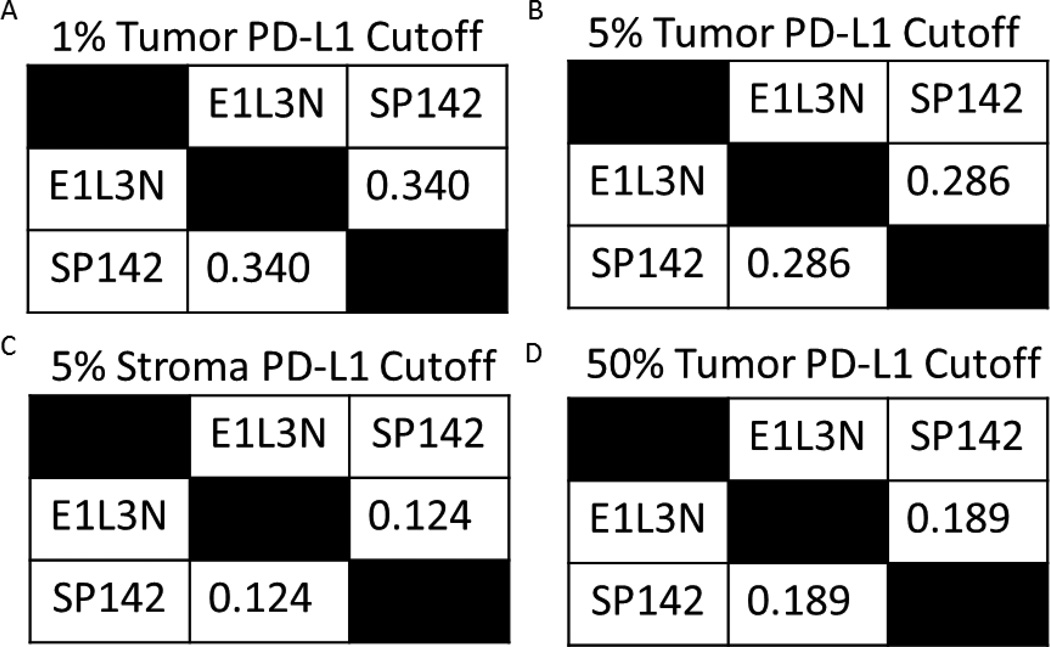
Results: Using chromogenic IHC, both antibodies showed fair to poor concordance. The PD-L1 antibodies showed poor concordance (Cohen κ range, 0.124-0.340) using conventional chromogenic IHC and showed intra-assay heterogeneity (E1L3N coefficient of variation [CV], 6.75%-75.24%; SP142 CV, 12.17%-109.61%) and significant interassay discordance using QIF (26.6%). Quantitative immunofluorescence showed that PD-L1 expression using both PD-L1 antibodies was heterogeneous. Using QIF, the scores obtained with E1L3N and SP142 for each tumor were significantly different according to nonparametric paired test (P < .001). Assessment of 588 serial section fields of view from whole tissue showed discordant expression at a frequency of 25%. Expression of PD-L1 was correlated with high TILs using both E1L3N (P = .007) and SP142 (P = .02).
Conclusions and relevance: Objective determination of PD-L1 protein levels in NSCLC reveals heterogeneity within tumors and prominent interassay variability or discordance. This could be due to different antibody affinities, limited specificity, or distinct target epitopes. Efforts to determine the clinical value of these observations are under way.
Conflict of interest statement
The other authors have no conflicts of interest to disclose.
Figures


Comment in
-
PD-L1 Expression as a Predictive Biomarker: Is Absence of Proof the Same as Proof of Absence?JAMA Oncol. 2016 Jan;2(1):54-5. doi: 10.1001/jamaoncol.2015.3782. JAMA Oncol. 2016. PMID: 26561922 No abstract available.
References
Publication types
MeSH terms
Substances
Grants and funding
LinkOut - more resources
Full Text Sources
Other Literature Sources
Medical
Research Materials