. 2015 Nov 12;11(11):e1005250.
doi: 10.1371/journal.ppat.1005250. eCollection 2015.

Cryptosporidium Lactate Dehydrogenase Is Associated with the Parasitophorous Vacuole Membrane and Is a Potential Target for Developing Therapeutics

Affiliations

Cryptosporidium Lactate Dehydrogenase Is Associated with the Parasitophorous Vacuole Membrane and Is a Potential Target for Developing Therapeutics

Haili Zhang et al. PLoS Pathog. .

Abstract

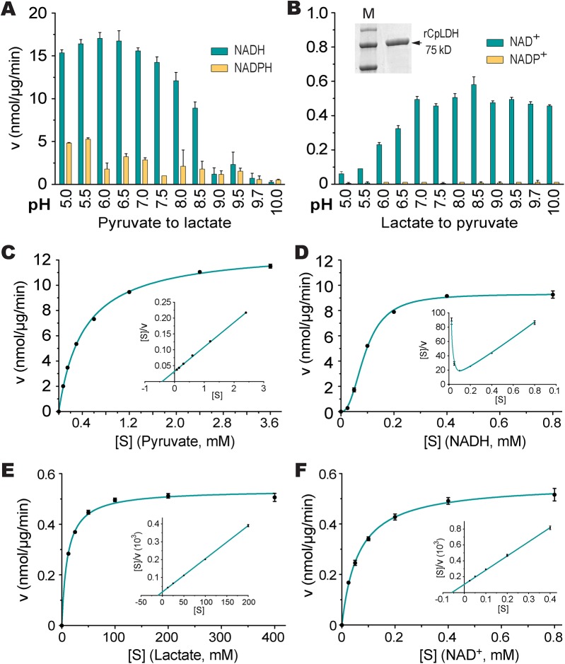
The apicomplexan, Cryptosporidium parvum, possesses a bacterial-type lactate dehydrogenase (CpLDH). This is considered to be an essential enzyme, as this parasite lacks the Krebs cycle and cytochrome-based respiration, and mainly-if not solely, relies on glycolysis to produce ATP. Here, we provide evidence that in extracellular parasites (e.g., sporozoites and merozoites), CpLDH is localized in the cytosol. However, it becomes associated with the parasitophorous vacuole membrane (PVM) during the intracellular developmental stages, suggesting involvement of the PVM in parasite energy metabolism. We characterized the biochemical features of CpLDH and observed that, at lower micromolar levels, the LDH inhibitors gossypol and FX11 could inhibit both CpLDH activity (Ki = 14.8 μM and 55.6 μM, respectively), as well as parasite growth in vitro (IC50 = 11.8 μM and 39.5 μM, respectively). These observations not only reveal a new function for the poorly understood PVM structure in hosting the intracellular development of C. parvum, but also suggest LDH as a potential target for developing therapeutics against this opportunistic pathogen, for which fully effective treatments are not yet available.


Conflict of interest statement

The authors have declared that no competing interests exist.

Figures

Fig 1
Fig 1. Lactate produced by C. parvum oocysts and free sporozoites.
Oocysts were removed from refrigeration (4°C) and incubated at 37°C for 1 and 4 h, respectively. Sporozoites (spz) were prepared by excystation as described, and then incubated at 37°C for 1 and 4 h, respectively. Lactate levels released from 107 oocysts or 4×10⁷ sporozoites are expressed in nanomolar amounts; means ± SD (n = 3) from one representative of three independent experiments.
Fig 2
Fig 2. Western blot detection of CpLDH.
The assays were performed using rabbit and rat polyclonal antibodies raised against a synthetic peptide or the recombinant CpLDH protein, respectively. Samples include recombinant maltose-binding protein (MBP), cleaved MBP-CpLDH fusion protein (MBP+CpLDH), and protein extracts from C. parvum-free sporozoites and HCT-8 cells infected with C. parvum for 18 h. Uninfected cells cultured in parallel were used as control. No immunoreactive bands are observed in samples probed with pre-immune sera. All antibodies, as well as pre-immune sera, were affinity purified as described in the Materials and Methods.
Fig 3
Fig 3. Immunofluorescence microscopic detection of CpLDH in different C. parvum life cycle stages.
(A) Cytosolic distribution CpLDH in the extracellular parasite stages, including oocysts, sporozoites (Sp), and merozoites (Mz), using rabbit anti-CpLDH antibody. (B) Association of CpLDH with the parasitophorous vacuole membrane (PVM) during the parasite intracellular developmental stages, including early and mature meronts, using rabbit anti-CpLDH antibody. (C) Confirmation of the specificity of CpLDH detection in the PVM using primary antibodies that were untreated (non-presoaked) or presoaked with either the maltose-binding protein (MBP), recombinant CpLDH (rCpLDH), or synthetic peptide antigen (Ag). Fluorescence signals were eliminated by presoaking antibody with rCpLDH and synthetic antigen (right panel), but not with MBP (lower left panel). Insets on the right panel showed over-exposed images (red channel). (D) Detection of CpLDH protein in intracellular parasites using rat anti-CpLDH antibodies that were untreated or presoaked with either MBP or recombinant CpLDH (rCpLDH). All antibodies, including pre-immune sera, were affinity-purified as described in the Materials and Methods. No fluorescence signals were observed using rabbit and rat pre-immune sera (S1 Fig and S2 Fig). DIC, differential interference microscopy; CpLDH+DAPI, superimposed images of CpLDH labeled with TRITC and nuclei counterstained with DAPI. Bar = 5 μm.
Fig 4
Fig 4. Subcellular distribution of CpLDH protein in intracellular C. parvum by immunoelectron microscopy (IEM).
(A) Electron micrographs showing the distribution of CpLDH in intracellular parasites. Affinity-purified rabbit and rat antibodies (Ab) were used as primary antibodies. Colloidal gold beads were mainly distributed along the inner side of the parasitophorous vacuole membrane (PVM), although some gold beads were observable in intracellular parasites (Cp). Minimal numbers of particles were present in the host cell (HC) and parasitophorous vacuolar space (PVS). Arrowheads indicate example gold beads. Regions from the lower panel are enlarged in the upper panels, as indicated by arrows. (B) Bar chart showing the distribution of gold particles distributed in four types of cellular structures. Gold particles were manually counted, converted to the number of particles/μm2 and expressed as the percentage of total in each dataset. Means ± SD (n = 3) were calculated from three IEM images for each dataset. Statistical significance was assessed by two-tailed Student t-test.
Fig 5
Fig 5. CpLDH enzyme kinetics.
(A, B) Effects of pH value on the activity of CpLDH in the forward and reverse reactions. CpLDH prefers using NADH/NAD+ to convert pyruvate to lactate at conditions near neutral pH. Inset shows SDS-PAGE gel image of recombinant CpLDH (rCpLDH) used in these assays. (C—F) Activities of CpLDH on substrates, including pyruvate and lactate, and utilizing the cofactors NADH and NAD+. Insets show the Hanes-Woolf plots of the same datasets. Kinetic parameters are summarized in Table 1. S, substrate as indicated; v, velocity of the reaction. Means ± SD (n≥3) from one representative of at least three independent experiments.
Fig 6
Fig 6. Inhibition constants of gossypol and FX11 against CpLDH.
Michaelis-Menten curves (A, C) and corresponding Lineweaver-Burk plots (B, D) to determine the inhibition constants of gossypol (A, B) and FX11 (C, D) on CpLDH in respect to NADH. The reactions were conducted in 50 mM Tris-HCl buffer (pH 7.0), 2.4 mM pyruvate and varying concentrations of NADH. Separate experiments were performed using fixed amounts of pyruvate (2.4 mM) and NADH (250 μM) in the presence of various concentrations of gossypol and FX11 for determining the IC50 values (S3 Fig). Means ± SD (n ≥3) were calculated from one representative of at least three independent experiments.
Fig 7
Fig 7. Efficacies of gossypol and FX11 on the growth of C. parvum in HCT-8 cells.
(A, B) Inhibition of C. parvum growth in vitro by gossypol and FX11 in a standard 44-h infection assay, in which intracellular parasites were treated for 3–44 h post-infection (hpi). Paromomycin at 140 μM was used as positive control. (C) Effects of gossypol (16 μM) and FX11 (60 μM) on the first and second asexual developmental cycles (merogony). Cultured parasites were treated with specified inhibitors for 3–20 h post-infection (hpi) and for 20–44 hpi, respectively, and parasite growth was evaluated at the end of each treatment. (D) Effects of pretreatment of free sporozoites (spz) and type I merozoites (mrz) with gossypol on their attachment and invasions into host cells (40 min pretreatment + 3 hpi assay). (E) Effect of pretreatment of host cells by gossypol on parasite infection, in which HCT-8 cells were pretreated with gossypol (16 μM, 24 h) followed by 3 h parasite infection in the absence of inhibitor. Anti-cryptosporidial activities were determined by a qRT-PCR-based assay. Means ± SD (n ≥3) from one representative of at least three independent experiments. An asterisk indicates statistically significant difference between the sample and corresponding control by two-tailed Student t-test (p<0.05).
Fig 8
Fig 8. Effects of LDH inhibitors on the growth of C. parvum in primary enterocytes.
Parasites were cultured in FHs 74 Int primary enterocytes for 44 h, and anti-cryptosporidial activities of gossypol (10 μM) and FX11 (60 μM) were determined by a qRT-PCR-based assay.
Fig 9
Fig 9. Relative mRNA levels of two human LDH isoforms in host cells.
Total RNA was isolated from HCT-8 cells to determine the levels of mRNA of HsLDH-A and HsLDH-B and those of 18S rRNA by qRT-PCR. The levels of HsLDH isoforms were expressed in relative to the host cell 18S rRNA.

References

    1. Checkley W, White AC Jr., Jaganath D, Arrowood MJ, Chalmers RM, Chen XM, et al. A review of the global burden, novel diagnostics, therapeutics, and vaccine targets for cryptosporidium. Lancet Infect Dis. 2015;15(1):85–94. Epub 2014/10/04. 10.1016/s1473-3099(14)70772-8 - DOI - PMC - PubMed
    1. Chen XM, Keithly JS, Paya CV, LaRusso NF. Cryptosporidiosis. New England J Med. 2002;346(22):1723–31. - PubMed
    1. Carpenter C, Fayer R, Trout J, Beach MJ. Chlorine disinfection of recreational water for Cryptosporidium parvum . Emerg Infect Dis. 1999;5(4):579–84. Epub 1999/08/25. 10.3201/eid0504.990425 - DOI - PMC - PubMed
    1. Peeters JE, Mazas EA, Masschelein WJ, Villacorta Martiez de Maturana I, Debacker E. Effect of disinfection of drinking water with ozone or chlorine dioxide on survival of Cryptosporidium parvum oocysts. Appl Environ Microbiol. 1989;55(6):1519–22. Epub 1989/06/01. - PMC - PubMed
    1. Kelly P. Treatment and prevention of cryptosporidiosis: what options are there for a country like Zambia? Parasitology. 2011;138(12):1488–91. 10.1017/S0031182011000035 . - DOI - PubMed

Publication types

MeSH terms

Substances