Review
. 2016 Feb;30(2):179-85.
doi: 10.1038/eye.2015.236. Epub 2015 Nov 13.

Why rods and cones?

Affiliations
Review

Why rods and cones?

T D Lamb. Eye (Lond). 2016 Feb.

Abstract

Under twenty-first-century metropolitan conditions, almost all of our vision is mediated by cones and the photopic system, yet cones make up barely 5% of our retinal photoreceptors. This paper looks at reasons why we additionally possess rods and a scotopic system, and asks why rods comprise 95% of our retinal photoreceptors. It considers the ability of rods to reliably signal the arrival of individual photons of light, as well as the ability of the retina to process these single-photon signals, and it discusses the advantages that accrue. Drawbacks in the arrangement, including the very slow dark adaptation of scotopic vision, are also considered. Finally, the timing of the evolution of cone and rod photoreceptors, the retina, and the camera-style eye is summarised.


Figures
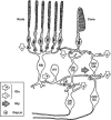
Figure 1
Figure 1
Simplified schematic of cone and rod pathways through the retina. Right-hand side shows the cone pathway and left-hand side shows the main (scotopic) rod pathway that provides input to the cone pathway. Chemical synapses are shown as arrowheads, with white fill denoting glutamate (Glu) synapses and grey fill denoting glycine (Gly) synapses; sign-conserving and sign-inverting synapses are indicated as ‘+' and ‘−' respectively; gap junctions are indicated as ‘⊕'. Cone ON pathway comprises cone photoreceptor to ON cone bipolar cell (ON CB) to ON ganglion cell. Cone OFF pathway comprises cone photoreceptor to OFF cone bipolar cell (OFF CB) to OFF ganglion cell. The scotopic pathway begins as: rod photoreceptor to rod bipolar cell (RB) to AII amacrine cell. The AII amacrine provides sign-conserving input via connexin-36 gap junctions onto ON cone bipolar cell terminals, as well as sign-inverting glycinergic input onto OFF cone bipolar cell terminals, thereby providing push–pull signals to ON and OFF ganglion cells. The sign-inverting glutamate synapses (from cones to cone ON bipolar cells, and from rods to rod bipolar cells) use a metabotropic postsynaptic mechanism involving a G-protein cascade, whereas the other chemical synapses use ionotropic mechanisms. Light hyperpolarises the photoreceptors, so that the sign-inverting synapse generates a depolarising light response in the ON cone bipolar cell and rod bipolar cell. Not shown in this diagram are surround mechanisms and lateral interactions mediated by horizontal cells and other classes of amacrine cell, or rod pathways used at mesopic levels (modified from Robson and Frishman; see Demb and Singer for recent review).
Figure 2
Figure 2
Psychophysical dark adaptation recovery for a normal human subject. The symbols plot measurements of log threshold elevation, following intense exposures that bleached from 0.2% to 98% of the rhodopsin (data from Pugh ). Horizontal dashed line indicates the cone plateau at 3.6 log units above the absolute scotopic threshold. Grey curves plot the predicted decline of log threshold elevation for a model in which opsin recombines with 11-cis retinal produced by a rate-limited enzymatic reaction (representing, eg, RDH5 or RPE65 activity). Exposures ranged from 4.7 to 7.6 log scotopic troland s, and were estimated to have bleached 0.17, 0.5, 1, 3.7, 14, 32, 53, 74, and 98% of the rhodopsin.
Figure 3
Figure 3
Evolution of vertebrates and the vertebrate camera-style eye. The origin of vertebrates is shown, over a timescale from roughly 700 to 400 millions of years ago (Mya). The red curve indicates our direct ancestors, beginning with early metazoans; dashed curves indicate extinct taxa of potential interest. By the time the ancestors of lampreys diverged at ∼500 Mya, they and our own ancestors were vertebrates. By ∼420 Mya, our own ancestors had evolved jaws and are referred to as jawed vertebrates or gnathostomes. Crucial events in the evolution of our camera-style eye occurred between the points marked 4 and 5: the divergence of tunicates at ∼600 Mya and the appearance of vertebrates at ∼500 Mya. Our last common ancestor with tunicates is presumed to have had no more than a simple eye-spot (ocellus), whereas our last common ancestor with lampreys is presumed to have had a camera-style eye. ‘2R' denotes the two rounds of whole-genome duplication that occurred before the radiation of vertebrates (from Lamb).

References

    1. 1Østerberg G. Topography of the layer of rods and cones in the human retina. Acta Ophthalmol 1935; 13(S6): 1–103.
    1. 2Curcio CA, Sloan KR, Kalina RE, Hendrickson AE. Human photoreceptor topography. J Comp Neurol 1990; 292: 497–523. - PubMed
    1. 3Curcio CA, Hendrickson AE. Organization and development of the primate photoreceptor mosaic. Prog Retinal Res 1991; 10: 89–120.
    1. 4Anderson SJ, Mullen KT, Hess RF. Human peripheral spatial resolution for achromatic and chromatic stimuli: limits imposed by optical and retinal factors. J Physiol 1991; 442: 47–64. - PMC - PubMed
    1. 5van Hateren JH, Lamb TD. The photocurrent response of human cones is fast and monophasic. BMC Neurosci 2006; 7: 34. - PMC - PubMed

LinkOut - more resources