

68Ga-DOTATATE PET/CT in the Localization of Head and Neck Paragangliomas Compared with Other Functional Imaging Modalities and CT/MRI
- PMID: 26564322
- PMCID: PMC4738157
- DOI: 10.2967/jnumed.115.161018
68Ga-DOTATATE PET/CT in the Localization of Head and Neck Paragangliomas Compared with Other Functional Imaging Modalities and CT/MRI
Abstract
Pheochromocytomas/paragangliomas overexpress somatostatin receptors, and recent studies have already shown excellent results in the localization of sympathetic succinate dehydrogenase complex, subunit B, mutation-related metastatic pheochromocytomas/paragangliomas using (68)Ga-DOTATATE PET/CT. Therefore, the goal of our study was to assess the clinical utility of this functional imaging modality in parasympathetic head and neck paragangliomas (HNPGLs) compared with anatomic imaging with CT/MRI and other functional imaging modalities, including (18)F-fluorohydroyphenylalanine ((18)F-FDOPA) PET/CT, currently the gold standard in the functional imaging of HNPGLs.
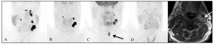
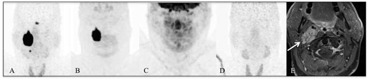
Methods: (68)Ga-DOTATATE PET/CT was prospectively performed in 20 patients with HNPGLs. All patients also underwent (18)F-FDOPA PET/CT, (18)F-FDG PET/CT, and CT/MRI, with 18 patients also undergoing (18)F-fluorodopamine ((18)F-FDA) PET/CT. (18)F-FDOPA PET/CT and CT/MRI served as the imaging comparators.
Results: Thirty-eight lesions in 20 patients were detected, with (18)F-FDOPA PET/CT identifying 37 of 38 and CT/MRI identifying 23 of 38 lesions (P < 0.01). All 38 and an additional 7 lesions (P = 0.016) were detected on (68)Ga-DOTATATE PET/CT. Significantly fewer lesions were identified by (18)F-FDG PET/CT (24/38, P < 0.01) and (18)F-FDA PET/CT (10/34, P < 0.01).
Conclusion: (68)Ga-DOTATATE PET/CT identified more lesions than other imaging modalities. With the results of the present study, and the increasing availability and use of DOTA analogs in the therapy of neuroendocrine tumors, we expect that (68)Ga-DOTATATE PET/CT will become the preferred functional imaging modality for HNPGLs in the near future.
Keywords: 18F-FDOPA; 68Ga-DOTATATE; head and neck paraganglioma.
© 2016 by the Society of Nuclear Medicine and Molecular Imaging, Inc.
Figures

Comment in
-
68Ga-DOTATATE PET/CT Versus MRI: Why the Comparison of 68Ga-DOTATATE PET/CT to an Appropriate MRI Protocol Is Essential.J Nucl Med. 2017 Jan;58(1):184-185. doi: 10.2967/jnumed.116.179960. Epub 2016 Jul 21. J Nucl Med. 2017. PMID: 27445293 No abstract available.
-
Reply: 68Ga-DOTATATE PET/CT Versus MRI: Why the Comparison of 68Ga-DOTATATE PET/CT to an Appropriate MRI Protocol Is Essential.J Nucl Med. 2017 Jan;58(1):185. doi: 10.2967/jnumed.116.180109. Epub 2016 Aug 4. J Nucl Med. 2017. PMID: 27493265 Free PMC article. No abstract available.
References
-
- Martin TP, Irving RM, Maher ER. The genetics of paragangliomas: a review. Clin Otolaryngol. 2007;32:7–11. - PubMed
-
- Sykes JM, Ossoff RH. Paragangliomas of the head and neck. Otolaryngol Clin North Am. 1986;19:755–767. - PubMed
-
- Piccini V, Rapizzi E, Bacca A, et al. Head and neck paragangliomas: genetic spectrum and clinical variability in 79 consecutive patients. Endocr Relat Cancer. 2012;19:149–155. - PubMed
Publication types
MeSH terms
Substances
Grants and funding
LinkOut - more resources
Full Text Sources
Other Literature Sources
Medical