. 2016 Feb;32(2):194-202.
doi: 10.1089/AID.2015.0211. Epub 2015 Dec 17.

A Simple Mouse Model for the Study of Human Immunodeficiency Virus

Affiliations

A Simple Mouse Model for the Study of Human Immunodeficiency Virus

Kang Chang Kim et al. AIDS Res Hum Retroviruses. 2016 Feb.

Abstract

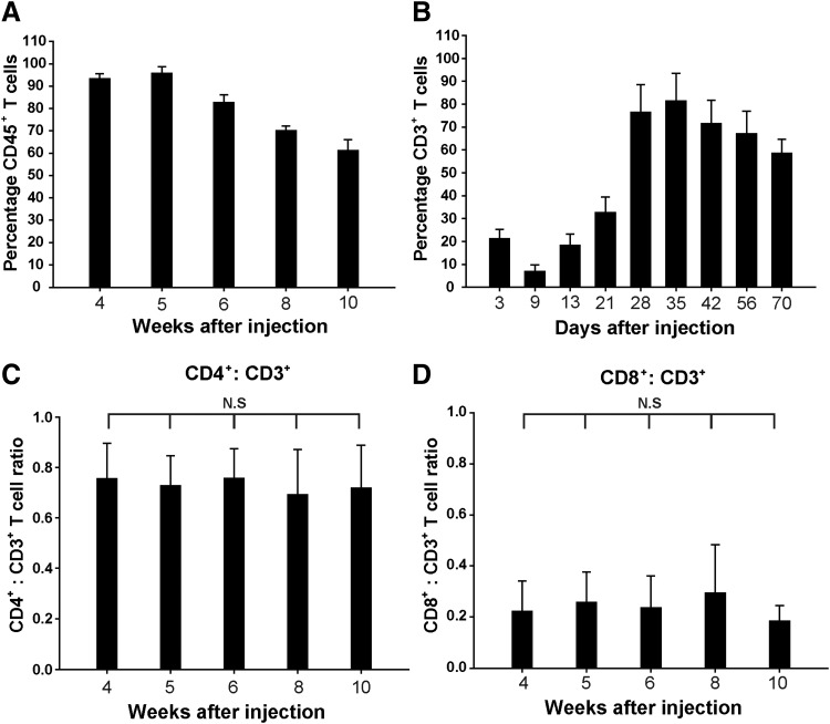
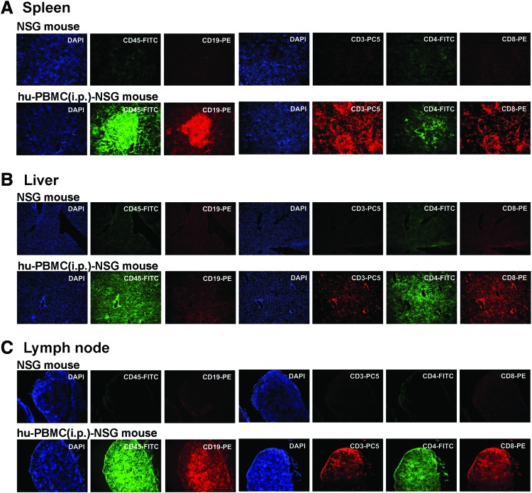
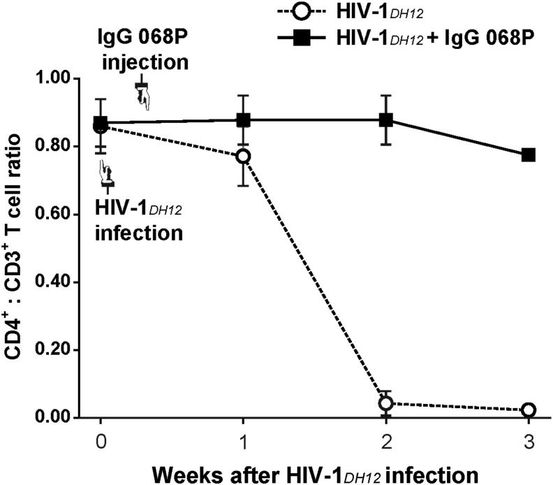
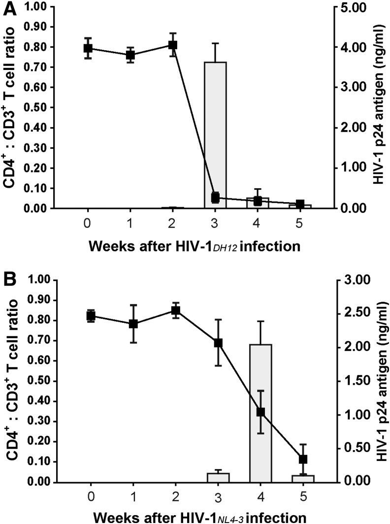
Humanized mouse models derived from immune-deficient mice have been the primary tool for studies of human infectious viruses, such as human immunodeficiency virus (HIV). However, the current protocol for constructing humanized mice requires elaborate procedures and complicated techniques, limiting the supply of such mice for viral studies. Here, we report a convenient method for constructing a simple HIV-1 mouse model. Without prior irradiation, NOD/SCID/IL2Rγ-null (NSG) mice were intraperitoneally injected with 1 × 10(7) adult human peripheral blood mononuclear cells (hu-PBMCs). Four weeks after PBMC inoculation, human CD45(+) cells, and CD3(+)CD4(+) and CD3(+)CD8(+) T cells were detected in peripheral blood, lymph nodes, spleen, and liver, whereas human CD19(+) cells were observed in lymph nodes and spleen. To examine the usefulness of hu-PBMC-inoculated NSG (hu-PBMC-NSG) mice as an HIV-1 infection model, we intravenously injected these mice with dual-tropic HIV-1DH12 and X4-tropic HIV-1NL4-3 strains. HIV-1-infected hu-PBMC-NSG mice showed significantly lower human CD4(+) T cell counts and high HIV viral loads in the peripheral blood compared with noninfected hu-PBMC-NSG mice. Following highly active antiretroviral therapy (HAART) and neutralizing antibody treatment, HIV-1 replication was significantly suppressed in HIV-1-infected hu-PBMC-NSG mice without detectable viremia or CD4(+) T cell depletion. Moreover, the numbers of human T cells were maintained in hu-PBMC-NSG mice for at least 10 weeks. Taken together, our results suggest that hu-PBMC-NSG mice may serve as a relevant HIV-1 infection and pathogenesis model that could facilitate in vivo studies of HIV-1 infection and candidate HIV-1 protective drugs.


Figures

<b>FIG. 1.</b>
FIG. 1.
Reconstitution of human immune cells in human peripheral blood mononuclear cells (hu-PBMC)-NSG mice. (A) The percentage of human CD45+ T cells in the peripheral blood of hu-PBMC-NSG mice (n = 20) reconstituted with human PBMCs 4 to 10 weeks after intraperitoneal injection. (B) The percentage of human CD3+ T cells in the peripheral blood of hu-PBMC-NSG mice increased significantly 28 days after intraperitoneal injection (p < 0.01). (C) CD4+/CD3+ and (D) CD8+/CD3+ T cell ratios in peripheral blood from NSG mice injected intraperitoneally with 1 × 107 human PBMCs (n = 20). Results are expressed as means ± standard deviation (error bars); n.s., not significant (p > 0.05). Mice were bled at the indicated time points after injection of human PBMCs, and PBMCs were isolated for quantification of cell subsets by flow cytometry.
<b>FIG. 2.</b>
FIG. 2.
Immunohistochemical analysis of human cells in hu-PBMC-NSG mouse tissues. Representative immunohistochemical analyses of human CD45+ cells (green), CD45+ T cells (orange), CD3+ T cells (red), CD4+ T cells (green), and CD8+ T cells (orange) in slices from spleen (A), liver (B), and lymph node (C) of hu-PBMC-NSG mice 4 weeks after intraperitoneal injection. Sections were counterstained with the nuclear dye DAPI (blue). Color images available online at www.liebertpub.com/aid
<b>FIG. 3.</b>
FIG. 3.
HIV-1DH12 infection and antiviral therapy in hu-PBMC-NSG mice. (A) Human CD4+ T cell depletion and HIV-1 p24 Ag detection in HIV-1DH12-infected hu-PBMC-NSG mice (HIV-1DH12, n = 5) and uninfected hu-PBMC-NSG mice (control, n = 5). (B) Quantification of human CD4+ T cell reconstitution and HIV-1 p24 levels in blood of HIV-1DH12-infected mice administered HAART for 5 weeks (HIV-1DH12 + HAART) and only 2 weeks (HIV-1DH12 + HAART for 2 weeks). Mice were infected with HIV-1DH12 (1 × 105 IU) for 2 days and then administered a HAART regimen consisting of 4.5 mg of indinavir, 1.2 mg of azidothymidine, and 0.4 mg of atazanavir per mouse. Lines show human CD4+/CD3+ T cell ratios; squares show HIV-1 p24 levels analyzed by ELISA. Results are expressed as means ± standard deviation (error bars).
<b>FIG. 4.</b>
FIG. 4.
Hu-PBMC-NSG mice as an in vivo HIV-1 neutralizing-antibody assay. Hu-PBMC-NSG mice (n = 5) were infected with 1 × 105 IU of HIV-1DH12 4 weeks after injection of human PBMCs. After 2 days, mice were injected with 50 μl of 068P antibody obtained from an HIV-1-infected and recovered macaque monkey (■) or 50 μl of phosphate-buffered saline (PBS) (○). Mice were bled at the indicated time points after infection with HIV-1DH12, and PBMCs were isolated for quantification of cell subsets by flow cytometry. Results are expressed as means ± standard deviation (error bars).
<b>FIG. 5.</b>
FIG. 5.
Comparison of viral kinetics in hu-PBMC-NSG mice infected with two different tropic HIV-1 strains, HIV-1DH12 and HIV-1NL4-3. (A) Human CD4+ T cell depletion (line) and HIV-1 p24 Ag detection (square) in hu-PBMC-NSG mice (n = 5) infected with 1 × 103 IU of HIV-1DH12 4 weeks after intraperitoneal injection of human PBMCs. (B) Human CD4+ T cell depletion (line) and HIV-1 p24 Ag detection (square) in hu-PBMC-NSG mice (n = 5) infected with 1 × 103 IU of HIV-1NL4-3 4 weeks after intraperitoneal injection of human PBMCs. Mice were bled at the indicated time points after infection with HIV-1DH12 or HIV-1NL4-3; PBMCs were isolated for quantification of cell subsets by flow cytometry, and HIV-1 p24 levels in blood were analyzed by ELISA. Results are expressed as means ± standard deviation (error bars).

References

    1. Namikawa R, Kaneshima H, Lieberman M, et al. : Infection of the SCID-hu mouse by HIV-1. Science 1988;242(4886):1684–1686 - PubMed
    1. McCune JM, Namikawa R, Kaneshima H, et al. : The SCID-hu mouse: Murine model for the analysis of human hematolymphoid differentiation and function. Science 1988;241(4873):1632–1639 - PubMed
    1. Mosier DE: Adoptive transfer of human lymphoid cells to severely immunodeficient mice: Models for normal human immune function, autoimmunity, lymphomagenesis, and AIDS. Adv Immunol 1991;50:303–325 - PubMed
    1. Torbett BE, Picchio G, and Mosier DE: hu-PBL-SCID mice: A model for human immune function, AIDS, and lymphomagenesis. Immunol Rev 1991;124:139–164 - PubMed
    1. Ruxrungtham K, Boone E, Ford H Jr, et al. : Potent activity of 2'-beta-fluoro-2',3'-dideoxyadenosine against human immunodeficiency virus type 1 infection in hu-PBL-SCID mice. Antimicrob Agents Chemother 1996;40(10):2369–2374 - PMC - PubMed

Publication types

MeSH terms