. 2016 Mar;37(3):415-22.
doi: 10.3174/ajnr.A4563. Epub 2015 Nov 12.

Utility and Significance of Gadolinium-Based Contrast Enhancement in Posterior Reversible Encephalopathy Syndrome

Affiliations

Utility and Significance of Gadolinium-Based Contrast Enhancement in Posterior Reversible Encephalopathy Syndrome

S J Karia et al. AJNR Am J Neuroradiol. 2016 Mar.

Abstract

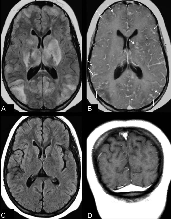
Background and purpose: Posterior reversible encephalopathy syndrome is a clinicoradiologic syndrome. Literature regarding associated factors and the prognostic significance of contrast enhancement in posterior reversible encephalopathy syndrome is sparse. This study set out to evaluate an association between the presence of enhancement in posterior reversible encephalopathy syndrome and various clinical factors in a large series of patients with this syndrome.

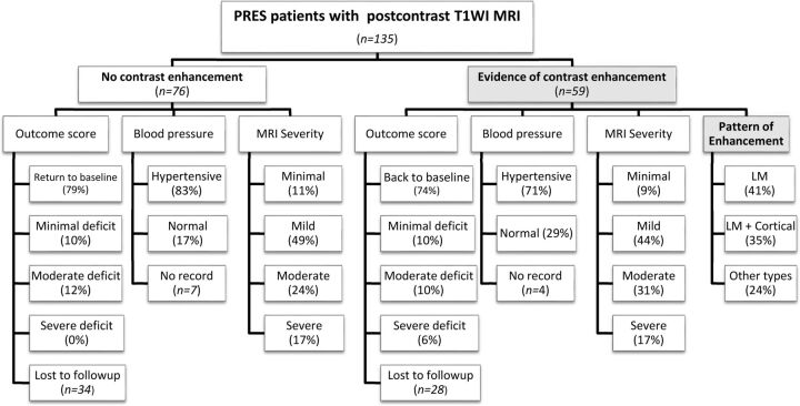
Materials and methods: From an MR imaging report search that yielded 176 patients with clinically confirmed posterior reversible encephalopathy syndrome between 1997 and 2014, we identified 135 patients who had received gadolinium-based contrast. The presenting symptoms, etiology, clinical follow-up, and maximum systolic and diastolic blood pressures within 1 day of MR imaging were recorded. MRIs were reviewed for parenchymal hemorrhage, MR imaging severity, and the presence and pattern of contrast enhancement. Statistical analyses evaluated a correlation between any clinical features and the presence or pattern of enhancement.

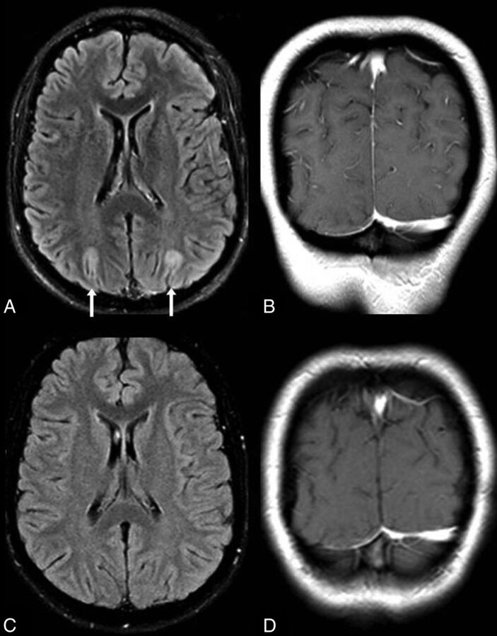
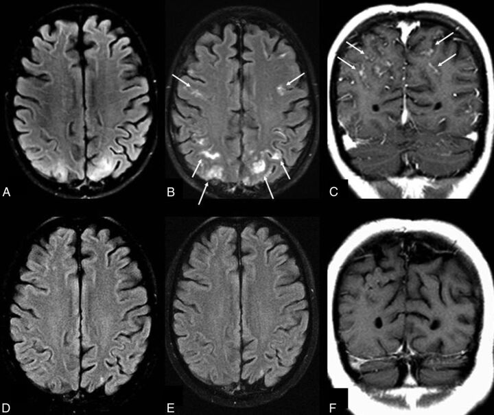
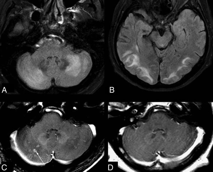
Results: Of 135 included patients (67.4% females; age range, 7-82 years), 59 (43.7%) had contrast enhancement on T1-weighted MR imaging, the most common pattern being leptomeningeal (n = 24, 17.8%) or leptomeningeal plus cortical (n = 21, 15.6%). Clinical outcomes were available in 96 patients. No significant association was found between the presence or pattern of enhancement and any of the variables, including sex, age, symptom, MR imaging severity, blood pressure, or outcome (all P > .05 after Bonferroni correction).

Conclusions: The presence or pattern of enhancement in posterior reversible encephalopathy syndrome is not associated with any of the tested variables. However, an association was found between MR imaging severity and clinical outcome.


Figures

Fig 1.
Fig 1.
Organizational chart of the makeup of the cohort for this study, including MR imaging severity and various clinical factors. LM indicates leptomeningeal.
Fig 2.
Fig 2.
No enhancement in “minimal” PRES. A 22-year-old woman with end-stage renal disease presented with seizure, with SBPmax and DBPmax of 201/119 mm Hg within 1 day of MR imaging. “Minimal” cortical edema is noted on FLAIR (arrows, A) in the occipital regions, without abnormal contrast enhancement on postcontrast coronal T1WI (B). On a 3-month follow-up 1.5T MR imaging, the edema has resolved on FLAIR (C), and there is no enhancement on T1WI (D).
Fig 3.
Fig 3.
Leptomeningeal enhancement pattern in “mild” PRES. A 19-year-old woman with a history of systemic lupus erythematosus and pancytopenia presented with a seizure (blood pressure unavailable). A 1.5T MR imaging demonstrates mild parieto-occipital edema on FLAIR (A), with moderate leptomeningeal enhancement (thin arrows) on both gadolinium-enhanced FLAIR (B) and T1WI (C). D–F, A follow-up MR imaging 2 months later shows that both the mild cortical and subcortical edema on FLAIR (D) has resolved as well as the leptomeningeal enhancement on gadolinium-enhanced FLAIR (E) and T1WI (F). While gadolinium-enhanced FLAIR was not used to score the degree of edema or enhancement, the use of postcontrast FLAIR in this example demonstrates how enhancement can occur in areas lacking edema on noncontrast FLAIR, perhaps due to transient blood-brain barrier injury.
Fig 4.
Fig 4.
Nodular enhancement pattern in “moderate” PRES. A 58-year-old man, on a multiple chemotherapy regimen for metastatic renal cancer with sepsis, developed seizures. The patient was hypotensive, with SBPmax and DBPmax of 92/67 mm Hg. The initial 3T MR imaging demonstrates moderate edema from PRES, graded moderate due to the degree of cerebellar and parieto-occipital edema on FLAIR (A and B); there is also nodular enhancement on postcontrast T1WI (C), demonstrated in multiple planes. D, On a follow-up MR imaging 7 days later, the enhancing cerebellar nodular lesions have resolved.
Fig 5.
Fig 5.
Leptomeningeal and cortical enhancement pattern in “severe” PRES. A 14-year-old girl with systemic lupus erythematosus presented with seizures. SBPmax and DBPmax were normal (136/83 mm Hg). The initial 1.5T MR imaging demonstrates severe edema on FLAIR (A), involving the basal ganglia, thalami, and brain stem (not shown), while there is diffusely thickened leptomeningeal enhancement (thin arrows) and cortical and parenchymal enhancement (dashed arrows) on postcontrast T1WI (B). Vessels are seen both within and outside of areas of vasogenic edema and remain an important consideration for these appearances, perhaps due to slow flow or endothelial dysfunction. On a follow-up 1.5T MR imaging 5 months later, the findings have entirely resolved on FLAIR (C) and postcontrast T1WI (D).

Comment in

  • Reply.
    Karia SJ, McKinney AM, Rykken JB, Roshan SK, Tore H. Karia SJ, et al. AJNR Am J Neuroradiol. 2016 Sep;37(9):E59. doi: 10.3174/ajnr.A4872. Epub 2016 Jun 16. AJNR Am J Neuroradiol. 2016. PMID: 27313133 Free PMC article. No abstract available.
  • Utility and Significance of Gadolinium-Based Contrast Enhancement in Posterior Reversible Encephalopathy Syndrome.
    Bhatia V, Gupta V, Khandelwal N. Bhatia V, et al. AJNR Am J Neuroradiol. 2016 Sep;37(9):E58. doi: 10.3174/ajnr.A4847. Epub 2016 Jun 16. AJNR Am J Neuroradiol. 2016. PMID: 27313134 Free PMC article. No abstract available.
  • Reply.
    Roshan SK, McKinney AM, Karia SJ, Tore H, Rykken JB. Roshan SK, et al. AJNR Am J Neuroradiol. 2016 Sep;37(9):E57. doi: 10.3174/ajnr.A4876. Epub 2016 Jun 30. AJNR Am J Neuroradiol. 2016. PMID: 27365327 Free PMC article. No abstract available.
  • The Clinical Outcome of Posterior Reversible Encephalopathy Syndrome.
    Gao B, Lerner A, Law M. Gao B, et al. AJNR Am J Neuroradiol. 2016 Sep;37(9):E55-6. doi: 10.3174/ajnr.A4853. Epub 2016 Jun 30. AJNR Am J Neuroradiol. 2016. PMID: 27365328 Free PMC article. No abstract available.

References

    1. Bartynski WS. Posterior reversible encephalopathy syndrome, part 1: fundamental imaging and clinical features. AJNR Am J Neuroradiol 2008;29:1036–42 10.3174/ajnr.A0928 - DOI - PMC - PubMed
    1. Hinchey J, Chaves C, Appignani B, et al. A reversible posterior leukoencephalopathy syndrome. N Engl J Med 1996;334:494–500 10.1056/NEJM199602223340803 - DOI - PubMed
    1. Covarrubias DJ, Luetmer PH, Campeau NG. Posterior reversible encephalopathy syndrome: prognostic utility of quantitative diffusion-weighted MR images. AJNR Am J Neuroradiol 2002;23:1038–48 - PMC - PubMed
    1. McKinney AM, Kieffer SA, Paylor RT, et al. Acute toxic leukoencephalopathy: potential for reversibility clinically and on MRI with diffusion-weighted and FLAIR imaging. AJR Am J Roentgenol 2009;193:192–206 10.2214/AJR.08.1176 - DOI - PubMed
    1. Larsson HB, Stubgaard M, Frederiksen JL, et al. Quantitation of blood-brain barrier defect by magnetic resonance imaging and gadolinium-DTPA in patients with multiple sclerosis and brain tumors. Magn Reson Med 1990;16:117–31 10.1002/mrm.1910160111 - DOI - PubMed