. 2015 Nov;201(3):815-42.
doi: 10.1534/genetics.115.183392.

Genetics on the Fly: A Primer on the Drosophila Model System

Affiliations

Genetics on the Fly: A Primer on the Drosophila Model System

Karen G Hales et al. Genetics. 2015 Nov.

Abstract

Fruit flies of the genus Drosophila have been an attractive and effective genetic model organism since Thomas Hunt Morgan and colleagues made seminal discoveries with them a century ago. Work with Drosophila has enabled dramatic advances in cell and developmental biology, neurobiology and behavior, molecular biology, evolutionary and population genetics, and other fields. With more tissue types and observable behaviors than in other short-generation model organisms, and with vast genome data available for many species within the genus, the fly's tractable complexity will continue to enable exciting opportunities to explore mechanisms of complex developmental programs, behaviors, and broader evolutionary questions. This primer describes the organism's natural history, the features of sequenced genomes within the genus, the wide range of available genetic tools and online resources, the types of biological questions Drosophila can help address, and historical milestones.

Keywords: Drosophila; comparative genomics; development; model organism.


Figures

Figure 1
Figure 1
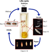
Life cycle of D. melanogaster. D. melanogaster are cultured in vials with food in the bottom and a cotton, rayon, or foam plug at the top. The pictured vial shows each major stage of the life cycle, which is completed in 9–10 days when flies are maintained at 25°. Embryos hatch from the egg after ∼1 day and spend ∼4 days as larvae in the food. Around day 5, third instar larvae crawl out of the food to pupate on the side of the vial. During days 5–9, metamorphosis occurs, and the darkening wings within the pupal case indicate that maturation is nearly complete. Adult flies eclose from pupal cases around days 9–10.
Figure 2
Figure 2
Sex determination. The number of X chromosomes in D. melanogaster is determined by an X chromosome counting mechanism. In XX females, early expression of the RNA-processing gene Sex lethal (Sxl) later results in female-specific processing of its own transcript. Sxl then begins a cascade of alternative splicing events that ultimately result in generation of the female-specific isoform of Dsx (DsxF). Note that Fru is not shown here for clarity. In males, the absence of early Sxl expression results in default processing of Sxl and tra transcripts that contain an early stop codon. Dsx pre-mRNA is then processed for a male-specific isoform of Dsx (DsxM). These Dsx isoforms then promote expression of downstream genes that govern sex-specific decisions related to morphology and behavior.
Figure 3
Figure 3
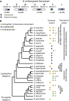
Genome organization and phylogeny. (A) Organization of the Drosophila melanogaster genome. D. melanogaster has two metacentric autosome arms (chromosomes 2 and 3; Muller elements B and C and D and E), a small autosome (chromosome 4; Muller element F) and a pair of sex chromosomes (chromosome X—Muller element A—and chromosome Y). The approximate sizes and division of heterochromatin/euchromatin are shown. (B) Comparative genomics resources. Phylogeny of Drosophila species whose genomes were sequenced either by a large consortium (i.e., Drosophila 12 Genomes Consortium et al. 2007 or modENCODE https://www.hgsc.bcm.edu/drosophila-modencode-project) or other community or individual lab sequencing project. While likely not an exhaustive list, it highlights the power to do comparative genomics in Drosophila. The tree topology is derived from several sources (Drosophila 12 Genomes Consortium et al. 2007; Gao et al. 2007; Seetharam and Stuart 2013), as the phylogenetic relationships between some of these species are not well resolved. The references for each genome are as follows: (1) Hu et al. (2013); (2) Garrigan et al. (2012); (3) Nolte et al. (2013); (4) Adams et al. (2000); (5) Rogers et al. (2014); (6) http://genomics.princeton.edu/AndolfattoLab/Dsantomea_genome.html; (7) Chiu et al. (2013); (8) Richards et al. (2005); (9) Kulathinal et al. (2009); (10) McGaugh et al. (2012); (11) Zhou and Bachtrog (2012); (12) Palmieri et al. (2014); (13) Fonseca et al. (2013); (14) Guillen et al. (2014); (15) Zhou et al. (2012); and (16) Zhou and Bachtrog (2015).
Figure 4
Figure 4
Generalized scheme for a forward genetic screen using chemical mutagenesis. (A) Male flies eat food laced with ethane methyl sulfonate (EMS), an alkylating agent which typically causes point mutations. (B) Different mutations occur in each cell of the feeding flies, including sperm (indicated by pink, yellow, and green sperm cells). (C) Outcrossing the mutagenized flies to untreated females yields (D) offspring that each potentially have a different new mutations throughout their bodies, indicated schematically by body colors corresponding to the sperm cells above. (E) Outcrossing these flies individually and (F) inbreeding each set of offspring gives a population of flies for each new mutation. (G) Researchers then test homozygous flies (darker pink and green) for the phenotype of interest. In some cases, adult homozygotes are not viable (as in the yellow population) and so researchers interested in earlier developmental steps may examine embryos and larvae within these populations to find dying homozygotes.
Figure 5
Figure 5
GAL4/UAS system for modular expression of transgenes in specific tissues. To express a transgene or RNAi construct in a particular tissue, one needs flies carrying (A) a “driver” with a tissue-specific promoter/enhancer placed 5′ of the gene encoding the yeast GAL4 transcription factor (left) and (right) the gene of interest placed 3′ of the upstream activating sequence (UAS), which is activated by GAL4. (B) Transgenic flies carrying either of the two constructs alone (top) do not express the gene of interest, but when crossed into the same fly, the tissue-specific promoter (a wing promoter in this example) drives expression of GAL4, which turns on the gene of interest (here indicated by green) in the specified tissue. The system can also be used to express a hairpin RNA to knock down a gene in the target tissue.
Figure 6
Figure 6
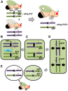
Clonal analysis in somatic tissue. (A) Schematic diagram of a genetic cross used to create homozygous somatic clones of a mutation of interest within a heterozygous background. In this example, the clone is a patch of mutant cells within a wild-type wing. One parent is homozygous for a chromosome that carries the FLP recombinase recognition site, FRT (triangles) and a distal GFP marker that is being expressed under the control of a wing promoter. All of the cells of this fly’s wings will express GFP and appear green under a fluorescent microscope. This fly is also homozygous on another chromosome (not drawn) for the FLP recombinase gene, which is being expressed under the control of a wing promoter. The other parental fly is homozygous for a mutation of interest (red star) on the same FRT-carrying chromosome. Progeny from this cross will be transheterozygous for the GFP-marked chromosome and the mutant chromosome, and they will be heterozygous for FLP. During development, FLP-mediated mitotic recombination in the developing wing will produce patches of unmarked homozygous mutant cells (white patch). Panels B–E show the mechanics of clone production through mitotic recombination in the progeny. (B) Cells in mitotic G2 have replicated chromosomes with sister chromatids. In some wing cells, the FLP recombinase triggers recombination between FRT sites on nonsister chromatids, and (C) one copy each of the GFP marker and the mutation of interest will switch between homologous chromosomes. (D) One of two possible chromatid alignments at mitotic metaphase for the cell pictured in C. (The other alignment, not shown, leads to two heterozygous daughter cells.) The black arrows show the direction of sister chromatid separation during the completion of mitotic division. (E) Daughter cells produced upon completion of cell division for the cell pictured in D. One daughter cell is homozygous for the mutation of interest and will subsequently divide to give rise to a homozygous patch of cells, which can be identified for phenotypic analysis based on loss of the GFP marker. The other daughter cell is homozygous for the GFP marker and will blend into the surrounding heterozygous cells that also fluoresce.
Figure 7
Figure 7
Stages of embryonic development. D. melanogaster development begins in a syncytium characterized by nuclear divisions without cytokinesis (stage 2). After 10 synchronized rounds of division, nuclei migrate to the periphery where they become partially encapsulated by actin-based furrow canals (stage 3/4). True cellularization occurs in stage 5, followed by gastrulation (stage 8), which determines the three germ layers. Dramatic morphogenetic movements then reshape the body plan as cells from the posterior migrate toward the anterior in germband extension (stage 9) followed by later retraction to the posterior (germband retraction; stage 12). Epithelial cells then migrate toward the dorsal midline in dorsal closure (stage 13), and head structures begin to mature (head involution; stage 15). Finally the larva reaches its mature state (stage 17) and hatches from the eggshell. Images adapted from the Atlas of Drosophila Development (Hartenstein 1993) and used with permission. In each panel, anterior is to the right and dorsal is up.

References

    1. Adams M. D., Celniker S. E., Holt R. A., Evans C. A., Gocayne J. D., et al. , 2000. The genome sequence of Drosophila melanogaster. Science 287: 2185–2195. - PubMed
    1. Adoutte A., Balavoine G., Lartillot N., Lespinet O., Prud’homme B., et al. , 2000. The new animal phylogeny: reliability and implications. Proc. Natl. Acad. Sci. USA 97: 4453–4456. - PMC - PubMed
    1. Adryan B., Teichmann S. A., 2006. FlyTF: a systematic review of site-specific transcription factors in the fruit fly Drosophila melanogaster. Bioinformatics 22: 1532–1533. - PubMed
    1. Armknecht S., Boutros M., Kiger A., Nybakken K., Mathey-Prevot B., et al. , 2005. High-throughput RNA interference screens in Drosophila tissue culture cells. Methods Enzymol. 392: 55–73. - PubMed
    1. Arnone M. I., Davidson E. H., 1997. The hardwiring of development: organization and function of genomic regulatory systems. Development 124: 1851–1864. - PubMed