. 2015;11(12):2142-52.
doi: 10.1080/15548627.2015.1108508.

Reactivation of autophagy by spermidine ameliorates the myopathic defects of collagen VI-null mice

Affiliations

Reactivation of autophagy by spermidine ameliorates the myopathic defects of collagen VI-null mice

Martina Chrisam et al. Autophagy. 2015.

Abstract

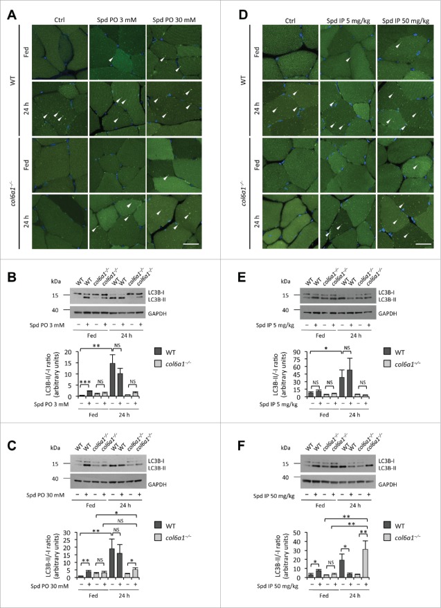
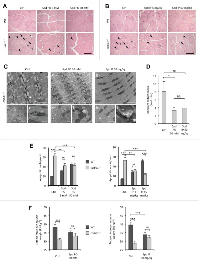
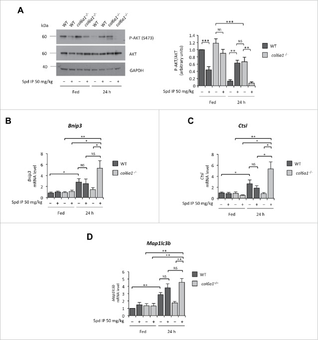
Autophagy is a self-degradative process responsible for the clearance of damaged or unnecessary cellular components. We have previously found that persistence of dysfunctional organelles due to autophagy failure is a key event in the pathogenesis of COL6/collagen VI-related myopathies, and have demonstrated that reactivation of a proper autophagic flux rescues the muscle defects of Col6a1-null (col6a1(-/-)) mice. Here we show that treatment with spermidine, a naturally occurring nontoxic autophagy inducer, is beneficial for col6a1(-/-) mice. Systemic administration of spermidine in col6a1(-/-) mice reactivated autophagy in a dose-dependent manner, leading to a concurrent amelioration of the histological and ultrastructural muscle defects. The beneficial effects of spermidine, together with its being easy to administer and the lack of overt side effects, open the field for the design of novel nutraceutical strategies for the treatment of muscle diseases characterized by autophagy impairment.

Keywords: autophagy; collagen VI; muscular dystrophies; skeletal muscle; spermidine.


Figures

Figure 1 (see previous page).
Spermidine induces autophagy in wild-type and COL6-deficient tibialis anterior muscles. (A) Detection of fluorescent puncta in cross sections of tibialis anterior muscles from untreated (Ctrl) and per os spermidine-treated (Spd PO) wild-type and col6a1−/− mice bearing a GFP-LC3 reporter construct. Mice were analyzed under fed conditions or after 24 h fasting. White arrowheads point at some GFP-LC3-positive puncta. Nuclei were stained with Hoechst. Scale bar: 25 μm. (B, C) Western blot analysis of LC3B lipidation in protein extracts from tibialis anterior muscles of untreated (–) and per os spermidine-treated (+) wild-type and col6a1−/− mice. Densitometric quantifications of LC3B-II vs LC3B-I ratio, as determined by at least 3 independent western blot experiments, are shown in the lower panels (n = 3 or 4; *, P< 0.05; **, P < 0.01; ***, P < 0.001; NS, not significant). (D) Detection of fluorescent puncta in cross sections of tibialis anterior muscles from untreated (Ctrl) and i.p. spermidine-treated (Spd IP) wild-type and col6a1−/− mice. Mice were analyzed under fed conditions or after 24 h fasting. White arrowheads point at some GFP-LC3-positive puncta. Nuclei were stained with Hoechst. Scale bar: 25 μm. (E, F) Western blot analysis of LC3B lipidation in protein extracts from tibialis anterior muscles of untreated (–) and i.p. spermidine-treated (+) wild-type and col6a1−/− mice. Densitometric quantifications of LC3B-II vs LC3B-I ratio, as determined by at least 3 independent western blot experiments, are shown in the lower panels (n = 3 or 4; *, P < 0.05; **, P < 0.01; ***, P < 0.001; NS, not significant). 24 h, twenty-four hour-long starvation; Ctrl, untreated control condition; Spd IP, spermidine i.p.; Spd PO, spermidine per os; WT, wild-type.
Figure 2 (see previous page).
Spermidine treatment ameliorates the myopathic phenotype of col6a1−/− mice. (A, B) Haematoxylin-eosin staining of cross sections of tibialis anterior muscles from untreated (Ctrl) and per os (A) or i.p. (B) spermidine-treated wild-type and col6a1−/− mice. In col6a1−/− mice, centrally nucleated fibers (black arrowheads) and atrophic fibers (white arrowheads) appear reduced after spermidine treatment. Scale bar: 50 μm. (C) Representative electron micrographs of diaphragm thin sections from untreated (a to c) and per os (d to f) or i.p. (g to i) spermidine-treated col6a1−/− mice. Low-power views (upper panels) reveal improvement of muscle ultrastructure following spermidine treatment. High-power magnifications (lower panels) show dilated sarcoplasmic reticulum (SR) and swollen mitochondria (mit) in untreated col6a1−/− mice (b,c), and the presence of autophagic vacuoles (AV) in spermidine-treated col6a1−/− mice (f,i) together with a marked amelioration of organelle morphology (e,h). Scale bar: 1 μm (upper panels) or 500 nm (lower panels). (D) Percentage of morphologically altered mitochondria in the diaphragm of untreated (Ctrl) and spermidine-treated (PO 30, per os 30 mM; IP 50, i.p. 50 mg/kg) col6a1−/− mice (n = 3 or 4; P< 0.05; NS, not significant.) (E) Quantification of apoptosis by TUNEL assay in diaphragm muscles of untreated (Ctrl) and per os (left panel) or i.p. (right panel) spermidine-treated wild-type and col6a1−/− mice (n = 3, each group; **, P < 0.01; ***, P < 0.001). (F) In vivo tetanic force measurements in gastrocnemius muscle of untreated (Ctrl) and per os or i.p. spermidine-treated wild-type and col6a1−/− mice. The histograms report the normalized strength at a stimulation frequency of 100 Hz (n = 5, each group; ***, P<0.001; NS, not significant). Ctrl, untreated control condition; Spd IP, spermidine i.p.; Spd PO, spermidine per os. WT, wild-type.
Figure 3.
Figure 3.
Spermidine modulates the AKT-FOXO axis in skeletal muscle. (A) Western blot analysis of AKT phosphorylation in protein extracts from tibialis anterior muscles of untreated (–) and i.p. spermidine-treated (+) wild-type and col6a1−/− mice. Densitometric quantifications of P-AKT vs AKT, as determined by 3 independent western blot experiments, are shown on the right panel (n = 3; **, P< 0.01; ***, P < 0.001). (BE) Real-time RT-PCR analysis of mRNA levels for Bnip3 (B), Ctsl (C) and Map1lc3b (E) in tibialis anterior muscles of untreated (–) and i.p. spermidine-treated (+) wild-type and col6a1−/− mice (n = 3 or 4; ***, P < 0.001; ** P < 0.01; *, P < 0.05; NS, not significant). 24 h, twenty-four hour-long starvation; Spd IP, spermidine i.p.; Spd PO, spermidine per os. WT, wild-type.

References

    1. Mizushima N. The pleiotropic role of autophagy: from protein metabolism to bactericide. Cell Death Differ 2005; 12 Suppl 2:1535-41; PMID:16247501; http://dx.doi.org/ 10.1038/sj.cdd.4401728 - DOI - PubMed
    1. Cecconi F, Levine B. The role of autophagy in mammalian development: cell makeover rather than cell death. Dev Cell 2008; 15:344-57; PMID:18804433; http://dx.doi.org/ 10.1016/j.devcel.2008.08.012 - DOI - PMC - PubMed
    1. Levine B, Kroemer G. Autophagy in the pathogenesis of disease. Cell 2008; 132:27-42; PMID:18191218; http://dx.doi.org/ 10.1016/j.cell.2007.12.018 - DOI - PMC - PubMed
    1. Rogov V, Dötsch V, Johansen T, Kirkin V. Interactions between autophagy receptors and ubiquitin-like proteins form the molecular basis for selective autophagy. Mol Cell 2014; 53:167-78; PMID:24462201; http://dx.doi.org/ 10.1016/j.molcel.2013.12.014 - DOI - PubMed
    1. Sandri M, Coletto L, Grumati P, Bonaldo P. Misregulation of autophagy and protein degradation systems in myopathies and muscular dystrophies. J Cell Sci 2013; 126:5325-33; PMID:24293330; http://dx.doi.org/ 10.1242/jcs.114041 - DOI - PubMed

Publication types