. 2015 Nov 13;9(11):e0004058.
doi: 10.1371/journal.pntd.0004058. eCollection 2015 Nov.

Proteasome Inhibition Suppresses Dengue Virus Egress in Antibody Dependent Infection

Affiliations

Proteasome Inhibition Suppresses Dengue Virus Egress in Antibody Dependent Infection

Milly M Choy et al. PLoS Negl Trop Dis. .

Abstract

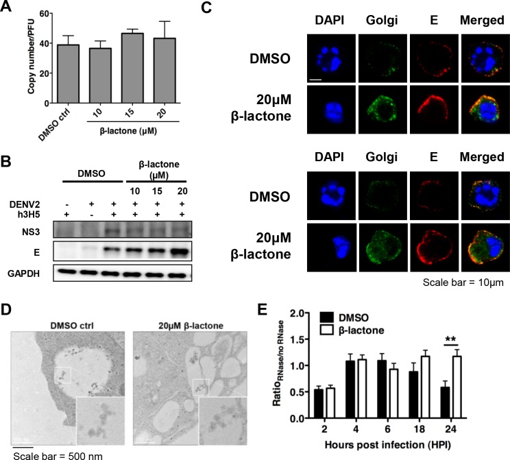
The mosquito-borne dengue virus (DENV) is a cause of significant global health burden, with an estimated 390 million infections occurring annually. However, no licensed vaccine or specific antiviral treatment for dengue is available. DENV interacts with host cell factors to complete its life cycle although this virus-host interplay remains to be fully elucidated. Many studies have identified the ubiquitin proteasome pathway (UPP) to be important for successful DENV production, but how the UPP contributes to DENV life cycle as host factors remains ill defined. We show here that proteasome inhibition decouples infectious virus production from viral RNA replication in antibody-dependent infection of THP-1 cells. Molecular and imaging analyses in β-lactone treated THP-1 cells suggest that proteasome function does not prevent virus assembly but rather DENV egress. Intriguingly, the licensed proteasome inhibitor, bortezomib, is able to inhibit DENV titers at low nanomolar drug concentrations for different strains of all four serotypes of DENV in primary monocytes. Furthermore, bortezomib treatment of DENV-infected mice inhibited the spread of DENV in the spleen as well as the overall pathological changes. Our findings suggest that preventing DENV egress through proteasome inhibition could be a suitable therapeutic strategy against dengue.


Conflict of interest statement

The authors have declared that no competing interests exist.

Figures

Fig 1
Fig 1. Proteasome inhibition decouples infectious DENV2 production from viral RNA replication in THP-1 cells.
(A) No cytotoxicity was observed in THP-1 cells in the presence of β-lactone and genistein for 48 h using a cell viability assay. Mean ± SD. N = 4. (B) Using flow cytometry, β-lactone does not block uptake of DENV2 immune complexes. Genistein, an inhibitor of Fc receptor-mediated entry blocked uptake. (C) Alexa Fluor 647 labeled DENV2 opsonized with h3H5 were internalized in cells pre-treated with DMSO or β-lactone, while genistein prevented the uptake of virus at 2 hpi. (D) Proteasome inhibition showed a dose-dependent decrease in plaque titers 48 hpi. Mean ± SD. N = 4. Student’s t test, **p<0.01, ***p<0.001. (E) DENV2 RNA copy number per GAPDH showed no significant difference between β-lactone treated and DMSO control 48 hpi. (F) Ratio of infectious DENV2 to genomic copies showed a dose-dependent decrease after β-lactone treatment. Mean ± SD. N = 4. Student’s t test, *p < 0.05, **p<0.01.
Fig 2
Fig 2. DENV2 egress is dependent on proteasome function.
(A) Ratio of viral genomic RNA from cell culture supernatant to infectious DENV2 showed no significant differences after β-lactone treatment at different drug concentrations, compared to DMSO control, suggesting that the treatment with β-lactone did not result in reduced DENV2 maturation. Mean ± SD. N = 4. (B) A dose-dependent accumulation of E protein in cells is observed with no difference in the levels of NS3 at 24 hpi. (C) Confocal analysis showed accumulation of structural proteins (prM and E, in red) in β-lactone treated compared to DMSO treated cells at 24 hpi with strong co-localization with Golgi (in green), suggesting that egress of viral particles were impaired in presence of UPP inhibition (Scale bar = 10 μm). (D) Accumulation of viral particles in intra-cytoplasmic vacuoles (50 nm) in β-lactone compared to DMSO treated cells was observed using electron microscopy, indicating that DENV2 RNA could replicate and be packaged with structural proteins (Scale bar = 500 nm). (E) Ratio of RNase to non-RNase treated cells after β-lactone treatment is significantly higher compared to the DMSO control 24 hpi. Mean ± SD. N = 4. Student’s t test, **p<0.01.
Fig 3
Fig 3. Proteasome inhibition increases eIF2α phosphorylation and represses translation of EXOC7, TC10 and EXOC1.
Effects of proteasome inhibition on the expression levels of EXOC7, its effector, TC10, and its interacting partner, EXOC1 were assessed. (A) Transcript levels of EXOC7, TC10 and EXOC1 remained constant. Mean ± SD. N = 4. (B) Protein levels of EXOC7, TC10 and EXOC1 decreased in both uninfected and DENV2-infected β-lactone treated cells. (C) Using flow cytometry, the mean fluorescence intensity of EXOC7 and TC10 decreased significantly i7n β-lactone treated infected cells compared to DMSO treated infected cells, indicating a decrease in the protein levels of EXOC7 and TC10 after β-lactone treatment. Mean ± SD. N = 4. Student’s t test, *p<0.05, **p<0.01. (D) Effect of proteasome inhibition on eIF2α phosphorylation was assessed in DMSO or β-lactone treated infected THP-1 cells. Increased phosphorylation of eIF2α was observed in β-lactone treated infected cells 4 hpi. Up-regulation of the resident ER chaperone protein BiP in DENV2-infected β-lactone treated cells was also observed. (E) ER stress inducer, thapsigargin, reduced protein levels of EXOC7 and TC10 in a dose-dependent manner. BiP, an ER stress signature, increases with increasing thapsigargin concentration. (F) Similar to DENV2-infected β-lactone treated cells, DENV2-infected thapsigargin treated THP-1 cells showed the same decoupling effect of infectious particles to RNA copy number. Viral RNA genome was detected in thapsigargin treated cells but no infectious DENV2 was detected in cell culture supernatant using plaque assay. Mean ± SD. N = 4.
Fig 4
Fig 4. Bortezomib decouples infectious DENV production from viral RNA replication in primary monocytes.
Viral RNA genome was detected using qRT-PCR, but no infectious DENV2 was detected using plaque assay in the supernatant of cells treated with higher concentrations of bortezomib for (A) DENV1, (B) DENV2, (C) DENV3 and (D) DENV4. Mean ± SD. N = 4.
Fig 5
Fig 5. Bortezomib inhibits infectious DENV production in primary monocytes.
Infectious DENV in cell culture supernatant was measured using plaque assay, and the percent inhibition of infectious DENV production after bortezomib treatment compared to DMSO treatment was calculated. At doses of bortezomib that is minimally toxic to primary monocytes, a significant dose-dependent decrease in virus titers was observed for different strains of all 4 dengue serotypes, (A) DENV1, (B) DENV2, (C) DENV3 and (D) DENV4, after drug treatment in primary monocytes. The identity of the clinical isolate is indicated next to a distinct colored dot. The bortezomib concentration that inhibited 50% of virus replication (in parentheses) was less than 20 nM for DENV1, 2, 3 and 4, respectively. (E) Bortezomib also inhibited 50% of virus production of the attenuated strain of yellow fever virus, YF17D, at a concentration of 0.5 nM. (F) Epoxomicin, another proteasome inhibitor, was able to reduce DENV titers by plaque assay in a dose-dependent manner for all 4 dengue serotypes. Mean ± SD. N = 4.
Fig 6
Fig 6. Bortezomib reduced viral load and signs of dengue pathology in C57BL/6 mice.
WT mice infected intraperitonealy with DENV2 were treated with bortezomib 6 hpi and analyzed at 24, 48 and 72 hpi for further analysis. (A) For quantification of NS3+ cells, the mean cell count in 20 alternate microscopic high-power fields (x400) was measured. Quantification was done only in the red pulp of spleen. Bortezomib treated mice showed significantly reduced number of DENV infected cells in the red pulp of the spleen at 24 and 48 hpi compared to vehicle control. Mean ± SEM. N = 4–5. Student’s t test, **p<0.01. (B) Representative serial sections from spleen of each group of mice stained with anti-DENV NS3 antibody at 24, 48 and 72 hpi. Multiple sections of each tissue were examined for staining (60x magnification). The inset is a representation of the spleen from mock infected mice without (top panel) or with (bottom panel) bortezomib treatment. The top panel shows DENV-infected spleen without bortezomib treatment 24, 48 and 72 hpi. The bottom panel shows DENV-infected spleen with bortezomib treatment 24, 48 and 72 hpi. Positive staining for NS3 is brown while hematoxylin counterstaining is blue. Bortezomib treated mice also showed significantly reduced number of DENV infected cells in the red pulp of the spleen at 24 and 48 hpi compared to vehicle control. (C) RNA copy number in the spleen was reduced in bortezomib treated mice compared to vehicle control at 48 hpi. Mean ± SEM. N = 4–5. Student’s t test, *p<0.05. (D-E) The vehicle control experienced a drop in platelet count over the first 48 hours of infection before recovering by day 3, and experienced a significant rise in hematocrit values that peaked 24 hpi. On the other hand, no significant changes were observed for the platelet count 24–72 hpi, and hematocrit levels 0–72 hpi in mice after bortezomib treatment, suggesting the efficiency of bortezomib in alleviating disease symptoms. Mean ± SEM. N = 4–5. Student’s t test, *p<0.05, ****p<0.0001. (F-G) DENV2-infected vehicle control displayed an increased systemic level of MCPT1, indicative of mast cell activation, in mouse serum and spleen when compared to bortezomib treated mice at all time-points. (H) Levels of TNF-α were decreased in bortezomib treated mice 24 hpi when compared to the vehicle control. Mean ± SEM. N = 4–5. Student’s t test, *p<0.05.

References

    1. Bhatt S, Gething PW, Brady OJ, Messina JP, Farlow AW, et al. (2013) The global distribution and burden of dengue. Nature 496: 504–507. 10.1038/nature12060 - DOI - PMC - PubMed
    1. Villar L, Dayan GH, Arredondo-Garcia JL, Rivera DM, Cunha R, et al. (2015) Efficacy of a tetravalent dengue vaccine in children in Latin America. N Engl J Med 372: 113–123. 10.1056/NEJMoa1411037 - DOI - PubMed
    1. Capeding MR, Tran NH, Hadinegoro SR, Ismail HI, Chotpitayasunondh T, et al. (2014) Clinical efficacy and safety of a novel tetravalent dengue vaccine in healthy children in Asia: a phase 3, randomised, observer-masked, placebo-controlled trial. Lancet 384: 1358–1365. 10.1016/S0140-6736(14)61060-6 - DOI - PubMed
    1. Guzman MG, Kouri G, Valdes L, Bravo J, Vazquez S, et al. (2002) Enhanced severity of secondary dengue-2 infections: death rates in 1981 and 1997 Cuban outbreaks. Rev Panam Salud Publica 11: 223–227. - PubMed
    1. Kliks SC, Nimmanitya S, Nisalak A, Burke DS (1988) Evidence that maternal dengue antibodies are important in the development of dengue hemorrhagic fever in infants. Am J Trop Med Hyg 38: 411–419. - PubMed

Publication types

MeSH terms

LinkOut - more resources