. 2015 Nov 3;13(5):915-23.
doi: 10.1016/j.celrep.2015.09.060. Epub 2015 Oct 22.

The Etv2-miR-130a Network Regulates Mesodermal Specification

Affiliations

The Etv2-miR-130a Network Regulates Mesodermal Specification

Bhairab N Singh et al. Cell Rep. .

Abstract

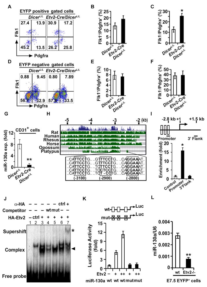
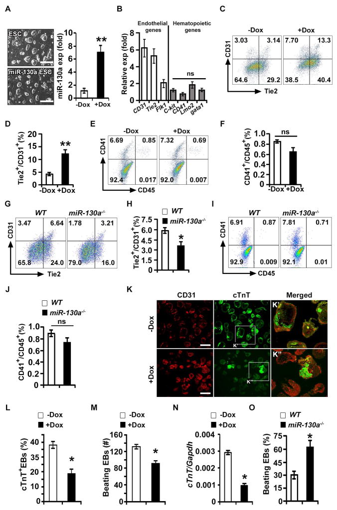
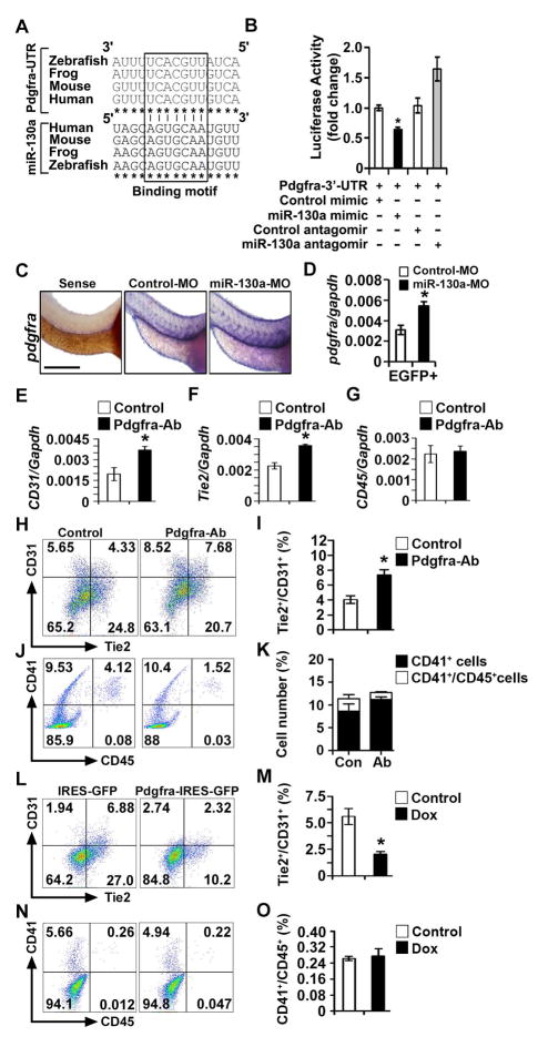
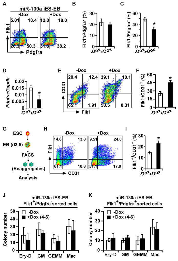
MicroRNAs (miRNAs) are known to regulate critical developmental stages during embryogenesis. Here, we defined an Etv2-miR-130a cascade that regulates mesodermal specification and determination. Ablation of Dicer in the Etv2-expressing precursors resulted in altered mesodermal lineages and embryonic lethality. We identified miR-130a as a direct target of Etv2 and demonstrated its role in the segregation of bipotent hemato-endothelial progenitors toward the endothelial lineage. Gain-of-function experiments demonstrated that miR-130a promoted the endothelial program at the expense of the cardiac program without impacting the hematopoietic lineages. In contrast, CRISPR/Cas9-mediated knockout of miR-130a demonstrated a reduction of the endothelial program without affecting hematopoiesis. Mechanistically, miR-130a directly suppressed Pdgfra expression and promoted the endothelial program by blocking Pdgfra signaling. Inhibition or activation of Pdgfra signaling phenocopied the miR-130a overexpression and knockout phenotypes, respectively. In summary, we report the function of a miRNA that specifically promotes the divergence of the hemato-endothelial progenitor to the endothelial lineage.

Keywords: Etv2; dicer; lineage specification; miR-130a; microRNA.


Figures

Figure 1
Figure 1. Etv2 modulates miR-130a expression in the endothelial progenitors
(A–F) Representative FACS profiles (A, D) and quantification (B, C, E, F) of mesodermal populations in Etv2-EYFP:Etv2-Cre;DicerL/L embryos at E7.5 from EYFP+ and EYFP compartments. (G) qPCR analysis of miR-130a in CD31+ cells sorted from DicerL/L and Etv2-Cre;DicerL/L embryos at E9.5. (H) Evolutionary conservation of the 5.0 kb upstream fragment of the miR-130a gene. (I) Top: Schematic of the 2.8 kb upstream region of the miR-130a promoter. Bottom: ChIP analysis of d4 Dox-inducible HA-Etv2 EBs using an HA antibody. ChIP assay for the Gapdh promoter (Control) and a non-specific locus (miR-130a 3′ UTR region: 3′ Flank) are shown as controls. (J) EMSA showing Etv2 bound to the Ets binding site in the miR-130a promoter region. (K) Luciferase reporter constructs using the miR-130a promoter (-1.0 kb) harboring wild-type (wt; open box) or mutant (mut; crossed box) Etv2 binding sites. (L) qPCR analysis of miR-130a using EYFP+ sorted cells from Etv2 wild-type and mutant embryos at E7.5. Error bars indicate SEM (n = 4; *p<0.05; **p < 0.005) (see also Figure S1).
Figure 2
Figure 2. miR-130a promotes the endothelial lineage
(A) Left panel: phase contrast images of wild-type E14 (ESCs) and Dox-inducible miR-130a ES cell (miR-130a iES) colonies. Right panel: qPCR analysis of miR-130a in the absence (−Dox) and presence of Dox (+Dox). (B) qPCR analyses for endothelial and hematopoietic markers using −Dox and +Dox EBs at d6 of differentiation (ratio shown as +Dox/−Dox). (C–F) FACS profiles (C, E) and quantification (D, F) of endothelial [Tie2 and CD31 (C, D)]; and hematopoietic [CD41 and CD45 (E, F)] markers in −Dox and +Dox conditions. (G–J) Representative FACS profiles (G, I) and quantification (H, J) of endothelial (G, H) and hematopoietic (I, J) markers in WT and miR130a−/− EBs. (K) Sections of −Dox and +Dox miR-130a iES/EBs immunostained with CD31 (red) and cTnT (green) at d10 of EB differentiation. Boxed regions are shown in panel K′ and K″. (L) Quantitative analysis of cTnT+ EBs in −Dox (n = 123) and +Dox (n = 142) EBs. (M) Contractility assay using −Dox (n = 146) and +Dox (n = 135) EBs. (N) qPCR analyses of a cardiac marker (cTnT) from −Dox and +Dox miR-130a iES/EBs. (O) Cardiogenic assay using WT and miR-130a−/− embryoid bodies. Error bars indicate SEM (n = 5; *p < 0.05). Scale bar: 200 μm, (see also Figure S2).
Figure 3
Figure 3. miR-130a targets Pdgfra and miR-130a-Pdgfra pathway modulates mesodermal progenitors
(A) ClustalW multiple sequence alignment of Pdgfra 3′ UTR and miR-130a. (B) Luciferase activity of Luc-Pdgfra-3′-UTR reporter constructs in the presence of miR-130a mimic and miR-130a antagomir. (C) Whole-mount in situ hybridization images of control and miR-130a morphants using pdgfra probes at 48 hpf. (D) qPCR analysis of pdgfra transcripts using fli1-EGFP+ sorted cells from control and miR-130a morphants at 48 hpf. (E–G) qPCR analysis of CD31, Tie2 and CD45 transcripts from control and Pdgfra neutralizing antibody-treated EBs. (H–K) Representative FACS profile (H, J) and quantification (I, K) of endothelial lineages (H, I) and hematopoietic lineages (J, K) in control and Pdgfra neutralizing antibody-treated EBs. (L–O) Representative FACS profile (L, N) and quantification (M, O) of endothelial lineages (L, M) and hematopoietic lineages (N, O) using d6 EB in control and Pdgfra over-expression conditions. Error bars indicate SEM (n = 5; *p < 0.05). Scale bar: 200 μm (see also Figure S3).
Figure 4
Figure 4. miR-130a targets Pdgfra in vivo and promotes lateral plate mesodermal lineage
(A–C) FACS profile (A) and quantification (B, C) of mesodermal populations in miR-130a iES/EB differentiation in −Dox and +Dox conditions. (D) qPCR analysis of Pdgfra in −Dox and +Dox conditions using d4 EBs. (E, F) FACS profile and quantification (F) of endothelial (Flk1 and CD31) markers during miR-130a iES/EB differentiation in −Dox and +Dox conditions. (G) Schematic of the experiment to determine the ability of miR-130a to re-specify mesoderm. (H, I) FACS profiles (H) and quantification (I) of endothelial markers at d6. (J, K) Hematopoietic colony forming assay from Flk1+/Pdgfra (J) and Flk+/Pdgfra+ (K) sorted cells using d3.5 miR-130a iES/EB system. Error bars indicate SEM (n = 3; *p < 0.05).

References

    1. Bernstein E, Kim SY, Carmell MA, Murchison EP, Alcorn H, Li MZ, Mills AA, Elledge SJ, Anderson KV, Hannon GJ. Dicer is essential for mouse development. Nat Genet. 2003;35:215–217. - PubMed
    1. Chen Y, Gorski DH. Regulation of angiogenesis through a microRNA (miR-130a) that down-regulates antiangiogenic homeobox genes GAX and HOXA5. Blood. 2008;111:1217–1226. - PMC - PubMed
    1. De Val S, Chi NC, Meadows SM, Minovitsky S, Anderson JP, Harris IS, Ehlers ML, Agarwal P, Visel A, Xu SM, et al. Combinatorial regulation of endothelial gene expression by ets and forkhead transcription factors. Cell. 2008;135:1053–1064. - PMC - PubMed
    1. Ding G, Tanaka Y, Hayashi M, Nishikawa S, Kataoka H. PDGF receptor alpha+ mesoderm contributes to endothelial and hematopoietic cells in mice. Dev Dyn. 2013;242:254–268. - PMC - PubMed
    1. Ferdous A, Caprioli A, Iacovino M, Martin CM, Morris J, Richardson JA, Latif S, Hammer RE, Harvey RP, Olson EN, et al. Nkx2-5 transactivates the Ets-related protein 71 gene and specifies an endothelial/endocardial fate in the developing embryo. Proc Natl Acad Sci USA. 2009;106:814–819. - PMC - PubMed

Publication types

MeSH terms

LinkOut - more resources