. 2015 Nov 3;13(5):1016-32.
doi: 10.1016/j.celrep.2015.09.049. Epub 2015 Oct 22.

Matrix Remodeling Promotes Pulmonary Hypertension through Feedback Mechanoactivation of the YAP/TAZ-miR-130/301 Circuit

Affiliations

Matrix Remodeling Promotes Pulmonary Hypertension through Feedback Mechanoactivation of the YAP/TAZ-miR-130/301 Circuit

Thomas Bertero et al. Cell Rep. .

Abstract

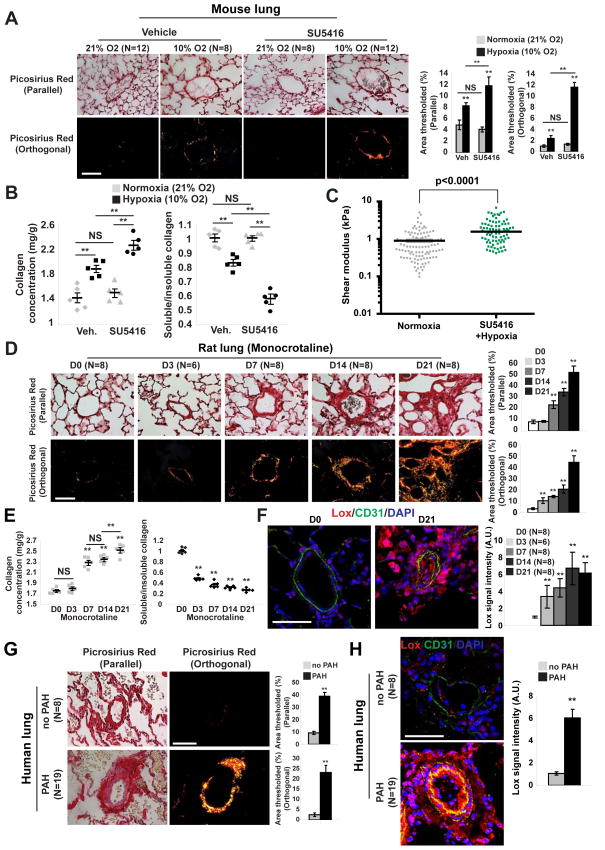
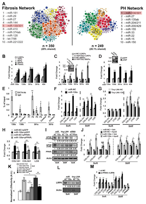
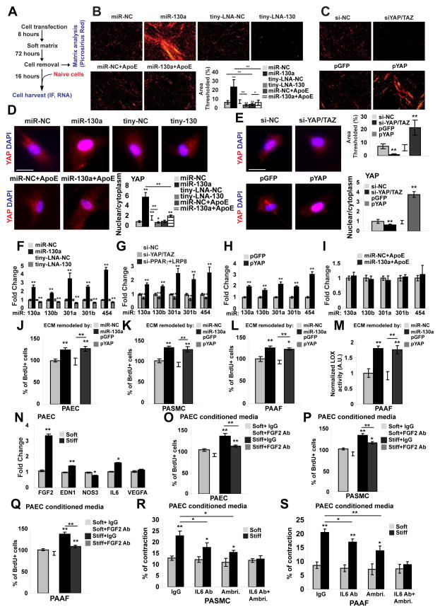
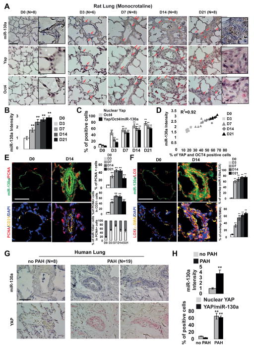
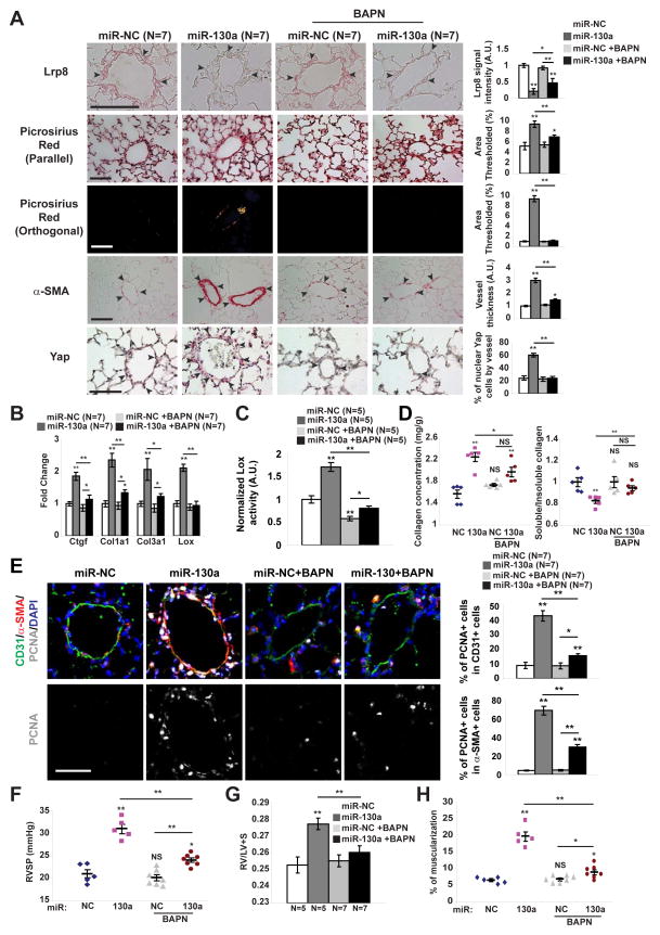
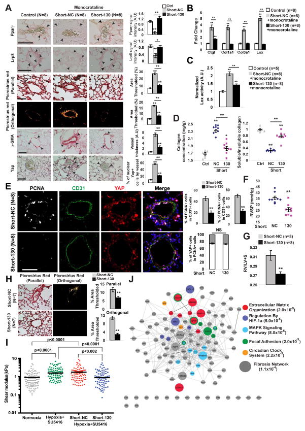
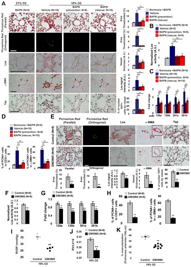
Pulmonary hypertension (PH) is a deadly vascular disease with enigmatic molecular origins. We found that vascular extracellular matrix (ECM) remodeling and stiffening are early and pervasive processes that promote PH. In multiple pulmonary vascular cell types, such ECM stiffening induced the microRNA-130/301 family via activation of the co-transcription factors YAP and TAZ. MicroRNA-130/301 controlled a PPAR?-APOE-LRP8 axis, promoting collagen deposition and LOX-dependent remodeling and further upregulating YAP/TAZ via a mechanoactive feedback loop. In turn, ECM remodeling controlled pulmonary vascular cell crosstalk via such mechanotransduction, modulation of secreted vasoactive effectors, and regulation of associated microRNA pathways. In vivo, pharmacologic inhibition of microRNA-130/301, APOE, or LOX activity ameliorated ECM remodeling and PH. Thus, ECM remodeling, as controlled by the YAP/TAZ-miR-130/301 feedback circuit, is an early PH trigger and offers combinatorial therapeutic targets for this devastating disease.


Figures

Figure 1
Figure 1. Pulmonary arteriolar ECM remodeling and stiffening is an early hallmark of PH
A–C) Mice were exposed to hypoxia +/− SU5416 (10% O2 for 3 weeks) in order to induce PH. A) Picrosirius Red stain of mouse lung tissues was imaged in parallel light to display total collagen content (top) or orthogonal light to display fibrillar collagen (bottom) (<100μm vessel diameter; 10 vessels/animal). B) Increased total collagen and fibrillar collagen (decreased Soluble/Insoluble ratio) in PH lung were demonstrated by Sircol assay. C) Atomic force microscopy revealed increased pulmonary arteriolar (<50 μm) stiffness in PH lung (n=3 mice) versus untreated (n=4 mice); horizontal lines denotes median; symbols denote individual PA measurements. P-value calculated by Mann Whitney U testing. D–F) Monocrotaline was administered to rats (3 weeks, n=6–8/time point) to induce PH. Picrosirius Red stain (D) and Sircol assay (E) revealed increased arteriolar fibrillar collagen (starting at D3) evident prior to hemodynamic disease (see Fig. S1K). Increased Lox in intimal and media of rat PH pulmonary arterioles was demonstrated by immunofluorescence (F). G) By Picrosirius Red stain of human lung, quantification of < 200μm vessels (10 vessels/patient) revealed increased collagen deposition and fibrillar collagen expression in PAH. H) Lox was increased in intima and media of human PAH lung arterioles, shown by immunofluorescence. Data are expressed as mean ± SEM (*P<0.05; ** P<0.01, *** P<0.001, **** P<0.0001). Scale bars 50 μm. See also Fig. S1.
Figure 2
Figure 2. ECM stiffening induces miR-130/301 for downstream modulation of collagen deposition and remodeling through a PPARγ-ApoE-LRP8 axis
A) A fibrosis network, composed of known fibrotic genes and their closest first degree interactors (left, Tables S2–S3) shares a large portion of its members with a PH disease network [right, as previously described [(Bertero et al., 2014b)]. Color-coding denotes architectural network clusters. Enlarged nodes are shared by both networks (70 nodes), and encircled genes are miR-130/301 direct targets (per Targetscan 6.2), thus highlighting a prominent fibrotic component among the miR-130/301 targets. miR-130/301 was ranked among the top five miRNA by “spanning score” (Bertero et al., 2014b) in both network contexts, reflecting the functional overlap and this miRNA family’s shared, systems-level control over both networks. B–C) miR-130/301 expression was quantified in human PAAFs cultured in hydrogel of varying stiffness (B) and transfected with siRNAs (YAP/TAZ versus si-NC control) (C). D) miR-130/301 was increased in human PAAFs overexpressing YAP (pYAP) versus a control vector (pGFP). E) ChIP-qPCR confirmed the presence of OCT4 binding sites (P1-4) in distinct miR-130/301 family member promoter regions near the transcription start site (proximal promoter 1Kb). Results are expressed as percent of total input DNA prior to IP. F–G) PAAFs were transfected with miR-NC, miR-130a, tiny-LNA-NC or tiny-LNA-130 and cultivated in soft or stiff matrix. Forced miR-130a expression or stiff matrix increased collagen transcripts, LOX, and CTGF, a marker of ECM stiffening and fibrosis (F). In high ECM stiffness, tiny-LNA-130 decreased this fibrotic gene cohort (G). H) By RT-qPCR in PAAFs, constitutive PPARγ (pPPARγ) reversed the miR-130a-induced up-regulation of collagen and LOX. I) By immunoblot, miR-130a, matrix stiffening, and PPARγ knockdown decreased ApoE while inhibition of miR-130/301 (tiny-LNA-130) increased ApoE in stiff conditions. J) PAAFs exposed to exogenous ApoE were resistant to miR-130a-induced up-regulation of collagen, CTGF, and LOX. K) Lox activity was measured in PAAFs cultured in soft or stiff matrix and treated with miR-130a +/− exogenous ApoE or miR-130/301 inhibitor (tiny-LNA-130). L) By immunoblot, LRP8 was reduced by miR-130a or matrix stiffening and preserved by miR-130/301 inhibition. M) siRNA knockdown of both LRP8 and PPARγ, increased collagen and CTGF. In all panels, mean expression in control groups (0.2 KPa, miR-NC, Tiny-LNA-NC or si-NC in soft matrix) was assigned a fold change of 1, to which relevant samples were compared. Data are expressed as mean ± SD (*P<0.05; ** P<0.01). See also Fig. S2–S3.
Figure 3
Figure 3. A YAP/TAZ-miR-130/301 feedback loop promotes ECM modification to induce dysregulated pulmonary vascular crosstalk
A) Schema of the experimental procedure. B) ECM staining by Picrosirius Red revealed that miR-130a increased fibrillar collagen, while miR-130/301 inhibition (tiny-LNA-130) or ApoE reversed such effects. C) Similarly, YAP (pYAP) increased fibrillar collagen as compared with control (pGFP), while knockdown of YAP/TAZ (siYAP/TAZ) decreased fibrillar collagen. D–E) In that context, immunofluorescence for YAP (red) and nuclei (DAPI, blue) was performed on naïve PAAFs plated on ECM remodeled by the indicated conditions. Nuclear stain relative to cytosolic stain of YAP was quantified (n=3 experiments with ten 20x fields analyzed per experiment). F–H) RT-qPCR revealed that ECM remodeled by miR-130a (F) or by siPPARγ+LRP8 (G) or by YAP (H) up-regulated miR-130/301 in naïve cells plated on remodeled ECM. Conversely, ECM remodeled by tiny-LNA-130 (F) or by si-YAP/TAZ (G) down-regulated miR-130/301 in naïve cells. I) miR-130/301 induction in naïve cells cultured on matrix remodeled by miR-130a was reversed by ApoE. J–M) Direct cellular effects of miR-130a/YAP-dependent stiffness were demonstrated by increased proliferation (BrdU-staining) of PAECs (J), PASMCs (K), and PAAFs (L) when culturing in ECM remodeled by PAAFs overexpressing miR-130a or YAP. Such ECM also increased LOX activity in PAAFs (M). N–S) Pulmonary vascular cell crosstalk dependent on ECM remodeling was revealed by modulation of a panel of vasoactive genes in PAECs via stiff ECM (N). Conditioned media from PAECs cultured in such stiff matrix increased proliferation in naïve PAECs (O), PASMCs (P), and PAAFs (Q), and was reversed by a FGF2 blocking antibody (FGF2 Ab). Conditioned media from PAECs cultured in stiff ECM increased contraction of PASMCs (R) and PAAFs (S). Contraction was partially reversed by ambrisentan or an IL-6 antibody (IL6 Ab) alone, and more robustly reversed by their combination. In all panels, mean expression in control groups (miR-NC, Tiny-LNA-NC, or si-NC in soft matrix) was assigned a fold change of 1, to which relevant samples were compared. Data are expressed as mean ± SD (*P<0.05; ** P<0.01). See also Fig. S4.
Figure 4
Figure 4. The YAP/TAZ-miR-130/301 molecular circuit is active in rodent and human examples of PH
A–F) Monocrotaline was administered to rats (3 weeks, n=6–8/time point) to induce PH (see Fig. 1). In serial sections of pulmonary arterioles (A), quantification of stain intensity (B) and percentage of positively stained cells (C) revealed a correlation between miR-130a, Yap, and Oct4 expression (D). E–F) Immunofluorescence stain confirmed an increase of PCNA (E) and Lox (F) expression in miR-130a positive cells G–H) In serial sections of human pulmonary arterioles, a similar relationship was observed between increased miR-130a and YAP in PAH. Data are expressed as mean ± SD (*P<0.05; ** P<0.01). Scale bars 50 μm. See also Fig. S5.
Figure 5
Figure 5. miR-130a induces YAP/TAZ-miR-130/301 to promote pulmonary vascular ECM remodeling in a LOX-dependent manner
Along with SU5416, mice received 4 weekly administrations of miR-NC or miR-130a and were treated either with daily BAPN or vehicle. A) In situ staining of mouse lung demonstrated that BAPN blunted miR-130a-specific induction of LRP8, ECM remodeling (Picrosirius Red), and medial thickening (α– SMA), thus leading to decreased YAP nuclear localization. B) By RT-qPCR, fibrillar collagen, Lox, and YAP-dependent gene expression (i.e., CTGF), were increased in miR-130a-diseased lung, but were blunted by BAPN. C–E) Lox activity (C), biochemical indices of collagen remodeling (D), and proliferation (PCNA staining in arteriolar CD31+ and α-SMA+ cells) (E) were increased in diseased lung, but blunted by BAPN. F–H) BAPN decreased the miR-130-mediated increase in PH severity, as quantified by RVSP (F), Fulton index (RV/LV+S) (G), and pulmonary arteriolar muscularization (H). Data are expressed as mean ± SEM (*P<0.05; ** P<0.01). Normalized values are expressed as arbitrary units (A.U.) in (A, C). Scale bars 50 μm.
Figure 6
Figure 6. miR-130/301 inhibition disrupts YAP/TAZ-miR-130/301 signaling and reverses a program of ECM remodeling and PH
A–G) Following monocrotaline exposure, rats were treated with Short-NC or Short-130. A) By in situ stain, Short-130 reversed monocrotaline-mediated changes in target genes Pparγ and Lrp8, decreased collagen deposition and remodeling (Picrosirius Red), and medial thickening (α-SMA). In turn, Yap nuclear localization decreased. B) RT-qPCR demonstrated a decrease in fibrillar collagen isoforms, Lox, and Ctgf in Short-130-treated lung. C) Short-130 decreased Lox activity in PH lung. D) Short-130 decreased monocrotaline-induced collagen deposition (left) and fibrillar collagen content (right) in PH lung. E) Co-immunofluorescence microscopy revealed YAP-positive proliferating cells (PCNA/YAP double-positive stain) in diseased pulmonary arterioles (Short-NC). Short-130 reduced the number of PCNA/YAP double-positive cells in CD31+ and CD31− compartments. F–G) Short-130 decreased the miR-130/301-mediated increase in PH severity, as quantified by RVSP (F) and right ventricular hypertrophy (G). H–I) After two weeks of PH induction with hy-poxia+SU5416, mice were serially injected with Short-NC or Short-130 along with hypox-ia+SU5416 for two more weeks. Short-130 decreased collagen deposition and fibrillar collagen (Picrosirius Red) (H). Atomic force microscopy revealed increased pulmonary arteriolar (<50 μm) stiffness in PH, but Short-130 blunted this alteration (I). Black lines denote median; symbols denote individual PA measurements (n=4 normoxic mice vs n=3/hypoxic group). P-values were calculated by Kruskal-Wallis testing followed by Mann Whitney U post-hoc analysis; a significance cutoff of <0.008 was based on Bonferroni correction. (J) Transcriptomic analyses of whole mouse lung were performed after exposure to hypoxia+SU5416 treated with Short-NC (n=3) or Short-130 (n=3) versus mice in normoxia+SU5416 (Control; n=3). Genes from this transcriptomic analysis were identified according to modulation by both hypoxia and miR-130/301 inhibition. Pathway enrichment identified ECM modification as the pathway with the lowest hypergeometric p-value. Genes are color-coded according to membership in the five pathways with lowest p-values. Enlarged genes are shared by the fibrosis network (see Fig. 2A). Data are expressed as mean ± SEM (*P<0.05; ** P<0.01). Scale bars 50 μm. See also Fig. S5.
Figure 7
Figure 7. Pharmacologic inhibition of LOX or activation of APOE disrupts YAP/TAZ-miR-130/301 signaling to reduce vascular ECM remodeling and PH
A–D) Mice were treated with BAPN either simultaneously with hypoxia (prevention) or after hypoxic disease induction (reversal). In prevention and rescue experiments, BAPN blunted hypoxia-mediated increases of vascular Lox, collagen remodeling (Picrosirius Red), medial thickening (α-SMA) (A), and Lox activity (B). In correlation with these ECM modifications, BAPN decreased Yap nuclear localization (A), miR-130/301 (C), and proliferation (PCNA-positive) of arteriolar CD31+ and α-SMA+ cells as compared with diseased controls (D). E–K) Hypoxic mice were treated with the LXR agonist GW3965 by prevention protocol. Similar to BAPN, the LXR agonist GW3965 blunted hypoxia-mediated increases of Lox, collagen remodeling (Picrosirius Red), medial thickening (α-SMA) (E), and Lox activity (F). Consistent with such ECM alterations, GW3965 decreased Yap nuclear localization (E), miR-130/301 (G), and downstream pulmonary vascular proliferation (PCNA stain in arteriolar CD31+ and α-SMA+ cells) compared with diseased controls (H). Consequently, GW3965 ameliorated PH severity, as quantified by RVSP (I), right ventricular hypertrophy (Fulton index, RV/LV+S) (J), and arteriolar muscularization (K). Data are expressed as mean ± SEM (*P<0.05; ** P<0.01). Normalized values are expressed as arbitrary units (A.U.) in (A, B, E, F). Scale bars 50 μm. See also Fig. S6.

References

    1. Bertero T, Cottrill K, Krauszman A, Lu Y, Annis S, Hale A, Bhat B, Waxman AB, Chau BN, Kuebler WM, et al. The microRNA-130/301 family controls vasoconstriction in pulmonary hypertension. J Biol Chem. 2014a;290:2069–2085. - PMC - PubMed
    1. Bertero T, Lu Y, Annis S, Hale A, Bhat B, Saggar R, Saggar R, Wallace WD, Ross DJ, Vargas SO, et al. Systems-level regulation of microRNA networks by miR-130/301 promotes pulmonary hypertension. J Clin Invest. 2014b;124:3514–3528. - PMC - PubMed
    1. Bi R, Bao C, Jiang L, Liu H, Yang Y, Mei J, Ding F. MicroRNA-27b plays a role in pulmonary arterial hypertension by modulating peroxisome proliferator-activated receptor gamma dependent Hsp90-eNOS signaling and nitric oxide production. Biochem Biophys Res Commun. 2015;460:469–475. - PubMed
    1. Boutet K, Montani D, Jais X, Yaici A, Sitbon O, Simonneau G, Humbert M. Therapeutic advances in pulmonary arterial hypertension. Ther Adv Respir Dis. 2008;2:249–265. - PubMed
    1. Chan SY, Loscalzo J. Pathogenic mechanisms of pulmonary arterial hypertension. J Mol Cell Cardiol. 2008;44:14–30. - PMC - PubMed

Publication types

MeSH terms