

Panretinal Photocoagulation vs Intravitreous Ranibizumab for Proliferative Diabetic Retinopathy: A Randomized Clinical Trial
- PMID: 26565927
- PMCID: PMC5567801
- DOI: 10.1001/jama.2015.15217
Panretinal Photocoagulation vs Intravitreous Ranibizumab for Proliferative Diabetic Retinopathy: A Randomized Clinical Trial
Erratum in
-
Coordinating Clinical Site Omitted.JAMA. 2016 Mar 1;315(9):944. doi: 10.1001/jama.2016.1591. JAMA. 2016. PMID: 26934270 No abstract available.
-
Errors in Analysis of Study of Photocoagulation vs Ranibizumab for Diabetic Retinopathy.JAMA. 2019 Mar 12;321(10):1008. doi: 10.1001/jama.2019.0265. JAMA. 2019. PMID: 30860547 Free PMC article. No abstract available.
Abstract
Importance: Panretinal photocoagulation (PRP) is the standard treatment for reducing severe visual loss from proliferative diabetic retinopathy. However, PRP can damage the retina, resulting in peripheral vision loss or worsening diabetic macular edema (DME).
Objective: To evaluate the noninferiority of intravitreous ranibizumab compared with PRP for visual acuity outcomes in patients with proliferative diabetic retinopathy.
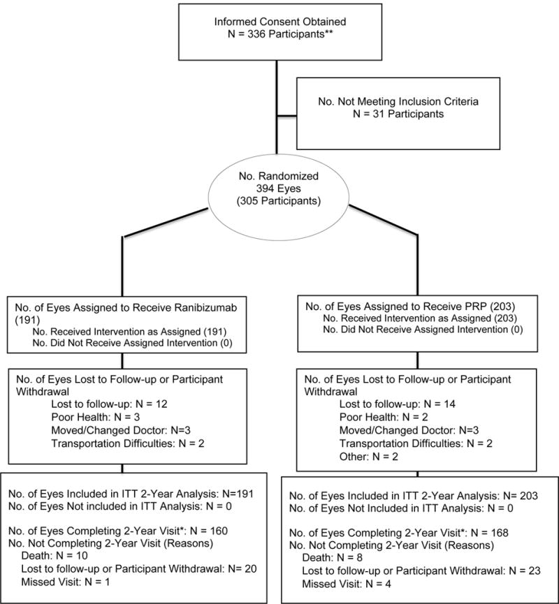
Design, setting, and participants: Randomized clinical trial conducted at 55 US sites among 305 adults with proliferative diabetic retinopathy enrolled between February and December 2012 (mean age, 52 years; 44% female; 52% white). Both eyes were enrolled for 89 participants (1 eye to each study group), with a total of 394 study eyes. The final 2-year visit was completed in January 2015.
Interventions: Individual eyes were randomly assigned to receive PRP treatment, completed in 1 to 3 visits (n = 203 eyes), or ranibizumab, 0.5 mg, by intravitreous injection at baseline and as frequently as every 4 weeks based on a structured re-treatment protocol (n = 191 eyes). Eyes in both treatment groups could receive ranibizumab for DME.
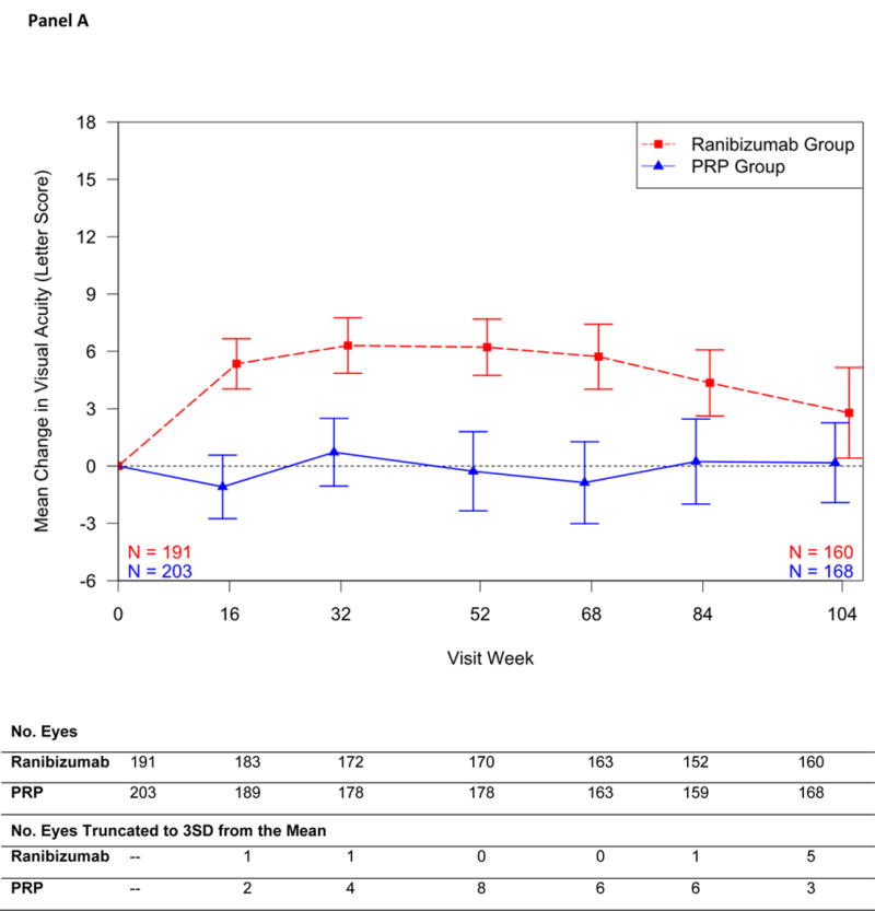
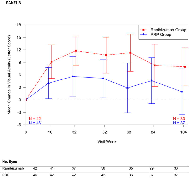
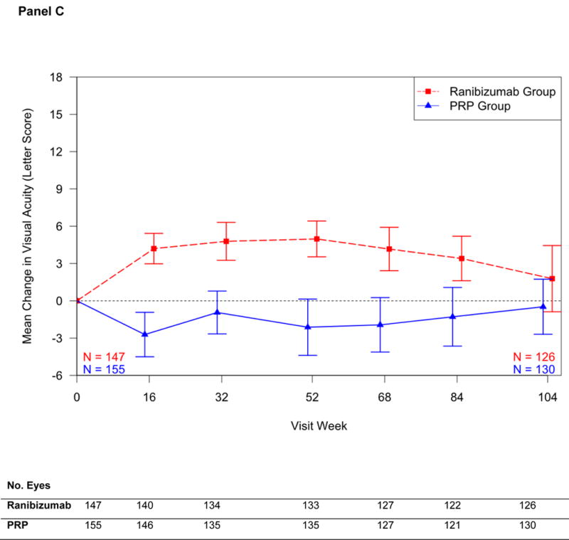
Main outcomes and measures: The primary outcome was mean visual acuity change at 2 years (5-letter noninferiority margin; intention-to-treat analysis). Secondary outcomes included visual acuity area under the curve, peripheral visual field loss, vitrectomy, DME development, and retinal neovascularization.
Results: Mean visual acuity letter improvement at 2 years was +2.8 in the ranibizumab group vs +0.2 in the PRP group (difference, +2.2; 95% CI, -0.5 to +5.0; P < .001 for noninferiority). The mean treatment group difference in visual acuity area under the curve over 2 years was +4.2 (95% CI, +3.0 to +5.4; P < .001). Mean peripheral visual field sensitivity loss was worse (-23 dB vs -422 dB; difference, 372 dB; 95% CI, 213-531 dB; P < .001), vitrectomy was more frequent (15% vs 4%; difference, 9%; 95% CI, 4%-15%; P < .001), and DME development was more frequent (28% vs 9%; difference, 19%; 95% CI, 10%-28%; P < .001) in the PRP group vs the ranibizumab group, respectively. Eyes without active or regressed neovascularization at 2 years were not significantly different (35% in the ranibizumab group vs 30% in the PRP group; difference, 3%; 95% CI, -7% to 12%; P = .58). One eye in the ranibizumab group developed endophthalmitis. No significant differences between groups in rates of major cardiovascular events were identified.
Conclusions and relevance: Among eyes with proliferative diabetic retinopathy, treatment with ranibizumab resulted in visual acuity that was noninferior to (not worse than) PRP treatment at 2 years. Although longer-term follow-up is needed, ranibizumab may be a reasonable treatment alternative, at least through 2 years, for patients with proliferative diabetic retinopathy.
Trial registration: clinicaltrials.gov Identifier: NCT01489189.
Figures
Comment in
-
Anti-VEGF Pharmacotherapy as an Alternative to Panretinal Laser Photocoagulation for Proliferative Diabetic Retinopathy.JAMA. 2015 Nov 24;314(20):2135-6. doi: 10.1001/jama.2015.15409. JAMA. 2015. PMID: 26565713 No abstract available.
-
Lucentis offers treatment alternative for diabetic retinopathy, trial finds.BMJ. 2015 Nov 16;351:h6145. doi: 10.1136/bmj.h6145. BMJ. 2015. PMID: 26577167 No abstract available.
-
Diabetes: Intravitreous ranibizumab for proliferative diabetic retinopathy.Nat Rev Endocrinol. 2016 Mar;12(3):130-1. doi: 10.1038/nrendo.2016.1. Epub 2016 Jan 22. Nat Rev Endocrinol. 2016. PMID: 26794438 No abstract available.
-
Errors in Analysis of Mean Deviations in Study of Photocoagulation vs Ranibizumab for Diabetic Retinopathy.JAMA. 2019 Mar 12;321(10):1007-1008. doi: 10.1001/jama.2019.0374. JAMA. 2019. PMID: 30860555 No abstract available.
References
-
- Antonetti DA, Klein R, Gardner TW. Diabetic retinopathy. N Engl J Med. 2012;366(13):1227–39. - PubMed
-
- Aiello LP. Angiogenic pathways in diabetic retinopathy. N Engl J Med. 2005;353:839–41. - PubMed
-
- Centers for Disease Control and Prevention. National diabetes fact sheet: general information and national estimates on diabetes in the United States. 2007 http://www.cdc.gov/diabetes/pubs/pdf/ndfs_2007.pdf. Accessed 10-13-09.
-
- Photocoagulation treatment of proliferative diabetic retinopathy. Clinical application of Diabetic Retinopathy Study (DRS) findings, DRS Report Number 8. The Diabetic Retinopathy Study Research Group. Ophthalmology. 1981;88(7):583–600. - PubMed
-
- Silva PS, Cavallerano JD, JK S, et al. Proliferative Diabetic Retinopathy. In: Ryan SJ, editor. Retina. 5th. Elsevier; Philadelphia, PA: 2013.
Publication types
MeSH terms
Substances
Associated data
Grants and funding
LinkOut - more resources
Full Text Sources
Other Literature Sources
Medical
Research Materials
Miscellaneous
