Review
. 2015:31:171-99.
doi: 10.1146/annurev-cellbio-100814-125211.

Bacterial chromosome organization and segregation

Affiliations
Review

Bacterial chromosome organization and segregation

Anjana Badrinarayanan et al. Annu Rev Cell Dev Biol. 2015.

Abstract

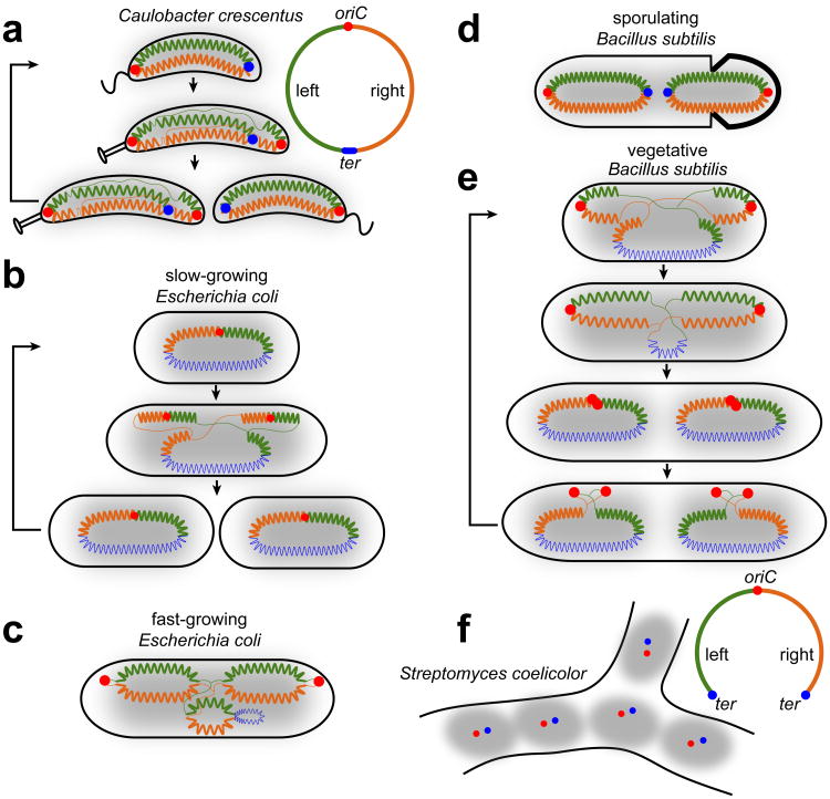
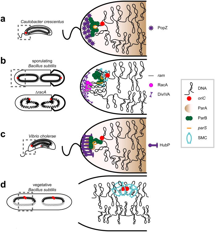
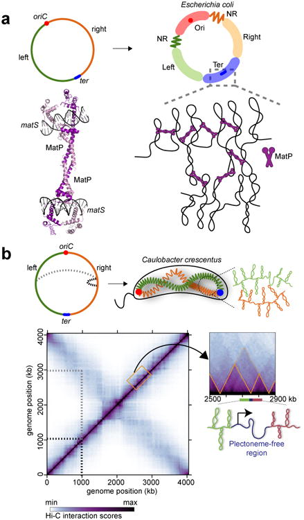
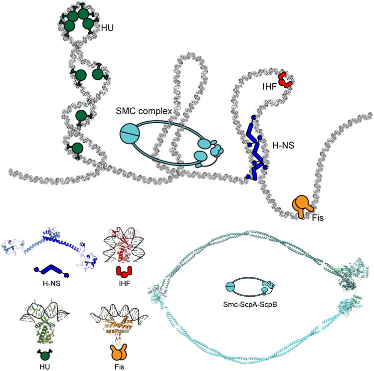
If fully stretched out, a typical bacterial chromosome would be nearly 1 mm long, approximately 1,000 times the length of a cell. Not only must cells massively compact their genetic material, but they must also organize their DNA in a manner that is compatible with a range of cellular processes, including DNA replication, DNA repair, homologous recombination, and horizontal gene transfer. Recent work, driven in part by technological advances, has begun to reveal the general principles of chromosome organization in bacteria. Here, drawing on studies of many different organisms, we review the emerging picture of how bacterial chromosomes are structured at multiple length scales, highlighting the functions of various DNA-binding proteins and the impact of physical forces. Additionally, we discuss the spatial dynamics of chromosomes, particularly during their segregation to daughter cells. Although there has been tremendous progress, we also highlight gaps that remain in understanding chromosome organization and segregation.

Keywords: Hi-C; ParA-ParB-parS; macrodomains; nucleoid-associated proteins; supercoiling; transcription.


Figures

Figure 1
Figure 1. The global organization of bacterial chromosomes
For the organism indicated in each panel, the schematics represent the origin of replication (oriC) as a red dot and terminus (ter) as blue dot or line. The left and right arms of the chromosome are colored green and orange, respectively. Thick zigzag lines denote compacted parts of the chromosome, while newly-synthesized DNA and hypothetically less-organized DNA are illustrated as thin lines. Overall nucleoid distribution is illustrated by grey shading. Black arrows indicate the progression of the global chromosome organization through a cell cycle.
Figure 2
Figure 2. Protein-based systems that anchor specific DNA regions
Schematics of polar anchoring complexes are shown for (a) C. crescentus, (b) sporulating B. subtilis, (c) V. cholerae and (d) vegetative B. subtilis. The likely global chromosome organization defect of a B. subtilis strain lacking RacA is shown in panel b. Specific DNA elements and proteins common to each organism are represented as shown in the legend (right), with species-specific factors indicated adjacent to each panel. Note that Soj/ParA is not represented in B. subtilis; although Soj/ParA is required for the bipolar localization of origins, its own localization is complex (Murray & Errington 2008), and precisely how localization impacts function is unclear. Schematics derived from similar drawings in (Wang & Rudner 2014).
Figure 3
Figure 3. Macrodomains and chromosomal-interaction domains
(a) Macrodomain-organization of E. coli chromosome, shown as in Figure 1 (left) or with the four macrodomains, Ori, Ter, Left and Right, and the two non-structured regions (NR) (right). MatP (colored purple) organizes the Ter macrodomain. The crystal structure of two MatP dimers, each bound to a matS recognition site is shown (PDB: 4D8J). (b) Chromosome conformation capture assay (5C and Hi-C) and computational modeling have revealed the organization of the C. crescentus chromosome. The Hi-C heat map (Le et al. 2013) indicates frequency of DNA-DNA interactions across the genome using the color scale shown. The most prominent diagonal indicates frequent interactions within a chromosomal arm (black dotted lines), while the other, less prominent diagonal shows interactions between the two arms (grey dotted lines). Orange triangles in the inset (right) indicate CIDs, or chromosomal-interaction domains (see text for details). Highly-transcribed genes are thought to create a less compacted, plectoneme-free region (blue) that serves to spatially insulate DNA (green and red) in adjacent domains, creating a CID boundary.
Figure 4
Figure 4. Nucleoid-associated proteins with DNA bridging, wrapping, or bending activities contribute to the organization of the chromosome
The functions of well-studied NAPs are schematized at the top, with the corresponding crystal structures below. H-NS dimers of dimers (blue) bridge DNA. The abundant HU (green) introduces ∼90° bending to DNA and may wrap DNA around itself, thereby promoting short-range DNA interactions. IHF (red) binding to DNA induces a dramatic U-turn on DNA that drastically changes the trajectory of the DNA backbone. Fis (orange) is another NAP with DNA-bending activity. SMC complexes (cyan) likely form a ring structure that can bring together and handcuff loci that are distal in primary sequence. The protein and protein:DNA complexes shown have PDB IDs: 1P78, 1IHF, and 3JRA for HU, IHF, and Fis, respectively. A hypothetical model of H-NS was constructed from PDB structures 3NR7 and 1HNR. A model of SMC-ScpA-ScpA was derived from PDB structures 4I98, 4I99 and 3ZGX.
Figure 5
Figure 5. Chromosome segregation
(a) Origin segregation in C. crescentus relies on the parABS system. ParB (green) binds parS sites located near the origin. Shortly after replication, one ParB:parS complex remains polarly localized while the second complex comes in contact with ATP-bound ParA (light brown). ParB stimulates ParA ATPase activity, resulting in the release of ParA from DNA (dark brown) and contraction of the cloud of ParA-ATP. The migrating ParB:parS complex can then move toward the retracting ParA-ATP and thus toward the opposite pole, eventually resulting in full segregation of the origin. PopZ (purple) influences ParAB activity directly or indirectly to promote origin segregation. (b) In vegetatively growing B. subtilis, chromosome organization oscillates between ori-ter and left-ori-right patterns. While ParA/Soj and ParB/Spo0J (green) ensure origin movement towards opposite poles, the SMC complex (cyan) relocates the origins to mid cell during the initial phase of DNA replication. This oscillation in chromosome organization may promote chromosome segregation by preventing entanglement of the chromosomes. (c) Origin segregation in E. coli. Unlike Caulobacter and B. subtilis, E. coli does not have a ParAB-like system for origin segregation. A distant relative of the SMC complex, MukBEF (cyan), localizes around the origin region and is thought to promote origin segregation and origin-proximal chromosome organization. MukBEF may also promote bulk chromosome segregation. (d) Circular chromosome replication can result in dimeric or catenated chromosomes, whose resolution requires the action of the DNA translocase FtsK (purple) and the tyrosine recombinase XerCD (blue-brown). Schematic shown is for E. coli. (e) Chromosome segregation in sporulating B. subtilis. Segregation of the origin region depends on RacA (pink) and Spo0J (green), with the rest of the chromosome pumped into the forespore by the DNA translocase SpoIIIE (blue). The origin is anchored to the cell pole by RacA and DivIVA (purple sticks).

References

    1. Adams D, Shekhtman E, Zechiedrich E, Schmid M, Cozzarelli N. The role of topoisomerase-IV in partitioning bacterial replicons and the structure of catenated intermediates in DNA replication. Cell. 1992;71(2):277–88. - PubMed
    1. Adams DW, Wu LJ, Errington J. Nucleoid occlusion protein Noc recruits DNA to the bacterial cell membrane. EMBO J 2015 - PMC - PubMed
    1. Auner H, Buckle M, Deufel A, Kutateladze T, Lazarus L, et al. Mechanism of transcriptional activation by FIS: role of core promoter structure and DNA topology. J Mol Biol. 2003;331(2):331–44. - PubMed
    1. Austin SJ, Mural RJ, Chattoraj DK, Abeles AL. Trans- and cis-acting elements for the replication of P1 miniplasmids. J Mol Biol. 1985;183(2):195–202. - PubMed
    1. Azam TA, Hiraga S, Ishihama A. Two types of localization of the DNA-binding proteins within the Escherichia coli nucleoid. Genes Cells. 2000;5(8):613–26. - PubMed

Publication types

MeSH terms

LinkOut - more resources