. 2016 Jan 1;76(1):50-61.
doi: 10.1158/0008-5472.CAN-14-3619. Epub 2015 Nov 13.

Radiotherapy Combined with Novel STING-Targeting Oligonucleotides Results in Regression of Established Tumors

Affiliations

Radiotherapy Combined with Novel STING-Targeting Oligonucleotides Results in Regression of Established Tumors

Jason R Baird et al. Cancer Res. .

Abstract

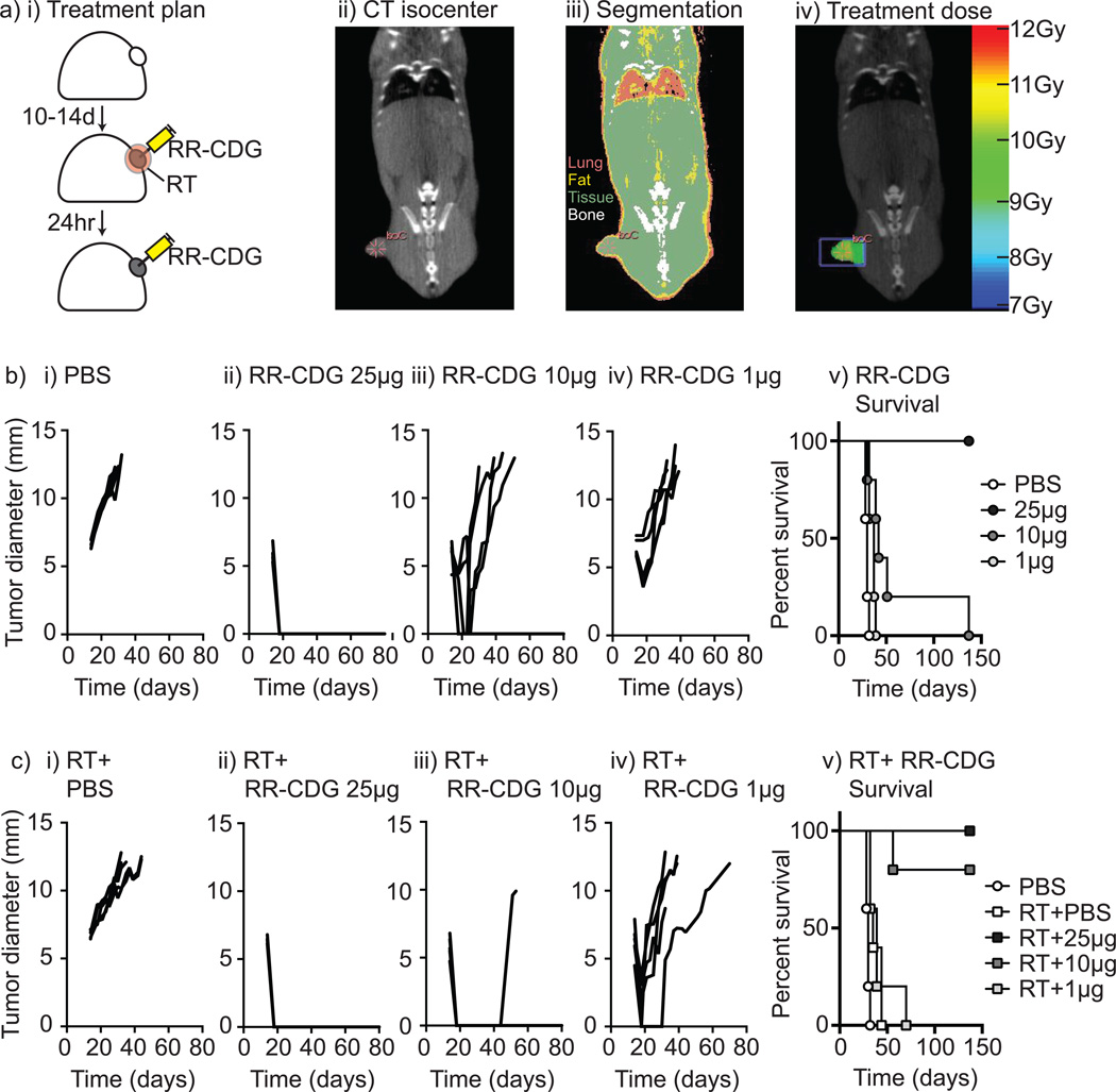
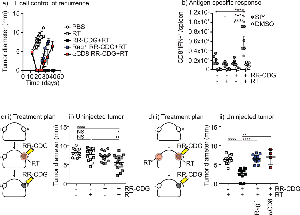
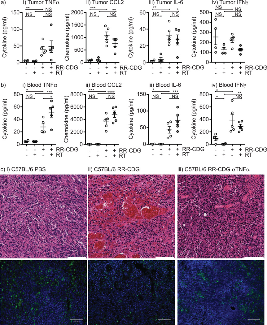
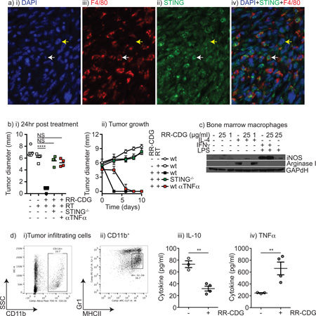
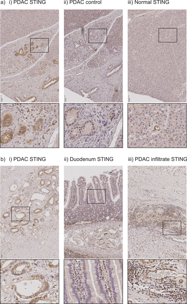
Cytotoxic therapies prime adaptive immune responses to cancer by stimulating the release of tumor-associated antigens. However, the tumor microenvironment into which these antigens are released is typically immunosuppressed, blunting the ability to initiate immune responses. Recently, activation of the DNA sensor molecule STING by cyclic dinucleotides was shown to stimulate infection-related inflammatory pathways in tumors. In this study, we report that the inflammatory pathways activated by STING ligands generate a powerful adjuvant activity for enhancing adaptive immune responses to tumor antigens released by radiotherapy. In a murine model of pancreatic cancer, we showed that combining CT-guided radiotherapy with a novel ligand of murine and human STING could synergize to control local and distant tumors. Mechanistic investigations revealed T-cell-independent and TNFα-dependent hemorrhagic necrosis at early times, followed by later CD8 T-cell-dependent control of residual disease. Clinically, STING was found to be expressed extensively in human pancreatic tumor and stromal cells. Our findings suggest that this novel STING ligand could offer a potent adjuvant for leveraging radiotherapeutic management of pancreatic cancer.


Conflict of interest statement

Conflict of interest:

Dr. Dubensky and Dr. Kanne are employees of Aduro Biotech, Inc., as listed in their author affiliations. Aduro Biotech did not finance or direct the study but did construct and provide the STING ligand as described. Dr. Crittenden has served as a consultant for or on the advisory board of Regeneron. No other conflicts are present.

Figures

Figure 1
Figure 1. RR-CDG synergizes with radiation therapy for durable tumor cures
Panc02 pancreatic adenocarcinoma tumors were established in immune competent C57BL/6 mice and a) randomized to remain untreated or to receive radiation therapy (RT) immediately followed by control (PBS) injections or 25, 10 or 1µg of RR-CDG into the tumor in matched volumes of PBS. ii) Tumor isocenters were identified by CT guidance. iii) CT images were used to determine tissue densities for iv) dosimetry using the SARRP to deliver 10Gy to isocenter with angled beams to minimize dose to radiosensitive organs. b) i–iv) growth of individual tumors in mice not treated with radiation therapy and v) overall survival. c) i–iv) growth of individual tumors in mice treated with radiation therapy and v) overall survival. Experiments incorporate 6–8 mice per group and the displayed experiment is representative of 3 independent repeats.
Figure 2
Figure 2. Radiation plus RR-CDG generates a T cell response capable of controlling distant tumors
C57BL/6 or Rag1−/− mice challenged with Panc02 were left untreated or treated with 10Gy focal radiation (RT) to the tumor followed immediately by intratumoral injection of 25µg RR-CDG or PBS with a repeat injection the following day. Comparison groups of C57BL/6 mice were treated with anti-CD8 depleting antibodies 1 day prior to RT and again 1 week later. a) Graphs show average tumor diameter. b) Panc02-SIY tumors were established and treated as in a) 7 days following treatment spleens were harvested and tested for SIY-peptide specific IFNγ production by intracellular cytokine staining. Graphs show the number of antigen-specific (SIY) or control (DMSO) CD8+IFNg+ cells per spleen. Each symbol represents one animal. c) i) Duplicate Panc02 tumors were established on each flank of C57BL/6 mice and only one tumor was treated as in a). ii) Graph shows tumor diameter of the untreated tumor 10 days following treatment. d) i) Duplicate Panc02 tumors were established on each flank of C57BL/6 or Rag1−/− mice and both tumors were treated with 10Gy RT, but only one tumor was treated with RR-CDG as in a). Additional groups were treated with anti-CD8 depleting antibodies 1 day prior to RT and again 1 week later. ii) Graph shows the tumor diameter of the uninjected tumor 10 days following treatment. Each symbol represents one mouse. Statistics calculated by ANOVA with Tukey’s multiple comparisons test: * p<0.05; ** p<0.01; *** p<0.005; **** p<0.001. The displayed experiment is representative of 2 independent repeats.
Figure 3
Figure 3. RR-CDG causes rapid vascular and macrophage reorganization in the tumor
a) Established Panc02 tumors in C57BL/6 mice were treated with intratumoral injection of i) PBS vehicle or ii) 25µg RR-CDG and harvested 6 hours later. At harvest 3 RR-CDG-treated tumors show macroscopic evidence of blood infiltrate. b) Histology of i) control and ii) RR-CDG-treated tumors at 6 hours. c) Immunofluorescence microscopy of i+iii) control and ii+iv) RR-CDG-treated tumors at 6 hours with staining for smooth muscle actin - red; CD3 - magenta; a DAPI nuclear stain - blue and either i–ii) CD31 - green or iii–iv) F4/80 - green. Experiments are representative of 2 independent replicates that include 4 or more mice per group. d) Pdx-Cre+/− Kras(G12D)+/− Trp53(R172H)+/− mice were treated with i) PBS or ii–iii) RR-CDG administered to established pancreatic masses via laparotomy. Images show representative sections of i–ii) tumor or iii) adjacent normal pancreas.
Figure 4
Figure 4. RR-CDG treatment results in cytokine secretion that causes early tumor regression
C57BL/6 mice challenged with Panc02 were left untreated or treated with 10Gy focal radiation (RT) to the tumor followed immediately by intratumoral injection of 25µg RR-CDG or PBS with a repeat injection the following day. Cytokine and chemokine levels were determined in the a) tumor or b) peripheral blood 6 hours following treatment by multiplex bead assay. Each symbol represents one mouse. Graphs additionally show mean plus SEM. c) C57BL/6 mice bearing established Panc02 tumors were treated with intratumoral injection of i) PBS, ii) 25µg RR-CDG or iii) 500µg anti-TNFα blocking antibody 6 hours prior to treatment with 25µg RR-CDG. Tumors were harvested 24 hours later for H&E staining in upper images or immunostained for CD45 (green) alongside DAPI (blue) in lower images. Scale bar (white) shows 100µM. Statistics calculated by ANOVA with Tukey’s multiple comparisons test: * p<0.05; ** p<0.01; *** p<0.005; **** p<0.001. Experiments incorporate 6–8 mice per group and the displayed experiment is representative of 2 independent repeats.
Figure 5
Figure 5. RR-CDG suppresses M2 differentiation and generates pro-inflammatory responses from tumor macrophages
a) Immune histology in Panc02 tumors showing i) DAPI, ii) F4/80, iii) STING and iv) a combined image. White arrow indicates a F4/80+ macrophage, yellow arrow a cancer cell. b) Wild-type C57BL/6 or STING−/− mice bearing established Panc02 tumors were left untreated or treated with 10Gy focal radiation along with concurrent intratumoral injection of PBS or 25µg RR-CDG and the injection was repeated 24 hours later. A control group of wild-type mice were treated with 500µg anti-TNFα blocking antibody 6 hours prior to treatment with 25µg RR-CDG and again 48 hours later. i) Average tumor diameter 24 hours following treatment. ii) tumor growth over time following treatment. c) Bone marrow macrophages were left untreated, treated with IL-4 to direct M2 differentiation, or IFNγ and LPS to direct M1 differentiation in the presence or absence of RR-CDG at 1 or 25µg/ml. 24 hours later macrophage differentiation was determined by western blotting for iNOS and Arginase I with GAPdH as a loading control. d) Tumor macrophages were sorted from Panc02 tumors by i) first gating for CD11b+ and ii) subgating on the MHCII+Gr1int population. c) sorted tumor macrophages were left untreated or treated with 25µg/ml RR-CDG for 6 hours, and secretion of i) IL-10 or ii) TNFα was determined by bead assay. Statistics calculated by ANOVA with Tukey’s multiple comparisons test: * p<0.05; ** p<0.01; *** p<0.005; **** p<0.001. Experiments incorporate 4–8 mice per group and the displayed experiment is representative of 2 independent repeats.
Figure 6
Figure 6. Expression of STING in pancreatic adenocarcinoma tumor and stroma
a) T3 pancreatic adenocarcinoma treated with neoadjuvant chemoradiation followed by resection showing i–ii) residual cancer or iii) residual normal regions of the tumor-bearing pancreas stained for i+iii) STING or ii) control staining. b) Resected tissue samples from i) untreated T3 pancreatic adenocarcinoma stained with STING; ii) adjacent normal duodenum resected as part of pancreaticoduodenectomy; iii) lymphoid aggregates in resected pancreas. Black scale bar shows 100µM. Boxes show origin of expanded images.

References

    1. Ludgate CM. Clinical cancer research : an official journal of the American Association for Cancer Research. 2012. Optimizing Cancer treatments to induce an acute immune response; radiation abscopal effects, PAMPS and DAMPS. - PubMed
    1. Apetoh L, Ghiringhelli F, Tesniere A, Obeid M, Ortiz C, Criollo A, et al. Toll-like receptor 4-dependent contribution of the immune system to anticancer chemotherapy and radiotherapy. Nature medicine. 2007;13(9):1050–1059. - PubMed
    1. Reits EA, Hodge JW, Herberts CA, Groothuis TA, Chakraborty M, Wansley EK, et al. Radiation modulates the peptide repertoire, enhances MHC class I expression, and induces successful antitumor immunotherapy. The Journal of experimental medicine. 2006;203(5):1259–1271. - PMC - PubMed
    1. Garnett CT, Palena C, Chakraborty M, Tsang KY, Schlom J, Hodge JW. Sublethal irradiation of human tumor cells modulates phenotype resulting in enhanced killing by cytotoxic T lymphocytes. Cancer Res. 2004;64(21):7985–7994. - PubMed
    1. Chakraborty M, Abrams SI, Coleman CN, Camphausen K, Schlom J, Hodge JW. External beam radiation of tumors alters phenotype of tumor cells to render them susceptible to vaccine-mediated T-cell killing. Cancer research. 2004;64(12):4328–4337. - PubMed

Publication types

MeSH terms