. 2015 Nov 11;18(5):549-59.
doi: 10.1016/j.chom.2015.10.013.

Filamentous Bacteriophage Promote Biofilm Assembly and Function

Affiliations

Filamentous Bacteriophage Promote Biofilm Assembly and Function

Patrick R Secor et al. Cell Host Microbe. .

Abstract

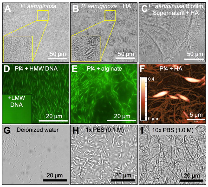
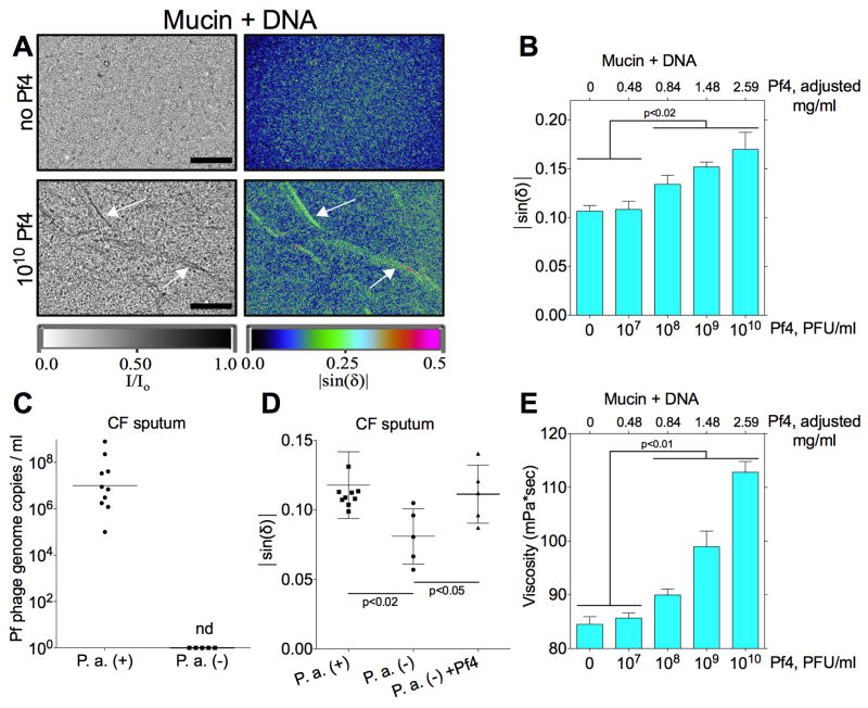
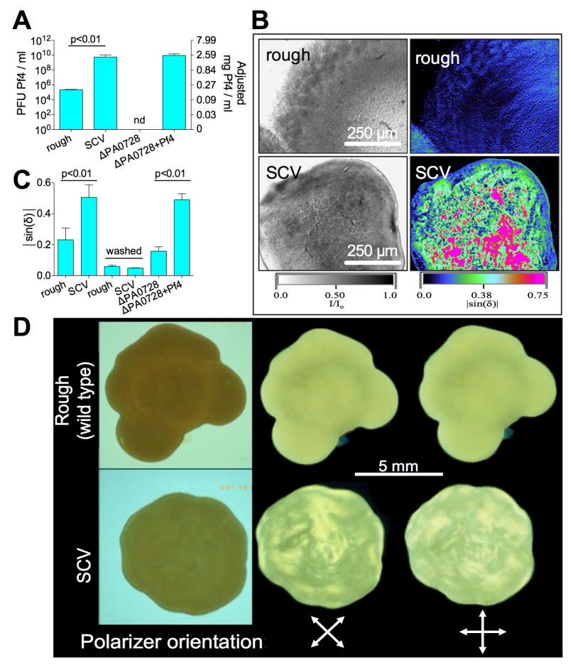
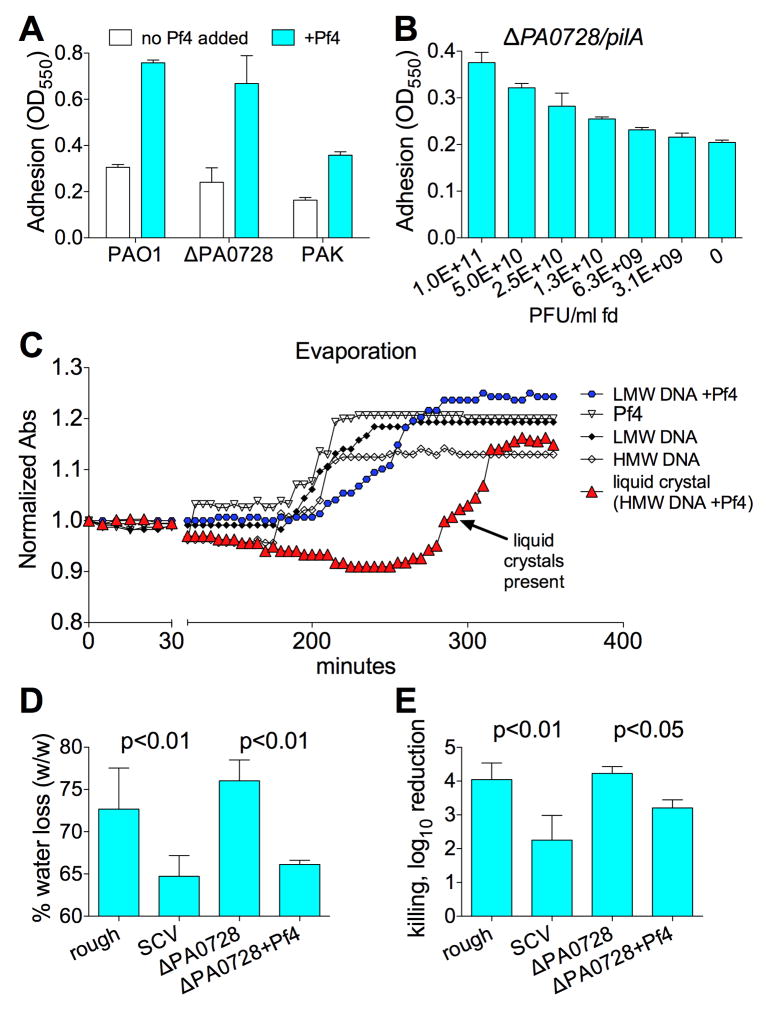
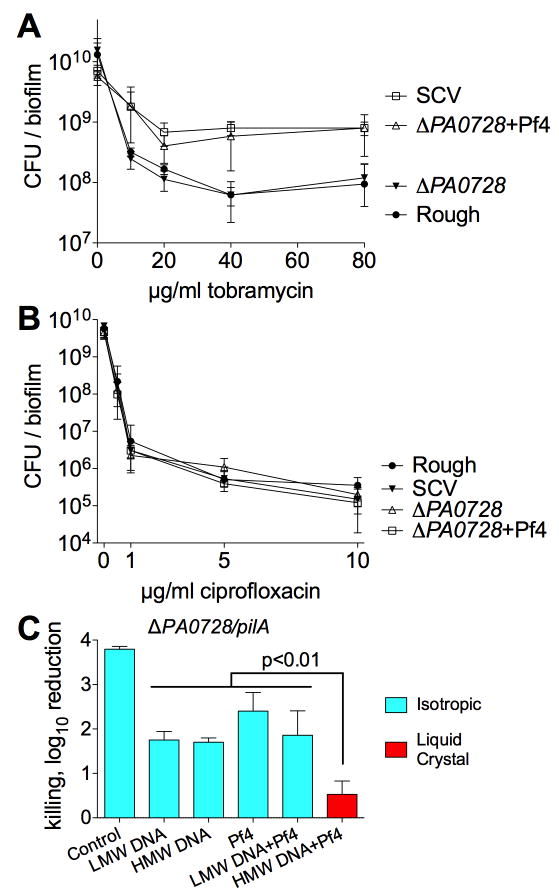
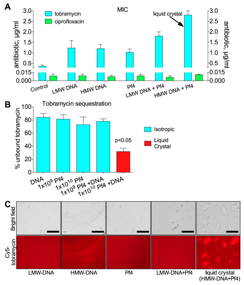
Biofilms-communities of bacteria encased in a polymer-rich matrix-confer bacteria with the ability to persist in pathologic host contexts, such as the cystic fibrosis (CF) airways. How bacteria assemble polymers into biofilms is largely unknown. We find that the extracellular matrix produced by Pseudomonas aeruginosa self-assembles into a liquid crystal through entropic interactions between polymers and filamentous Pf bacteriophages, which are long, negatively charged filaments. This liquid crystalline structure enhances biofilm function by increasing adhesion and tolerance to desiccation and antibiotics. Pf bacteriophages are prevalent among P. aeruginosa clinical isolates and were detected in CF sputum. The addition of Pf bacteriophage to sputum polymers or serum was sufficient to drive their rapid assembly into viscous liquid crystals. Fd, a related bacteriophage of Escherichia coli, has similar biofilm-building capabilities. Targeting filamentous bacteriophage or the liquid crystalline organization of the biofilm matrix may represent antibacterial strategies.


Conflict of interest statement

The authors declare no conflict of interest.

Figures

Figure 1
Figure 1. The Filamentous Phage Pf4 Interacts With Host and Microbial Polymers To Spontaneously Assemble Higher Order Structures
(A) P. aeruginosa forms a flat confluent biofilm in vitro. (B) P. aeruginosa supplemented with 5 mg/ml HA forms morphologically complex biofilms in vitro. (C) The addition of P. aeruginosa biofilm supernatant to HA (5 mg/ml) results in the spontaneous formation of higher order structures. (D) Purified, fluorescently labeled Pf4 (green, 8.8 × 10⁹ PFU/ml) mixed with 5 mg/ml DNA 2 kbp in size (HMW) forms large, interwoven structures while DNA <0.3 kbp in size (LMW) does not (inset). (E) Purified, fluorescently labeled Pf4 (green, 8.8 × 10⁹ PFU/ml) mixed with 5 mg/ml alginate forms large, interwoven structures. (F) Visualization of structures formed from Pf4 and HA by AFM semi-contact topography. The color scale indicates height. (G-I) Pf4 (8.8 × 10⁹ PFU/ml) and HA (5 mg/ml) were suspended in: DI water, 1× PBS, or 10× PBS. See also Figure S1 and S2.
Figure 2
Figure 2. Mixtures of Pf4 and Polymers Assemble Birefringent Liquid Crystals
(A) The birefringence of the indicated polymer (all 10 mg/ml) were quantified (|sin(δ)|) alone or in the presence of Pf4 (mixed 1:1 with 1×10¹⁰ PFU/ml). Scale bars, 10 μm. (B-D) Phase diagrams showing the relationship between polymer concentration, Pf4 concentration, and liquid crystal assembly. Note the negative slope of the phase boundary (dotted lines), indicative of depletion attraction. See text for details. See also Figure S3.
Figure 3
Figure 3. Pf4 Assembles Liquid Crystalline Structures in the Presence of Disease Relevant Polymers and Increases Viscosity
(A) Representative images of mixtures of mucin (8% solids) and DNA (HMW, 4 mg/ml) in the presence or absence of Pf4. Transmitted light is displayed as I/Io where I = intensity of light emerging from the sample and Io = intensity of incident light. Birefringence is displayed as |sin(δ)|. Arrows indicate filament assembly. Scale bars, 200 μm. (B) Birefringence was quantified as |sin(δ)| in mixtures of mucin and DNA described in (A) supplemented with increasing amounts of Pf4. Results are mean ± SD of 3 experiments. (C) Pf phage were quantified by qPCR in sputum collected from CF patients infected by P. aeruginosa (P. a. (+), n = 10) or patients not infected by P. aeruginosa (P. a. (-), n = 5). Results are plotted as the geometric mean, nd; not detected. (D) The birefringence (|sin(δ)|) of sputum samples described in (C) was quantified. The addition of 108 PFU/ml Pf4 to P. a. (-) sputum augmented birefringence. Results are mean ± SD of 3 experiments. (E) Changes in the viscosity (mPa*sec) of mucin + DNA samples described in (A) were measured after supplementation with Pf4. Results are mean ± SD of 4 experiments. See also Figure S4.
Figure 4
Figure 4. Pf4 Organizes the P. aeruginosa Biofilm Matrix Into a Liquid Crystalline Structure
(A) Pf4 production by colony biofilms formed from the indicated strains were enumerated as PFU/ml. Adjusted total Pf phage content are also plotted on the right axis, see text and Figure S4A. Results are mean ± SD of 3 experiments. (B) Representative images of rough and SCV colony biofilms showing transmitted light (displayed as I/Io) and birefringence (|sin(δ)|). (C) Birefringence (|sin(δ)|) of the indicated colony biofilms was quantified after normalizing for sample thickness. Birefringence was again measured after washing of the bacteria to remove the extracellular matrix. Results are mean ± SD of 4 experiments. (D) Representative images for SVC and “rough” colony biofilms (placed between glass plates) visualized through crossed polarizing lenses. Birefringence is visualized as bright areas where light passes through both polarizing lenses. The birefringence patterns change when the sample is rotated with respect to the polarizing lenses, revealing extended areas of birefringence. See also Figure S6.
Figure 5
Figure 5. P. aeruginosa Biofilms With Liquid Crystalline Matrices Display Increased Adhesion and Tolerance to Desiccation
(A) Biofilm adhesion after 24 hours of growth as measured by the crystal violet adhesion assay. Results are mean ± SD of 3 experiments. (B) Adhesion of biofilms formed by strain ΔPA0728/pilA in the setting of increasing concentrations of fd phage. (C) Evaporation of isotropic and liquid crystalline phases of Pf4 and DNA was monitored by absorbance (Abs, 600 nm). Absorbance was normalized to initial readings for each sample. As samples dried, the absorbance increased and stabilized once dried to completeness. In this way, the retention of water was monitored over time. Results are mean of 3 experiments; error bars are omitted for clarity. (D) Colony biofilms formed from the indicated strains were desiccated for 18-h at 37°C in an ambient incubator. The percent water loss was measured as the wet weight pre desiccation / wet weight post desiccation. Results are mean ± SD of 6 experiments for rough and SCV biofilms and 3 experiments for ΔPA0728 +/- Pf4 biofilms. (E) Killing by desiccation is represented as the log10 reduction of viable cells recovered from control biofilms compared to desiccated biofilms. Results are mean ± SD of 3 experiments. See also Figure S5.
Figure 6
Figure 6. The Liquid Crystalline Biofilm Matrix Protects Against Tobramycin but not Ciprofloxacin
(A and B) CFUs recovered from colony biofilms after treatment (90 minutes) with increasing concentrations of (A) tobramycin or (B) ciprofloxacin are plotted. Results are mean ± SD of 3 experiments. (C) To investigate whether or not Pf phage mediate tolerance to tobramycin through an extracellular mechanism, planktonic ΔPA0728/pilA, which cannot produce nor be infected by Pf4, was suspended in isotropic or liquid crystalline phases of Pf4 (1010 PFU/ml) and DNA (2.5 mg/ml). Killing is represented as the log10 reduction of viable cells recovered from cultures treated with tobramycin (10 μg/ml, 90 min) compared to untreated controls. Results are mean ± SD of 3 experiments. See also Figures S5 and S7.
Figure 7
Figure 7. The Liquid Crystalline Biofilm Matrix Increases Antibiotic Tolerance to Antibiotics by Enhancing Aminoglycoside Binding
(A) Antibiotic killing in the presence of DNA and Pf4 was investigated. Increasing concentrations of tobramycin or ciprofloxacin were added to isotropic or liquid crystal containing mixtures of Pf4 (1010 PFU/ml) and DNA (2.5 mg/ml). The arrow indicates the only sample with liquid crystals. E. coli was then added and samples were incubated overnight. The MIC for each antibiotic was plotted. Results are mean ± SD of 3 experiments. (B) Tobramycin (200 μg/ml) was mixed with the indicated amounts of Pf4 and DNA (2.5 mg/ml) and placed into a dialysis cassette. The diffusion of unbound tobramycin across the membrane was monitored by HPLC-MS. Results were normalized to controls containing no Pf4 or DNA. Results are mean ± SD of duplicate experiments. (C) Binding of fluorescent tobramycin (Cy5-tobramycin, 40 μg/ml) to isotropic and liquid crystalline phases of Pf4 (1010 PFU/ml) and HMW-DNA (2.5 mg/ml) was visualized by fluorescent microscopy. Scale bars, 20 μm. See also Figures S5 and S7.

Comment in

Similar articles

Cited by

References

    1. Asakura S, Oosawa F. Interaction between Particles Suspended in Solutions of Macromolecules. J Polym Sci. 1958;33:183–192.
    1. Boles BR, Singh PK. Endogenous oxidative stress produces diversity and adaptability in biofilm communities. Proc Natl Acad Sci U S A. 2008;105:12503–12508. - PMC - PubMed
    1. Brandt T, Breitenstein S, von der Hardt H, Tummler B. DNA concentration and length in sputum of patients with cystic fibrosis during inhalation with recombinant human DNase. Thorax. 1995;50:880–882. - PMC - PubMed
    1. Castang S, Dove SL. Basis for the essentiality of H-NS family members in Pseudomonas aeruginosa. J Bacteriol. 2012;194:5101–5109. - PMC - PubMed
    1. Costerton JW, Stewart PS, Greenberg EP. Bacterial biofilms: a common cause of persistent infections. Science. 1999;284:1318–1322. - PubMed

Publication types

MeSH terms

LinkOut - more resources