Review
. 2015 Dec;15(12):771-83.
doi: 10.1038/nri3919. Epub 2015 Nov 16.

IL-15 functions as a danger signal to regulate tissue-resident T cells and tissue destruction

Affiliations
Review

IL-15 functions as a danger signal to regulate tissue-resident T cells and tissue destruction

Bana Jabri et al. Nat Rev Immunol. 2015 Dec.

Abstract

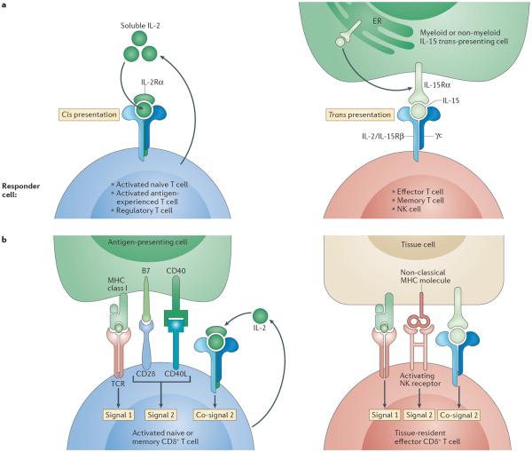
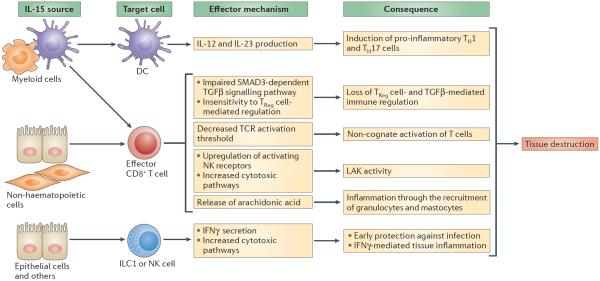
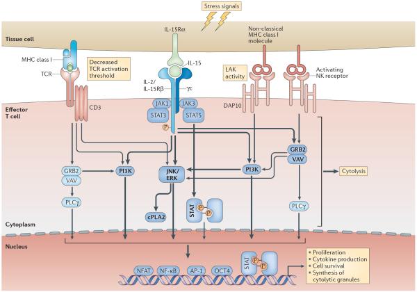
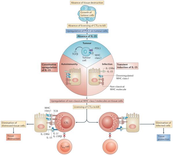
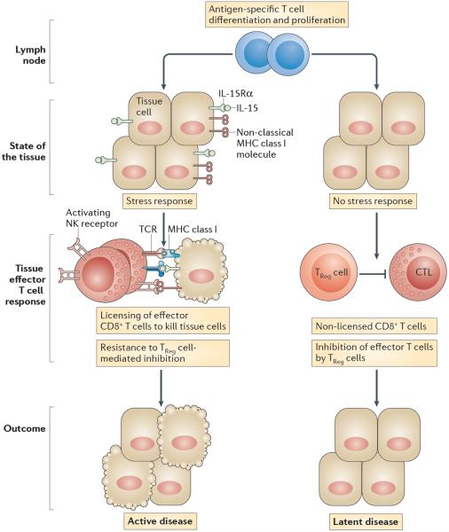
In this Opinion article, we discuss the function of tissues as a crucial checkpoint for the regulation of effector T cell responses, and the notion that interleukin-15 (IL-15) functions as a danger molecule that communicates to the immune system that the tissue is under attack and poises it to mediate tissue destruction. More specifically, we propose that expression of IL-15 in tissues promotes T helper 1 cell-mediated immunity and provides co-stimulatory signals to effector cytotoxic T cells to exert their effector functions and drive tissue destruction. Therefore, we think that IL-15 contributes to tissue protection by promoting the elimination of infected cells but that when its expression is chronically dysregulated, it can promote the development of complex T cell-mediated disorders associated with tissue destruction, such as coeliac disease and type 1 diabetes.


Figures

Figure 1
Figure 1. Models contrasting IL-15 and IL-2 signalling and the regulation of naive versus tissue-resident effector memory T cells
a | Interleukin-15 (IL-15) signalling compared with IL-2 signalling. The main mechanism by which IL-15 interacts with its receptor in vivo is trans-presentation. IL-15 is assembled as an IL-15–IL-15 receptor α-subunit (IL-15Rα) complex intracellularly in the endoplasmic reticulum (ER), then shuttled to the cell surface and presented by distressed cells in trans to responder cells expressing a heterodimer of the IL-2/IL-15 receptor β-chain (IL-2/IL-15Rβ) and the common cytokine receptor γ-chain (γc). This receptor is constitutively expressed by effector and memory T cells, as well as by natural killer (NK) cells. Unlike IL-15, IL-2 is mainly secreted as a soluble factor by T cells in response to co-stimulation. IL-2 can bind the IL-2/IL-15Rβ–γc receptor with low affinity and interacts with high affinity in an autocrine manner with the trimeric receptor IL-2Rα–IL-2/IL-15Rβ–γc. This trimeric receptor is only transiently expressed on all activated T cells and NK cells. b | Regulation of naive versus effector cytotoxic T lymphocytes (CTLs). Naive or memory CD8+ T cells require, in addition to T cell receptor (TCR) signals (signal 1), co-stimulation (signal 2) provided by CD28 and CD40 ligand (CD40L) — which recognize B7 and CD40, respectively, expressed by dendritic cells — to become activated and undergo differentiation. In the absence of co-stimulation, very little IL-2 is produced by T cells, and cells that receive a TCR signal die or become anergic. IL-2, which is induced in response to signal 2, promotes T cell proliferation and prevents anergy, and it therefore functions as a co-signal. By contrast, tissue-resident effector memory CD8+ T cells classically do not express CD28 and do not require signal 2 for survival. Furthermore, tissue cells do not express B7. However, we propose that a different form of co-stimulation is required for tissue effector CTLs to exert their effector function: signal 2 and co-signal 2 are provided by activating NK receptors recognizing non-classical MHC class I molecules and by IL-15, respectively, that are induced on tissue cells under conditions of stress and infection.
Figure 2
Figure 2. IL-15 has pleiotropic effects on tissue-resident cells that promote TH1 cell-mediated responses and tissue destruction
Interleukin-15 (IL-15), produced by cells of haematopoietic or non-haematopoietic origin, can act on dendritic cells (DCs) — possibly in an autocrine manner if the source of IL-15 is a DC — and endow them with the ability to secrete IL-12 and IL-23 and to promote the differentiation of T helper 1 (TH1) and TH17 cells. In addition, IL-15 blocks the ability of transforming growth factor-β (TGFβ) to suppress the activation of T cells by impairing SMAD3-dependent TGFβ-induced signalling. By activating the phosphoinositide 3-kinase (PI3K) pathway, IL-15 renders effector CD8+ T cells unresponsive to the suppressive effect of forkhead box P3 (FOXP3)+ regulatory T (TReg) cells. In addition to its ability to upregulate the expression of activating natural killer (NK) receptors such as natural killer group 2, member D (NKG2D), which endows cytotoxic T cells with lymphokine-activated killer (LAK) activity, IL-15 lowers the T cell receptor (TCR) activation threshold. IL-15 synergizes with the NKG2D cytolytic signalling pathway, leading to the activation of cytosolic phospholipase A2 (cPLA2), which in turn crucially regulates NKG2D-mediated degranulation and cytolysis and induces the release of arachidonic acid. Arachidonic acid can promote inflammation and the recruitment and activation of granulocytes. Finally, IL-15 promotes interferon-γ (IFNγ) production by group 1 innate lymphoid cells (ILC1s) and NK cells and cytotoxic pathways in NK cells. All of these IL-15-mediated immunological effects are directed towards promoting protection against intracellular pathogens but can also lead to tissue destruction.
Figure 3
Figure 3. IL-15, NKG2D and the TCR function in synergy to enable CTLs to kill distressed target cells
Interleukin-15 (IL-15) and non-classical MHC class I molecules are induced on tissue cells by cellular stress. IL-15-induced signalling in T cells activates phosphoinositide 3-kinase (PI3K), extracellular signal-regulated kinase (ERK), JUN N-terminal kinase (JNK) and cytosolic phospholipase A2 (cPLA2). Natural killer group 2, member D (NKG2D), which associates with the adaptor molecule DNAX-activation protein 10 (DAP10; containing a PI3K activation motif), additionally activates VAV–growth factor receptor-bound protein 2 (GRB2) and phospholipase Cγ (PLCγ). Both IL-15 and NKG2D can hence co-stimulate T cell receptor (TCR) signalling and enhance TCR-mediated effector functions and cell survival. As a result, IL-15-induced signalling in cytotoxic T lymphocytes (CTLs) substantially reduces the TCR activation threshold, enabling CTLs to recognize low-avidity antigens and acquire the potential for autoreactivity. Furthermore, by functioning as a co-stimulatory molecule for the NKG2D-mediated signalling pathway, IL-15 enables NKG2D to mediate direct cytolysis (that is, lymphokine-activated killer (LAK) activity), independently from signalling through the TCR. In addition, IL-15 activates the Janus kinase (JAK)–signal transducer and activator of transcription (STAT) pathway, leading to the phosphorylation of STAT3 and STAT5 and the formation of STAT dimers that traffic to the nucleus for transcriptional activation. How the JAK–STAT pathway intersects with TCR and NKG2D signalling remains to be determined. Arrows and words in bold highlight pathways and molecules activated by IL-15. γc, common cytokine receptor γ-chain; IL-2/IL-15Rβ, IL-2/IL-15 receptor β-subunit; IL-15Rα, IL-15 receptor α-subunit; NFAT, nuclear factor of activated T cells; NF-κB, nuclear factor-κB; NK, natural killer.
Figure 4
Figure 4. Proposed roles of IL-15 in tissue protection and tissue destruction
Intracellular microorganisms, in particular viruses, cause the downregulation of expression of MHC class I molecules as a mechanism of immune evasion to prevent the destruction of infected cells by cytotoxic T lymphocytes (CTLs). The host, in turn, upregulates expression by infected cells of interleukin-15 (IL-15) and the non-classical MHC class I molecules such as MHC class I polypeptide-related sequence A (MICA), which is recognized by the activating natural killer (NK) receptor NKG2D (natural killer group 2, member D). Together, this leads to a reduced T cell receptor (TCR) activation threshold and to lymphokine-activated killer (LAK) activity in CTLs. CTLs can hence destroy infected cells despite low levels of or absent MHC class I expression. Furthermore, effector CTLs are rendered resistant to the effects of forkhead box P3 (FOXP3)+ regulatory T (TReg) cells and transforming growth factor-β (TGFβ) in the presence of IL-15. Once the infected cells are eliminated and replaced by healthy cells, CTLs return to a `resting state' and again become sensitive to immune regulation. In the context of autoimmunity, MHC class I molecules are not downregulated, and the expression of IL-15 and MICA is constitutive for unknown reasons. This leads to the chronic activation of CTLs and ongoing tissue destruction. By contrast, tumours — in addition to expressing TGFβ and PDL1, which is the ligand for the inhibitory receptor PD1 (programmed cell death protein 1) — also lack surface expression of IL-15 and MICA. Hence, CTLs lack the necessary activating signals as well as being sensitive to the inhibitory signals present in the tumour environment, resulting in a lack of CTL-mediated killing of tumour cells. γc, common cytokine receptor γ-chain; IL-2/IL-15Rβ, IL-2/IL-15 receptor β-subunit; IL-15Rα, IL-15 receptor α-subunit.
Figure 5
Figure 5. Lack of IL-15 expression by tissue cells is associated with latent autoimmunity
Effector cytotoxic T lymphocytes (CTLs) residing in healthy tissue fail to receive the necessary signals to exert their effector functions and mediate tissue destruction. Only when these effector CTLs are in contact with non-haematopoietic tissue cells that upregulate expression of interleukin-15 (IL-15) and non-classical MHC class I molecules do they become licensed to kill the distressed tissue cells. Latent autoimmune diseases such as potential coeliac disease and latent autoimmune diabetes in adults (LADA) are characterized by the presence of a dysregulated immune response to gluten and β-islet self-antigens, respectively, with the preservation of functional tissue. Conspicuously, IL-15 upregulation is absent in the intestinal epithelial cells and β-islet cells of these patients, which supports the hypothesis that, to mediate tissue destruction, CTLs require signals that license them to kill their target cells. IL-15 upregulation in intestinal epithelial cells and β-islet cells is associated with the licensing of CTLs to promote tissue destruction and the development of active coeliac disease and overt type 1 diabetes, respectively. Licensing of CTLs comprises a reduction in the T cell receptor (TCR) activation threshold, the acquisition of lymphokine-activated killer activity and resistance to immune regulation. IL-15Rα, IL-15 receptor α-subunit; NK, natural killer; TReg cell, regulatory T cell.

References

    1. Cousens LP, et al. Two roads diverged: interferon α/β- and interleukin 12-mediated pathways in promoting T cell interferon γ responses during viral infection. J. Exp. Med. 1999;189:1315–1328. - PMC - PubMed
    1. Taylor GA, Feng CG, Sher A. Control of IFN-γ-mediated host resistance to intracellular pathogens by immunity-related GTPases (p47 GTPases) Microbes Infect. 2007;9:1644–1651. - PubMed
    1. Valentine L, Potts R, Premenko-Lanier M. CD8+ T cell-derived IFN-γ prevents infection by a second heterologous virus. J. Immunol. 2012;189:5841–5848. - PMC - PubMed
    1. Antonelli A, Ferrari SM, Corrado A, Di Domenicantonio A, Fallahi P. Autoimmune thyroid disorders. Autoimmun. Rev. 2015;14:174–180. - PubMed
    1. Jabri B, Sollid LM. Tissue-mediated control of immunopathology in coeliac disease. Nat. Rev. Immunol. 2009;9:858–870. - PubMed

Publication types

MeSH terms

Substances