. 2016 Jan;4(1):115-20.
doi: 10.1039/c5bm00325c.

Cancer cell-selective killing polymer/copper combination

Affiliations

Cancer cell-selective killing polymer/copper combination

Huacheng He et al. Biomater Sci. 2016 Jan.

Abstract

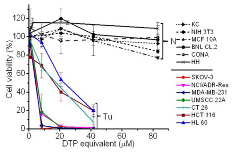
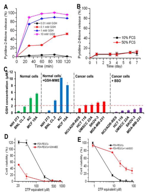
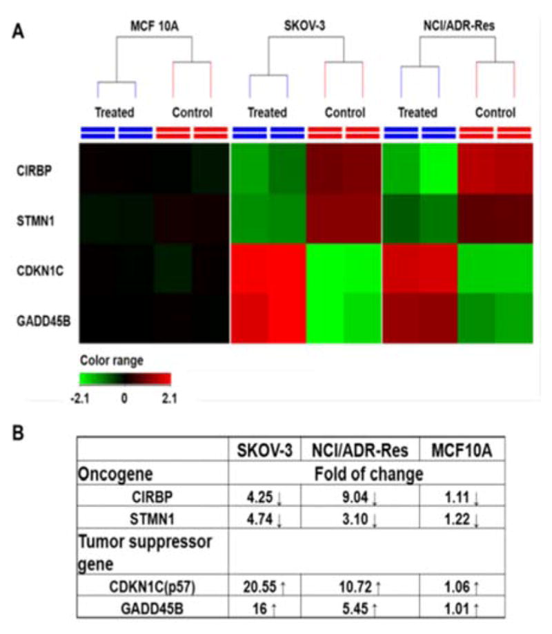
Chemotherapy has been adopted for cancer treatment for decades. However, its efficacy and safety are frequently compromised by the multidrug-resistance of cancer cells and the poor cancer cell selectivity of anticancer drugs. Hereby, we report a combination of a pyridine-2-thiol containing polymer and copper which can effectively kill a wide spectrum of cancer cells, including drug resistant cancer cells, while sparing normal cells. The polymer nanoparticle enters cells via an exofacial thiol facilitated route, and releases active pyridine-2-thiol with the help of intracellularly elevated glutathione (GSH). Due to their high GSH level, cancer cells are more vulnerable to the polymer/copper combination. In addition, RNA microarray analysis revealed that the treatment can reverse cancer cells' upregulated oncogenes (CIRBP and STMN1) and downregulated tumor suppressor genes (CDKN1C and GADD45B) to further enhance the selectivity for cancer cells.


Figures

Fig. 1
Fig. 1
TEM images of nanoparticle fabricated from PDA-PEG alone (A), and PDA-PEG/Cu2+ combination (B). Scale bars are 200 nm.
Fig. 2
Fig. 2
Cytotoxicity of PDA-PEG/Cu2+ combination for normal (N) and cancer (Tu) cells. Normal cells include KC (human keratinocyte), NIH 3T3 (murine fibroblast), MCF 10A (human breast epithelial cell), BNL CL.2 (murine liver cell), CONA (CCD 841 CoN, human colon cell), and HH (human hepatocyte). Data represent the means±SD, n=3.
Fig. 3
Fig. 3
Fluorescence images of cancer cells after culturing with different concentrations of PDA-PEG/Cu2+ combination. NIH 3T3, NCI/ADR-Res, SKOV-3 and UMSCC 22A were pre-stained with Cell Tracker deep red, blue, green CMFDA and orange CMTMR dye, respectively, and imaged 24 h after the treatment. The scale bars were 40 μm.
Fig. 4
Fig. 4
Flow cytometry spectra of SKOV-3 and NIH 3T3 cells (A) and confocal images of cellular uptake of nanoparticles in SKOV-3 cells (B). Cellular uptake assays were carried out 1 h after the addition of nanoparticles. Scale bars were 20 μm.
Fig. 5
Fig. 5
The release kinetic of pyridine-2-thiol liberating from PDA-PEG at different GSH levels (A) and in serum containing media (B), the GSH level in different cell lines and response to the addition of GSH-MME and BSO (C), GSH-MME effect on the cytotoxicity of PDA-PEG/Cu2+ for MCF10A cells (D), and BSO effect on the cytotoxicity of PDA-PEG/Cu2+ combination for NCI/ADR-Res cells (E). Data represent the means±SD, n=3.
Fig. 6
Fig. 6
RNA expression in response to PDA-PEG/Cu2+ treatment. Cells were treated with 41.58 μM of PDA-PEG/Cu2+ for 12 h. (A) Heatmap RNA level with and without drug treatment. Red: upregulation; green: downregulation; black: no change. (B) Genes alteration fold after treatment.
Fig. 7
Fig. 7
Cell cycle analysis of SKOV-3 cells after treated with 41.58 μM of PDA-PEG/Cu2+ for 12 h. Flow cytometry spectra (A) and quantitive analysis (B) of cell cycle. Data represent the means±SD, n=3. *p<0.01.

References

    1. Siegel R, Ma J, Zou Z, Jemal A. CA Cancer J Clin. 2014;64:9–29. - PubMed
    1. Holohan C, Van Schaeybroeck S, Longley DB, Johnston PG. Nat Rev Cancer. 2013;13:714–726. - PubMed
    2. Liang X-J, Chen C, Zhao Y, Wang P. In: Multi-Drug Resistance in Cancer. Zhou J, editor. Vol. 596. Humana Press; 2010. pp. 467–488. ch 21.
    1. Pauwels EK, Erba P, Mariani G, Gomes CM. Drug News Perspect. 2007;20:371–377. - PubMed
    1. Masui K, Gini B, Wykosky J, Zanca C, Mischel PS, Furnari FB, Cavenee WK. Carcinogenesis. 2013;34:725–738. - PMC - PubMed
    1. Herst PM, Davis JE, Neeson P, Berridge MV, Ritchie DS. Haematologica. 2009;94:928–934. - PMC - PubMed
    2. Mitchison TJ. Mol Biol Cell. 2012;23:1–6. - PMC - PubMed

Publication types

MeSH terms