. 2015 Nov 9;16(11):26813-31.
doi: 10.3390/ijms161125989.

Effect of Human Adipose Tissue Mesenchymal Stem Cells on the Regeneration of Ovine Articular Cartilage

Affiliations

Effect of Human Adipose Tissue Mesenchymal Stem Cells on the Regeneration of Ovine Articular Cartilage

Alessandro R Zorzi et al. Int J Mol Sci. .

Abstract

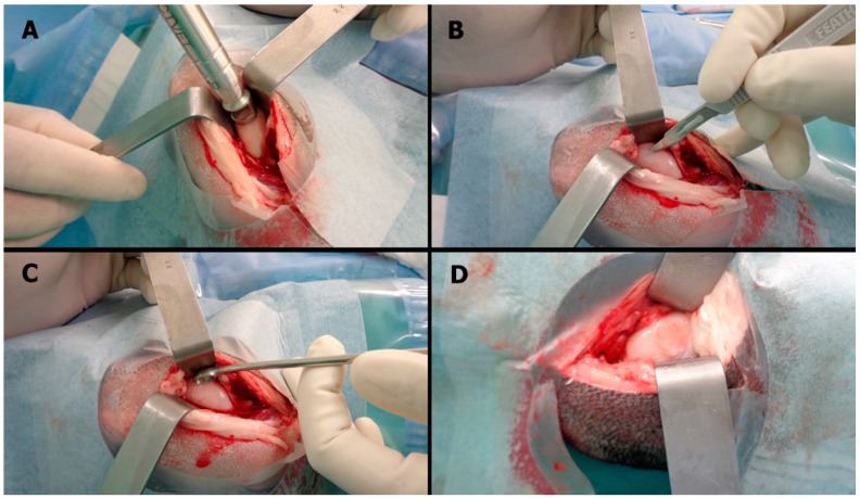
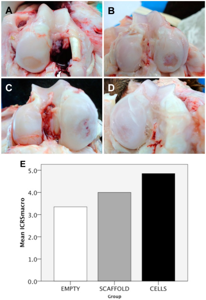
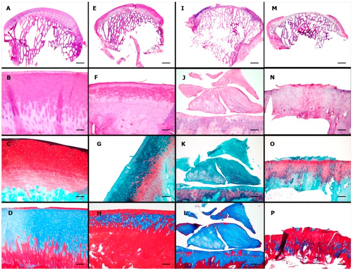
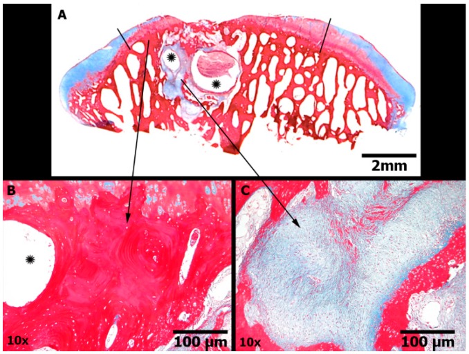
Cell therapy is a promising approach to improve cartilage healing. Adipose tissue is an abundant and readily accessible cell source. Previous studies have demonstrated good cartilage repair results with adipose tissue mesenchymal stem cells in small animal experiments. This study aimed to examine these cells in a large animal model. Thirty knees of adult sheep were randomly allocated to three treatment groups: CELLS (scaffold seeded with human adipose tissue mesenchymal stem cells), SCAFFOLD (scaffold without cells), or EMPTY (untreated lesions). A partial thickness defect was created in the medial femoral condyle. After six months, the knees were examined according to an adaptation of the International Cartilage Repair Society (ICRS 1) score, in addition to a new Partial Thickness Model scale and the ICRS macroscopic score. All of the animals completed the follow-up period. The CELLS group presented with the highest ICRS 1 score (8.3 ± 3.1), followed by the SCAFFOLD group (5.6 ± 2.2) and the EMPTY group (5.2 ± 2.4) (p = 0.033). Other scores were not significantly different. These results suggest that human adipose tissue mesenchymal stem cells promoted satisfactory cartilage repair in the ovine model.

Keywords: adult stem cell; biomaterials; cartilage; regenerative medicine; stem cell therapy; tissue engineering.


Figures

Figure 1
Figure 1
Results of flow cytometry analysis of the AT-MSCs. The cells were expanded to the fourth passage and analyzed by flow cytometry (10,000 cells were analyzed). Black histogram represents the expression of each molecule of IgG (negative control) and red histogram shows different markers investigated in the cell membrane of AT-MSCs.
Figure 2
Figure 2
Characterization of the isolated AT-MSCs (objective, ×10): (A) Spindle-shaped morphology on light microscopy; (B) AT-MSC differentiation into the adipogenic cell lineage (Oil Red O); (C) osteogenic cell lineage (Alizarin red); and (D) chondrogenic cell lineage (Sirius red, resorcin and fuchsine). Scale bars represent 100 μm.
Figure 3
Figure 3
Cell viability assay. Live/Dead Viability/Cytotoxicity Kit for mammalian cells (Invitrogen, Carlsbad, CA, USA). (A) One-day cell culture; (B) three-day cell culture; (C) seven-day cell culture; and (D) control scaffold without cells to exclude natural collagen fluorescence. Live cells are green (objective, ×4). Scale bars represent 200 μm.
Figure 4
Figure 4
Hematoxylin and Eosin (H&E) stained histological sections of the chitosan/collagen scaffold seeded with AT-MSCs after seven days in culture. (A,B) Panoramic view of AT-MSCs uniform distribution into the scaffold; (C) Arrow indicates the interaction of two cells on the scaffold; (D) Detail of AT-MSC adhered on the scaffold.
Figure 5
Figure 5
Surgical technique: (A) A ten-millimeter punch biopsy was used to demarcate a cylinder in the medial femoral condyle; (B) a number 11 blade scalpel was used to make serial parallel incisions in the cartilage surface followed by a new series of parallel incisions perpendicular to the first ones; (C) a surgical curette was used to remove the incised cartilage without evoking bleeding at the base of the lesion; and (D) the scaffold was fixed with 1 mL of fibrin glue for five minutes, and the stability was then checked with ten cycles of passive full range of motion.
Figure 6
Figure 6
Macroscopic results: (A) Completely empty defect (group EMPTY); (B) partially filled defect (group EMPTY); (C) completely filled defect, but with the presence of a gross vascularized tissue (group SCAFFOLD); (D) completely filled defect (group CELLS); and (E) International Cartilage Repair Society (ICRS) macroscopic score results presented as a bar chart (EMPTY = 3.4 ± 3.3; SCAFFOLD = 4.0 ± 2.2; CELLS = 4.9 ± 2.2; p = 0.738).
Figure 7
Figure 7
Microscopic results: first row, panoramic view; second row, H&E stain; third row, Safranin O (used to color in red the glycosaminoglycans); and fourth row, Masson’s trichrome (used to color in blue the collagens and new connective tissues). (A) Panoramic view by scanning; scale bar = 2 mm; (BD) objective, ×10; scale bars = 100 μm: normal ovine cartilage; (E) scanning; scale bar = 2 mm; (FH) objective, ×10; scale bars = 100 μm: group CELLS; a smooth continuous surface with complete horizontal filling and complete integration to the borders, but partial vertical filling of the defect and an heterogeneous Safranin O staining of the extracellular matrix; (I) scanning; scale bar = 2 mm; (JL) objective, ×10; scale bars = 100 μm: group SCAFFOLD; defect partially filled with fibrocartilage; (M) scanning; scale bar = 2mm; and (NP) objective, ×10: group EMPTY; defect unfilled. Scale bar = 100 μm.
Figure 8
Figure 8
Subchondral bone damage (Masson’s trichrome stain). Group SCAFFOLD. (A) Panoramic view of the articular surface in the region of the defect (delimited by black lines) showing cartilage border centripetal ingrowth. Evident cystic formations were noted (asterisk) in a localized area of subchondral bone, surrounded by loosely immature fibrous connective tissue and new sclerotic bone, similar to the findings of advanced degenerative joint disease; Scale bars represent 2 mm; (B) Sclerotic bone; Scale bars represent 100 μm; (C) Immature fibrous tissue; Scale bars represent 100 μm.

References

    1. Poole C.A. Articular cartilage chondrons: Form, function and failure. J. Anat. 1997;191:1–13. doi: 10.1046/j.1469-7580.1997.19110001.x. - DOI - PMC - PubMed
    1. Iwamoto M., Ohta Y., Larmour C., Enomoto-Iwamoto M. Towards regeneration of articular cartilage. Birth Defects Res. C Embryo Today. 2014;3:192–202. doi: 10.1002/bdrc.21042. - DOI - PMC - PubMed
    1. Hjelle K., Solheim E., Strand T., Muri R., Brittberg M. Articular cartilage defects in 1000 knee arthroscopies. Arthroscopy. 2002;18:730–734. doi: 10.1053/jars.2002.32839. - DOI - PubMed
    1. Widuchowski W., Widuchowski J., Koczy B., Szyluk K. Untreated asymptomatic deep cartilage lesions associated with anterior cruciate ligament injury: Results at 10- and 15-year follow-up. Am. J. Sports Med. 2009;37:688–692. doi: 10.1177/0363546508328104. - DOI - PubMed
    1. Widuchowski W., Widuchowski J., Faltus R., Lukasik P., Kwiatkowski G., Szyluk K., Koczy B. Long-term clinical and radiological assessment of untreated severe cartilage damage in the knee: A natural history study. Scand. J. Med. Sci. Sports. 2011;21:106–110. doi: 10.1111/j.1600-0838.2009.01062.x. - DOI - PubMed

Publication types

MeSH terms

LinkOut - more resources