Review
. 2015 Dec;7(12):1487-517.
doi: 10.1039/c5ib00221d. Epub 2015 Nov 16.

Re-membering the body: applications of computational neuroscience to the top-down control of regeneration of limbs and other complex organs

Affiliations
Review

Re-membering the body: applications of computational neuroscience to the top-down control of regeneration of limbs and other complex organs

G Pezzulo et al. Integr Biol (Camb). 2015 Dec.

Abstract

A major goal of regenerative medicine and bioengineering is the regeneration of complex organs, such as limbs, and the capability to create artificial constructs (so-called biobots) with defined morphologies and robust self-repair capabilities. Developmental biology presents remarkable examples of systems that self-assemble and regenerate complex structures toward their correct shape despite significant perturbations. A fundamental challenge is to translate progress in molecular genetics into control of large-scale organismal anatomy, and the field is still searching for an appropriate theoretical paradigm for facilitating control of pattern homeostasis. However, computational neuroscience provides many examples in which cell networks - brains - store memories (e.g., of geometric configurations, rules, and patterns) and coordinate their activity towards proximal and distant goals. In this Perspective, we propose that programming large-scale morphogenesis requires exploiting the information processing by which cellular structures work toward specific shapes. In non-neural cells, as in the brain, bioelectric signaling implements information processing, decision-making, and memory in regulating pattern and its remodeling. Thus, approaches used in computational neuroscience to understand goal-seeking neural systems offer a toolbox of techniques to model and control regenerative pattern formation. Here, we review recent data on developmental bioelectricity as a regulator of patterning, and propose that target morphology could be encoded within tissues as a kind of memory, using the same molecular mechanisms and algorithms so successfully exploited by the brain. We highlight the next steps of an unconventional research program, which may allow top-down control of growth and form for numerous applications in regenerative medicine and synthetic bioengineering.


Figures

Figure 1
Figure 1. Examples of dynamic pattern regulation
Large-scale patterning during regeneration and embryogenesis often exhibits flexible growth programs that work to achieve a specific target morphology. (A) Embryos of many species can be split in half but result in two perfectly normal individuals – monozygotic twins (photo by Oudeschool via Wikimedia Commons). (B) Similarly, mouse embryos can be joined together and yet re-pattern to give rise to a normal animal. (C) Salamander limbs can regenerate perfectly following amputation, and the process stops when a correct limb is rebuilt. (D) A tail grafted onto a flank of an amphibian slowly remodels into a limb – a structure more appropriate to its new anatomical position; this includes re-specification of the distal-most tip into fingers, showing that the process is non-local (because the immediate environment of the tail tip is its expected “tail” context, and it should have no reason to change unless it received long-range signals). (E) In some species of deer, damage at a particular spot on the invariant branched structure will result in an ectopic tine appearing in that same location next year after the antlers are shed and re-grow (used with permission ). (F) A tadpole modified during development such that its craniofacial organs are in the wrong positions nevertheless develops into a normal frog, showing the ability of morphogenesis to flexibly correct unexpected initial states towards the same anatomical outcome (frog image courtesy of Erin Switzer; tadpole image used with permission ). These examples illustrate the ability of biological systems to robustly pursue or maintain a goal state specified at the level of topological arrangement of organs – a capability we must learn to exploit, for transformative applications in synthetic bioengineering. We do not discuss plants, because though they often possess impressive powers of regeneration , they generally have no fixed target morphology at the level of the entire organism. Images in panel F are courtesy of Douglas Blackiston and Erin Switzer.
Figure 2
Figure 2. Non-neural cells use bioelectrical signaling for pattern formation
(A) Voltage-reporting fluorescent dyes reveal a rich pattern of bioelectrical communication among early frog embryo cells. (B) During later development in the frog embryo, a prepattern of hyperpolarization is seen (lighter cells) which establishes the prospective boundaries of craniofacial gene expression and the location of anatomical organs: in this way, bioelectric state information directly and functionally encodes the anatomy and structure of the face (used with permission ). If this bioelectric pattern is artificially perturbed, predictable changes in face morphology result. (C) Targeted changes of bioelectric state, by misexpression of ion channel mRNA in frog embryos in vivo, reprogram body regions at the level of organs: without having to specify the details, a portion of the gut can be re-specified to form a complete eye (red arrowhead; used with permission from ). (D) The process involves not only the cells whose voltage properties were changed (marked with blue lineage dye) but also recruits some of the host’s unaltered cells toward making a complete circular lens, revealing a non-local property of bioelectric organ induction.
Figure 3
Figure 3. Tools for perturbing bioelectrical networks
Much as in the nervous system, there are 2 basic options for experimentally modulating the activity of bioelectric networks in developmental contexts. Analogous to synaptic plasticity, the connectivity of the network can be modified, by blocking endogenous gap junctions (electrical synapses), either pharmacologically or via misexpression of a dominant negative connexin subunit, or introducing novel gap junctional connections by driving expression of wild-type connexins or connexin mutants with desired gating/permeability properties. Analogous to intrinsic plasticity, one can instead modify directly the bioelectrical state of specific cells. Pharmacological, genetically-encoded, or optogenetic strategies can be used to modify which channels are expressed in cells, or which are open/closed. Guided by the Goldman equation, these interventions can be designed to result in desired changes of resting potential in the targeted cells. Images in this figure were created by Jeremy Guay of Peregrine Creative.
Figure 4
Figure 4. Pattern memory encoded in bioelectric circuits
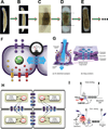
Planaria (A) can regenerate any body region, and their head-tail polarity is regulated in part by an endogenous voltage gradient. When the head and tail are removed and the middle fragment is treated with reagents that alter the topology of the bioelectric network (gene-specific RNAi targeting innexin proteins, or gap junction-targeting drugs that wash out in 24 hours, B), a 2-headed planarian results (C). Remarkably, weeks later, when these animals are cut and re-cut in plain water, 2-head worms continue to result (D,E) despite the animal’s normal genome and the fact that “epigenetically reprogrammed” tissues are removed at each round of cutting. This illustrates the distributed encoding of target morphology among all body regions, the storage of pattern information in bioelectrical properties distinct from genomic information, and the ability to alter the shape to which this animal repairs upon damage by changing network connectivity among cells long-term memory (all ideally mirrored by the known properties of long-term memory). Bioelectric circuits that could stably store such state information consist, much like neurons, of voltage potentials driven by ion channels (F, transcriptional changes in the expression of which are analogous to intrinsic plasticity in neuroscience) and of connectivity via highly tunable electric synapses – gap junctions (G, changes in which are analogous to synaptic plasticity). (H) Positive feedback loops between voltage states (an aggregate, systems property) and voltage-sensitive ion channel states allow stable attractors of distinct bioelectrical states. Together with known mechanisms of synaptic plasticity implemented by gap junctions, calcium, and neurotransmitters (I), these components should allow the creation of mechanistic models of pattern memory and the construction of synthetic bioengineered devices with memory and self-repair capabilities. Panels A-E used with permission . Images in panels F and G were created by Jeremy Guay of Peregrine Creative. Images in panel H used with permission .
Figure 5
Figure 5. Pattern formation and regeneration using the free energy principle (FEP)
(A) A sample computational model , in which undifferentiated cells self-organize to reach a target morphology, corresponding to a (simple) multicellular animal with head, body, and tail (e.g., a planarian). The target morphology is specified in such a way that, when it is achieved, all cells essentially sense the “right” electrochemical signals – a state in which no further remodeling (cellular activity) is necessary. However, the problem for the cells is ”finding their place” in this target morphology; because cells are initially undifferentiated, each can (in principle) become part of the head, body, or tail. This morphogenetic process is formulated as an inferential, FEP problem (B), where essentially the whole system undergoes a series of changes (e.g., in cellular position) until the target morphology is achieved. While changing their place, cells emit signals (chemical and/or physiological) that in turn guide the other cells, until a collective solution is found that corresponds to the state where the free energy of the whole system is minimized. Once the system has reached a stable solution, it can be perturbed, e.g., cut into two parts, (C) and this can lead to a new morphogenetic process with the regeneration of two organisms. Perturbing the system in more severe ways can lead to various forms of dysmorphogenesis (not shown, see ). Note that this self-organizing process is guided by an objective function (free energy minimization) and lends itself to top-down analysis, while able to accommodate known details of cellular signaling. Images reused according to the Creative Commons license from references , .
Figure 6
Figure 6. Formulating and solving a patterning problem via the free energy principle (FEP)
The figure schematizes a “methodological recipe” for formulating and solving a patterning problem using the free energy principle (FEP); see for one recent example where this approach has been successfully used. The methodology is composed of three steps. The first step (A) requires specifying mathematically the so-called generative model of the cells, or in other words their “internal states”, “active states”, “sensory states” and “external states”, along with their probabilistic dependencies and the prior knowledge (e.g., a previous, correct target morphology). The second step (B) requires specifying mathematically the exchanges (intercellular signalling) between the cells. Because the approach assumes that, for each cell, the behavior of (some or all) the other cells constitutes the “external state”, specifying the interactions between cells corresponds to specifying how the “active states” of one cell changes the “sensory states” or (some or all) the other cells. Intuitively, the active state of one cell might correspond to emitting chemical and/or physiological signals which can be sensed by other cells (the model requires specifying for example the gradients and concentrations that underlie this sort of intercellular signaling. The third and final step (C) corresponds to simulating the dynamics of the problem to find the solution that minimizes the free energy of the (collective) system. A MATLAB toolbox implementing a variational message passing scheme for free energy minimization is the SPM academic freeware (http://www.fil.ion.ucl.ac.uk/spm/); see also . Images reused according to the Creative Commons license from references , .
Figure 7
Figure 7. Cognitive neuroscience paradigms and their application to models of pattern formation
(A) The TOTE model of a cybernetic goal-directed process. Figure adapted from . Words in Italics represent the main processes composing the principle. Thin arrows represent information flows. The double-headed arrow represents a process of comparison between the desired and the actual state value. The process starts from a Test. If the Test fails (i.e. a mismatch is detected between desired and actual state) an action is triggered (dashed arrow) that causes a cascade of effects such as a change in the actual state that are sensed and used in the next Tests. When the Test succeeds, the process ends. (B) The same model applied to a regenerative context, in which comparison of current anatomical state to a stored target morphology generates signals for cell growth, differentiation, and movement that progressively restore pattern. Cognitive neuroscience is also an example of a field in which high-level information has causal power and is mechanistically integrated with low-level (molecular) details of its encoding and manipulation. (C) Changes of mental state (learning specific patterns for example) alters cell behaviour in the brain (taken with permission from ). (D) Manipulation of bioelectric states in the brain using optogenetic tools is able to insert specific cognitive content (false memories) . Credit: Collective Next. (E) Conversely, mental imagery can be read out by appropriate decoding of bioelectric state information from living brains (taken with permission from ). In complement to today’s models (formulated entirely bottom-up, in terms of molecular pathways), we suggest that successful top-down models of regeneration (in which organ-level topological pattern is represented within somatic cells and guides cell behavior) could be formulated by borrowing insights from cognitive neuroscience.
Figure 8
Figure 8. Applying free energy models to understanding cognition, a “primordial soup”, and dynamic morphogenesis
(A) A dynamical exchange between an agent and its environment as modeled in the active inference framework . Here, a discrepancy between the current sensory state and a goal state encoded in the internal state (reflecting some desired event or the homeostatic level of some variable) gives rise to interoceptive, proprioceptive, and exteroceptive prediction errors (red arrows). This produces a cascade of processes that ultimately enacts a sequence of actions (say, grasping and eating an apple). This process ceases when the interoceptive, proprioceptive, and/or exteroceptive feedback (e.g., the right gustatory sensations) matches the descending predictions (blue arrows) meaning that the organism has restored homeostasis through action. (B) A simulation of a “primordial soup” and the emergence of self-organization that is coherent with principles of active Bayesian inference; example from . Left part: This “soup” comprises an ensemble of dynamical subsystems (the dots) that represent macromolecules. The macromolecules have a physical state (representing e.g. their position) and an electrochemical state (representing e.g. concentrations) that change according to simplified Newtonian dynamics and electrochemical dynamics (modeled in using a Lorenz attractor). Crucially, the states have short-range interactions: they are coupled within and between the subsystems comprising an ensemble. Center part: as the system evolves over time, a structure self-organizes that separates subsystems that are conditionally dependent (called internal states) and independent (called external states). Formally, this structure is called a Markov blanket: a kind of “statistical boundary” (more formally the set of node’s parents, children, and its children’s other parents in a Bayesian network). Note the clear separations - after evolution - in the location of subsystems (macromolecules) with internal states (blue), their Markov blanket (magenta and red), and external or hidden states (azure). States in the Markov blanket can be further subdivided into two sets: those that depend on internal states (red) and those that do not (magenta), called active states and sensory states, respectively. As noticed in in this spatial configuration “the active subsystems support the sensory subsystems that are exposed to hidden environmental states. This is reminiscent of a biological cell with a cytoskeleton that supports some sensory epithelia or receptors within its membrane.” Importantly, active states change external states (but are not affected by them) and so they maintain the structural and functional integrity of the Markov blanket. Indeed, “lesioning” internal, sensory or active states (by decoupling them from the rest of the system) quickly leads to the disruption of the Markov blanket - not shown here, but see . Right part: These arguments suggest that a formal analogy can be established between active and sensory states and action and perception systems in living organisms, respectively. This speaks to an even more general interpretation of the self-organization process (shown in the Center part) in Bayesian terms, where the internal states are Bayesian models that infer/represent the hidden (azure) causes of sensory (magenta) states and cause these states through action (red). This can be verified if one considers that sensory states permit predicting external / hidden states - as shown in . (C) The same scheme can be applied now to regeneration, where the “internal” (biochemical) states essentially encode a target morphology that can be acquired through a learning process that obeys to free energy minimization processes (e.g., as shown in B) or using unsupervised learning in generative architectures as explained in the main text. Once the target morphology is acquired, the same error-correction mechanism explained in (A) permit to trigger (regenerative) actions that restore it when it is disrupted. Images reused according to the Creative Commons license from references ,

References

    1. Ingber DE, Levin M. What lies at the interface of regenerative medicine and developmental biology? Development. 2007;134:2541–2547. - PubMed
    2. Stoick-Cooper CL, Moon RT, Weidinger G. Advances in signaling in vertebrate regeneration as a prelude to regenerative medicine. Genes Dev. 2007;21:1292–1315. - PubMed
    1. Kamm RD, Bashir R. Creating living cellular machines. Ann. Biomed. Eng. 2014;42:445–459. - PMC - PubMed
    1. French V. Positional information around the segments of the cockroach leg. Journal of embryology and experimental morphology. 1980;59:281–313. - PubMed
    1. Oviedo NJ, Newmark PA, Sanchez Alvarado A. Allometric scaling and proportion regulation in the freshwater planarian Schmidtea mediterranea . Dev Dyn. 2003;226:326–333. - PubMed
    1. Farinella-Ferruzza N. The transformation of a tail into a limb after xenoplastic transformation. Experientia. 1956;15:304–305.

Publication types