. 2016 Jan;18(1):132-8.
doi: 10.1038/ncb3271. Epub 2015 Nov 16.

The leukodystrophy protein FAM126A (hyccin) regulates PtdIns(4)P synthesis at the plasma membrane

Affiliations

The leukodystrophy protein FAM126A (hyccin) regulates PtdIns(4)P synthesis at the plasma membrane

Jeremy M Baskin et al. Nat Cell Biol. 2016 Jan.

Abstract

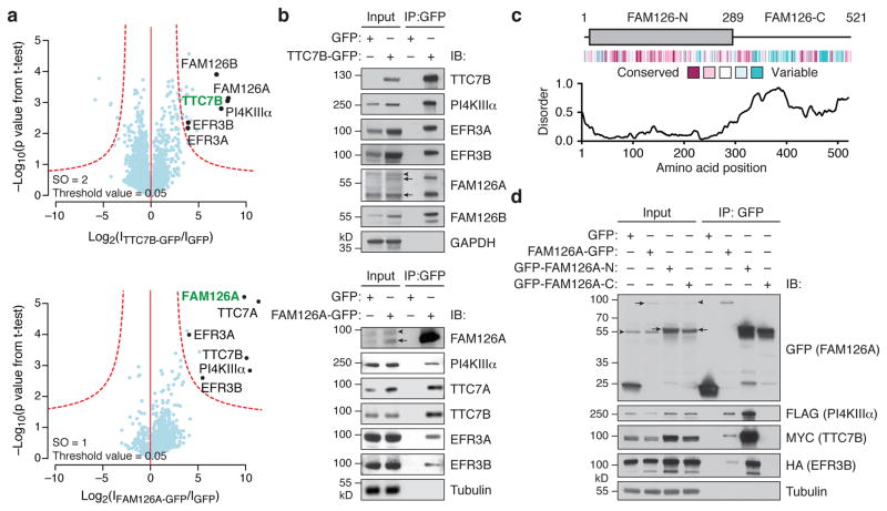
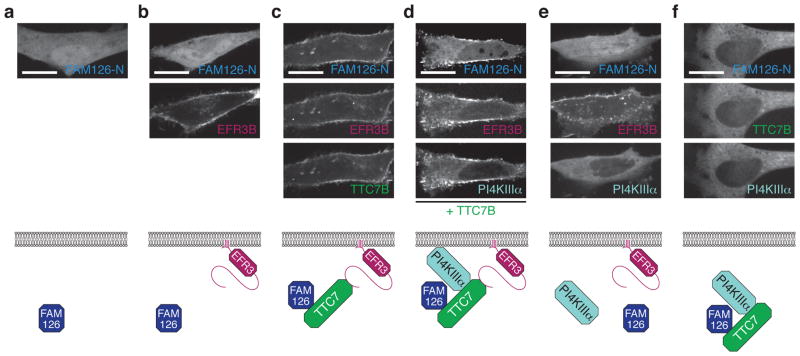
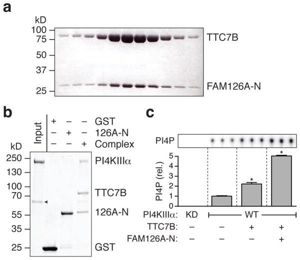
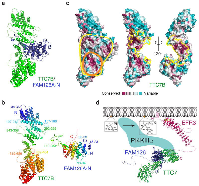
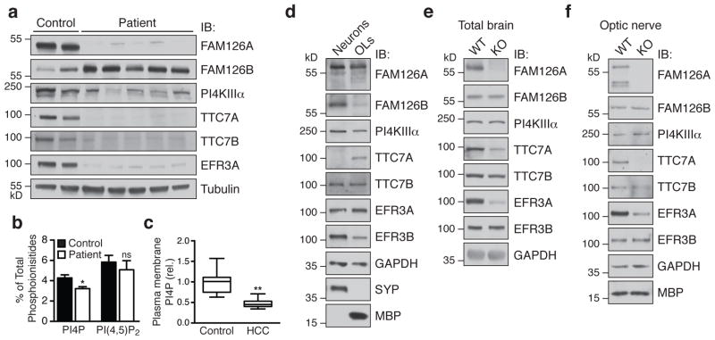
Genetic defects in myelin formation and maintenance cause leukodystrophies, a group of white matter diseases whose mechanistic underpinnings are poorly understood. Hypomyelination and congenital cataract (HCC), one of these disorders, is caused by mutations in FAM126A, a gene of unknown function. We show that FAM126A, also known as hyccin, regulates the synthesis of phosphatidylinositol 4-phosphate (PtdIns(4)P), a determinant of plasma membrane identity. HCC patient fibroblasts exhibit reduced PtdIns(4)P levels. FAM126A is an intrinsic component of the plasma membrane phosphatidylinositol 4-kinase complex that comprises PI4KIIIα and its adaptors TTC7 and EFR3 (refs 5,7). A FAM126A-TTC7 co-crystal structure reveals an all-α-helical heterodimer with a large protein-protein interface and a conserved surface that may mediate binding to PI4KIIIα. Absence of FAM126A, the predominant FAM126 isoform in oligodendrocytes, destabilizes the PI4KIIIα complex in mouse brain and patient fibroblasts. We propose that HCC pathogenesis involves defects in PtdIns(4)P production in oligodendrocytes, whose specialized function requires massive plasma membrane expansion and thus generation of PtdIns(4)P and downstream phosphoinositides. Our results point to a role for FAM126A in supporting myelination, an important process in development and also following acute exacerbations in multiple sclerosis.


Conflict of interest statement

The authors declare no competing financial interests.

Figures

Figure 1
Figure 1. FAM126 is a novel component of the PI4KIIIα complex
(a) Volcano plots of proteins associating with TTC7B-GFP vs. GFP only (top) and FAM126A-GFP vs. GFP only (bottom), from a label-free proteomics analysis of anti-GFP immunoprecipitates of HEK 293T cells stably expressing TTC7B-GFP, FAM126A-GFP, or GFP only. The logarithmic ratios of protein intensities are plotted against negative logarithmic p values of two-tailed Student’s t-test, equal variance, performed from n = 3 independent experiments. The red dotted line (significance, 0.05) separates specifically interacting proteins (top right portion of plot) from background. Selected top hits are indicated with black dots (bait is indicated in green), and all specific interactors are reported in Supplementary Table 1. (b) Immunoblot (IB) analysis of PI4KIIIα complex components in anti-GFP immunoprecipitates of HEK 293T cells stably expressing TTC7B-GFP (top), FAM126A-GFP (bottom), or GFP only. (c) Domain structure, evolutionary conservation, and predicted extent of disordered secondary structural elements of FAM126. (d) Immunoblot analysis of anti-GFP immunoprecipitates from HeLa cells transfected with 3xFLAG-PI4KIIIα, TTC7B-MYC, EFR3B-HA, and the indicated GFP-tagged construct. Arrows denote specific bands and arrowheads denote non-specific antibody bands.
Figure 2
Figure 2. FAM126A is recruited to the plasma membrane in a complex with TTC7B, PI4KIIIα, and EFR3B
Top, Confocal imaging of live HeLa cells transfected with GFP-FAM126A-N and the following additional plasmids: (a) none; (b) EFR3B-BFP; (c) EFR3B-BFP and TTC7B-mCherry; (d) EFR3B-BFP, PI4KIIIα-mCherry, and TTC7B-3xFLAG; (e) EFR3B-BFP and PI4KIIIα-mCherry; (f) TTC7B-BFP and PI4KIIIα-mCherry. Bottom, cartoon representation of the transfected constructs and interpretation of the results. Scale bars, 20 μm.
Figure 3
Figure 3. FAM126A binds directly to TTC7B, and both FAM126A and TTC7B bind to PI4KIIIα and stimulate its catalytic activity
(a) TTC7B and FAM126A-N co-migrate as a heterodimer on a sizing column (Superdex 200), as shown by SDS-PAGE analysis of elution fractions. Proteins were visualized using Coomassie blue staining. (b) TTC7B/GST-FAM126A-N pulls down PI4KIIIα in a protein-protein interaction experiment with purified proteins. The GST-tagged proteins (GST, GST-FAM126A-N (126A-N), or the GST-FAM126A-N/TTC7B dimer (Complex)) were immobilized on glutathione-conjugated sepharose and incubated with purified PI4KIIIα. Proteins were visualized using Coomassie blue staining. The experiment was performed in triplicate. Arrowhead denotes the chaperone Hsp70, which co-purified with PI4KIIIα. (c) Kinase activity assay using purified PI4KIIIα sub-complexes. Wild-type (WT) or kinase-dead (KD) PI4KIIIα was produced either alone or in complex with TTC7B or TTC7B/FAM126A-N as indicated (Supplementary Fig. 3a), and in vitro lipid kinase activity assays were performed using PI-containing liposomes and γ32P-labeled ATP. The extent of PI4P formation was assessed by TLC and autoradiography. Two-tailed Student’s t-test with unequal variance: *, p = 0.0001 (n = 3 independent experiments). Error bars represent standard deviation.
Figure 4
Figure 4. Crystal structure of a TTC7B/FAM126A co-complex reveals an unusually large protein-protein interface and a conserved binding surface for PI4KIIIα
(a) Ribbon diagram for the TTC7B/FAM126A-N complex. Note the FAM126A-N hairpin structure that wraps around TTC7B like an arm. Point mutations in FAM126A that underlie HCC are indicated in Supplementary Fig. 3c. (b) Ribbon diagrams for TTC7B and FAM126A-N colored from blue (N-terminus) to red (C-terminus). The “arm” in FAM126A is green. Disordered residues absent from the model are indicated by dotted lines. (c) Space-filling models of the TTC7B/FAM126A-N complex (left) and TTC7B (middle and right) colored by sequence conservation. Conserved surfaces on TTC7B/FAM126A-N (yellow outline) or TTC7B alone (orange outline) that may interact with PI4KIIIα are circled (left). The TTC7B surfaces at the interface with FAM126A-N are indicated by dotted yellow lines (middle and right). (d) Model for PI4KIIIα assembly at the plasma membrane. The EFR3 model is based on the structure of yeast Efr3 (PDBID 4N5A).
Figure 5
Figure 5. Loss of FAM126A leads to defects in the PI4KIIIα complex in HCC patient fibroblasts and FAM126A knockout mouse brain
(a) Immunoblot (IB) analysis of PI4KIIIα complex components in control and HCC patient fibroblasts. (b) HPLC analysis of total 3H-inositol-labeled phosphoinositide content in control and HCC patient fibroblasts. Two-tailed Student’s t-test, unequal variance, *, p = 0.0019, n = 4 independent experiments. Error bars represent standard deviation. (c) Quantification of the plasma membrane PI4P pool in control and HCC patient fibroblasts by immunofluorescence using an anti-PI4P antibody. Two-tailed Student’s t-test, unequal variance, **, p = 0.0001. Box plot demarcates 25th and 75th percentile (middle line is median), and bars represent the minimum and maximum values (n = 19 cells for control, n = 23 cells for patient, from a total of four independent experiments). Similar results were seen in a second HCC patient cell line. (d) Immunoblot (IB) analysis of PI4KIIIα complex components in wild-type primary cortical neuronal and oligodendrocyte cultures. (e) and (f) Immunoblot (IB) analysis of PI4KIIIα complex components in total brain (e) or optic nerve (f) from male WT or FAM126A knockout mice at age P30. See Supplementary Fig. 4a for quantification of the immunoblot data from a and d–f.

Similar articles

Cited by

References

    1. Aggarwal S, Yurlova L, Simons M. Central nervous system myelin: structure, synthesis and assembly. Trends in Cell Biology. 2011;21:585–593. - PubMed
    1. Perlman SJ, Mar S. Leukodystrophies. Adv Exp Med Biol. 2012;724:154–171. - PubMed
    1. Zara F, et al. Deficiency of hyccin, a newly identified membrane protein, causes hypomyelination and congenital cataract. Nat Genet. 2006;38:1111–1113. - PubMed
    1. Tan J, Brill JA. Cinderella story: PI4P goes from precursor to key signaling molecule. Crit Rev Biochem Mol Biol. 2014;49:33–58. - PubMed
    1. Nakatsu F, et al. PtdIns4P synthesis by PI4KIIIα at the plasma membrane and its impact on plasma membrane identity. J Cell Biol. 2012;199:1003–1016. - PMC - PubMed

Publication types

MeSH terms

Substances