Clinical Trial
. 2015 Dec;125(12):4483-96.
doi: 10.1172/JCI83178. Epub 2015 Nov 16.

A collagen VI-dependent pathogenic mechanism for Hirschsprung's disease

Clinical Trial

A collagen VI-dependent pathogenic mechanism for Hirschsprung's disease

Rodolphe Soret et al. J Clin Invest. 2015 Dec.

Abstract

Hirschsprung's disease (HSCR) is a severe congenital anomaly of the enteric nervous system (ENS) characterized by functional intestinal obstruction due to a lack of intrinsic innervation in the distal bowel. Distal innervation deficiency results from incomplete colonization of the bowel by enteric neural crest cells (eNCCs), the ENS precursors. Here, we report the generation of a mouse model for HSCR--named Holstein--that contains an untargeted transgenic insertion upstream of the collagen-6α4 (Col6a4) gene. This insertion induces eNCC-specific upregulation of Col6a4 expression that increases total collagen VI protein levels in the extracellular matrix (ECM) surrounding both the developing and the postnatal ENS. Increased collagen VI levels during development mainly result in slower migration of eNCCs. This appears to be due to the fact that collagen VI is a poor substratum for supporting eNCC migration and can even interfere with the migration-promoting effects of fibronectin. Importantly, for a majority of patients in a HSCR cohort, the myenteric ganglia from the ganglionated region are also specifically surrounded by abundant collagen VI microfibrils, an outcome accentuated by Down syndrome. Collectively, our data thus unveil a clinically relevant pathogenic mechanism for HSCR that involves cell-autonomous changes in ECM composition surrounding eNCCs. Moreover, as COL6A1 and COL6A2 are on human Chr.21q, this mechanism is highly relevant to the predisposition of patients with Down syndrome to HSCR.


Figures
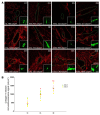
Figure 7
Figure 7. Myenteric ganglia of human patients with HSCR are surrounded by abundant collagen VI microfibrils.
(A) Representative single confocal sections of transverse cuts of human colonic muscles double labeled with antibodies against βIII-tubulin (green; in the insets) and collagen VI (red). Myenteric ganglia are delineated by green lines. The identification number for controls (C), patients with HSCR (H), and patients with Down syndrome and HSCR (D) is indicated in the top right corner of each image. Age at the time of tissue collection (indicated in days) as well as average levels of periganglionic collagen VI (n ≥ 5 ganglia per child’s sample; expressed in candelas per μm2) are indicated in the bottom left corner of each image. Scale bar: 50 μm (insets, 75 μm). (B) Quantification data of average periganglionic collagen VI levels for all tested human samples (data are presented as mean ± SEM; *P < 0.001 in comparison to controls; #P < 0.05 in comparison to patients with HSCR; 1-way ANOVA).
Figure 6
Figure 6. Collagen VI negatively impacts GDNF-stimulated eNCC migration ex vivo.
(A) Representative low-magnification views of E12.5 G4-RFP midgut explants cultured on the indicated coatings for 48 hours in GDNF-supplemented medium. FN, fibronectin; GEL, gelatin; Col I, collagen I; Col VI, collagen VI. Scale bar: 500 μm. (BD) Quantitative analysis of the net distance traveled, on average, by RFP-labeled eNCCs farthest from the edges of the explant (n = 8 cells per explants; 1 cell per quadrant) at the end of the 48-hour culture (data are presented as mean ± SEM; n ≥ 10 explants per condition from 4 independent experiments; *P < 0.01; 1-way ANOVA in B and C; Student’s t test in D). (B) Note that both collagens are inefficient in promoting eNCC migration and (C) that increased dosage of collagen VI inhibits the migration-promoting effects of fibronectin. (D) In addition, note that, in comparison to WT G4-RFP explants, eNCC migration on fibronectin is significantly less efficient when using HolTg/Tg G4-RFP explants.
Figure 5
Figure 5. Cell-autonomous defect in HolTg/Tg eNCC migration.
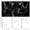
(A) Representative images of heterotopic E12.5 midgut-hindgut grafts after 24 hours of culture, showing that colonization of WT host tissues (aneural hindgut segments) by RFP-labeled eNCCs is much less efficient with mutant (HolTg/Tg G4-RFP) than with control (WT G4-RFP) midgut segments as donor tissues. Red arrowheads point to the location of the migration front at the end of the culture period. Scale bar: 150 μm. (B) Quantitative analysis of the extent of hindgut colonization by eNCCs coming from midgut tissues (data are presented as mean ± SEM; n = 5 grafts per combination; *P < 0.001; Student’s t test). (C) Representative images from 10-hour-long time-lapse recordings of eNCC movement at the migration front in WT and HolTg/Tg E13.5 intestines. The white arrows indicate the direction of colonization, whereas blue and orange arrowheads point to the location of the migration front at the start and end of recordings, respectively. Scale bar: 100 μm. (D) Quantification of the speed of individual cells at the tip of the migration front shows that HolTg/Tg eNCCs are significantly slower than WT eNCCs at both the E12.5 and E13.5 stages (data are presented as mean ± SEM; n represents the total number of cells from at least 3 intestines; *P < 0.01; Student’s t test).
Figure 4
Figure 4. Collagen VI protein levels are increased in E12.5 HolTg/Tg intestines.
(A) Single confocal sections in the plane of the developing myenteric plexus in wild-type and HolTg/Tg E12.5 intestines doubly labeled with anti–collagen VI (red) and anti–βIII-tubulin (green) antibodies. Scale bar: 20 μm. (B) Quantification of collagen VI immunofluorescence signals in candelas (cd) per μm2, showing that collagen VI protein levels are significantly increased in HolTg/Tg embryonic intestines colonized by neuronal-fated eNCCs (data are presented as mean ± SEM; n = 6 intestines per genotype; *P < 0.01; 1-way ANOVA).
Figure 3
Figure 3. The Holstein transgenic insertion upregulates Col6a4 expression in eNCCs.
(A) Schematic representation of the Holstein transgene insertion site based on whole-genome sequencing data and adapted from the Ensembl website (www.ensembl.org). Transgenic sequences are inserted in a 153-bp deletion (Δ 153 bp) between the Col6a4 and Glyctk genes on mouse Chr.9F1, which is syntenic to human Chr.3q22. Analysis of this region with the regulation track of Ensembl revealed the presence of multiple CTCF-binding motifs (green boxes) in the vicinity of the transgene insertion site, whereas the Comparative Genomics track (Genomic Evolutionary Rate Profiling [GERD] for 39 eutherian mammals; gray boxes) revealed that highly conserved noncoding sequences are not found close to the insertion site. (B) Example of PCR-based genotyping of Holstein animals using the oligos depicted in A (see Supplemental Table 2 for primers F/R1 and F/R2). (C and D) Analysis of Col6a4 transcript levels in E12.5 embryos via semiquantitative RT-PCR. (C) In contrast to that in whole intestines, a (D) robust allele dosage-dependent increase in Col6a4 gene expression is observed in FACS-recovered eNCCs. (E) Volcano plot of RNAseq-based comparative analysis of global gene expression between HolTg/Tg and wild-type eNCCs recovered by FACS from E12.5 intestines (n = 3 groups of 5–6 intestines per genotype). The log2 fold-change is on the x axis, while the DESeq P value is on the y axis.
Figure 2
Figure 2. Defective ENS formation in HolTg/Tg embryos is detected from E11.5 onward.
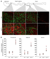
(A) Analysis of eNCC colonization in embryonic intestine at E11.5 (left; scale bar: 430 μm), E12.5 (middle; scale bar: 200 μm), and E15.5 (right; scale bar: 700 μm), using an anti–βIII-tubulin antibody. Arrows point to the most distal neuronal-fated eNCC of vagal origin, while arrowheads point to nonspecific staining of the meconium. SI, small intestine; Ce, cecum; Co, colon. (B) Quantification of the extent of eNCC colonization (at E11.5, E12.5, and E15.5) as measured from the base of cecum (white dashed lines in A) to the most distally located neuronal-fated eNCC of vagal origin. In comparison to that in wild-type tissues, the extent of bowel colonization by HolTg/Tg eNCCs is significantly decreased (data are presented as mean ± SEM; for each stage, n = 6 intestines per genotype; *P < 0.01; 1-way ANOVA).
Figure 1
Figure 1. The Holstein mouse line is a model for aganglionic megacolon.
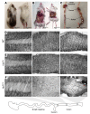
(A) Comparison between heterozygous (HolTg/+) and homozygous (HolTg/Tg) Holstein animals from a F2 litter at P20, showing allele dosage-dependent depigmentation. (B) HolTg/Tg mutants are smaller than their littermates and exhibit symptoms of aganglionic megacolon (asterisk). (C) These animals die due to blockage in the distal colon (arrow) and massive accumulation of fecal material (asterisks). (DF) Labeling of the myenteric plexus in the colons of (D) wild-type, (E) HolTg/+, and (F) HolTg/Tg P20 mice via staining of AchE activity. In comparison to wild-type mice (n = 8) and HolTg/+ controls (n = 7), the ENS network of HolTg/Tg animals (n = 12) is very slightly less dense in the proximal colon (left), is scarce and less interconnected in the mid-colon (middle), and is markedly absent in the distal colon (right), in which hypertrophic extrinsic nerve fibers typical of aganglionic megacolon as well as very rare isolated nerve cell bodies (red arrowhead) are seen. Scale bar: 1,000 μm.

Comment in

References

    1. Furness JB. The enteric nervous system and neurogastroenterology. Nat Rev Gastroenterol Hepatol. 2012;9(5):286–294. doi: 10.1038/nrgastro.2012.32. - DOI - PubMed
    1. Anderson RB, Newgreen DF, Young HM. Neural crest and the development of the enteric nervous system. Adv Exp Med Biol. 2006;589:181–196. doi: 10.1007/978-0-387-46954-6_11. - DOI - PubMed
    1. Heanue TA, Pachnis V. Enteric nervous system development and Hirschsprung’s disease: advances in genetic and stem cell studies. Nat Rev Neurosci. 2007;8(6):466–479. doi: 10.1038/nrn2137. - DOI - PubMed
    1. Obermayr F, Hotta R, Enomoto H, Young HM. Development and developmental disorders of the enteric nervous system. Nat Rev Gastroenterol Hepatol. 2013;10(1):43–57. - PubMed
    1. Bergeron KF, Silversides DW, Pilon N. The developmental genetics of Hirschsprung’s disease. Clin Genet. 2013;83(1):15–22. doi: 10.1111/cge.12032. - DOI - PubMed

Publication types

MeSH terms

Substances