. 2015 Nov 16;10(11):e0142991.
doi: 10.1371/journal.pone.0142991. eCollection 2015.

SS18-SSX, the Oncogenic Fusion Protein in Synovial Sarcoma, Is a Cellular Context-Dependent Epigenetic Modifier

Affiliations

SS18-SSX, the Oncogenic Fusion Protein in Synovial Sarcoma, Is a Cellular Context-Dependent Epigenetic Modifier

Sakura Tamaki et al. PLoS One. .

Abstract

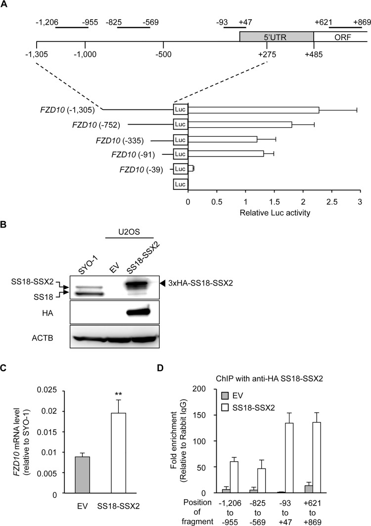
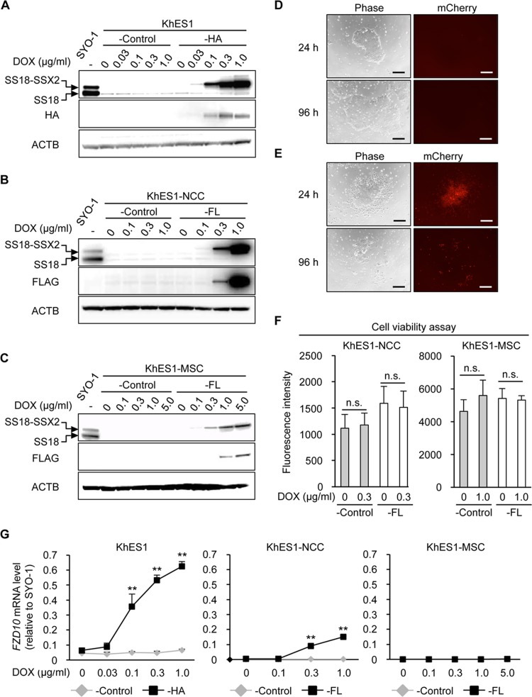
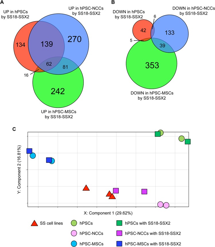
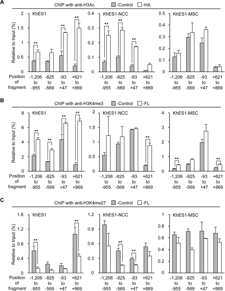
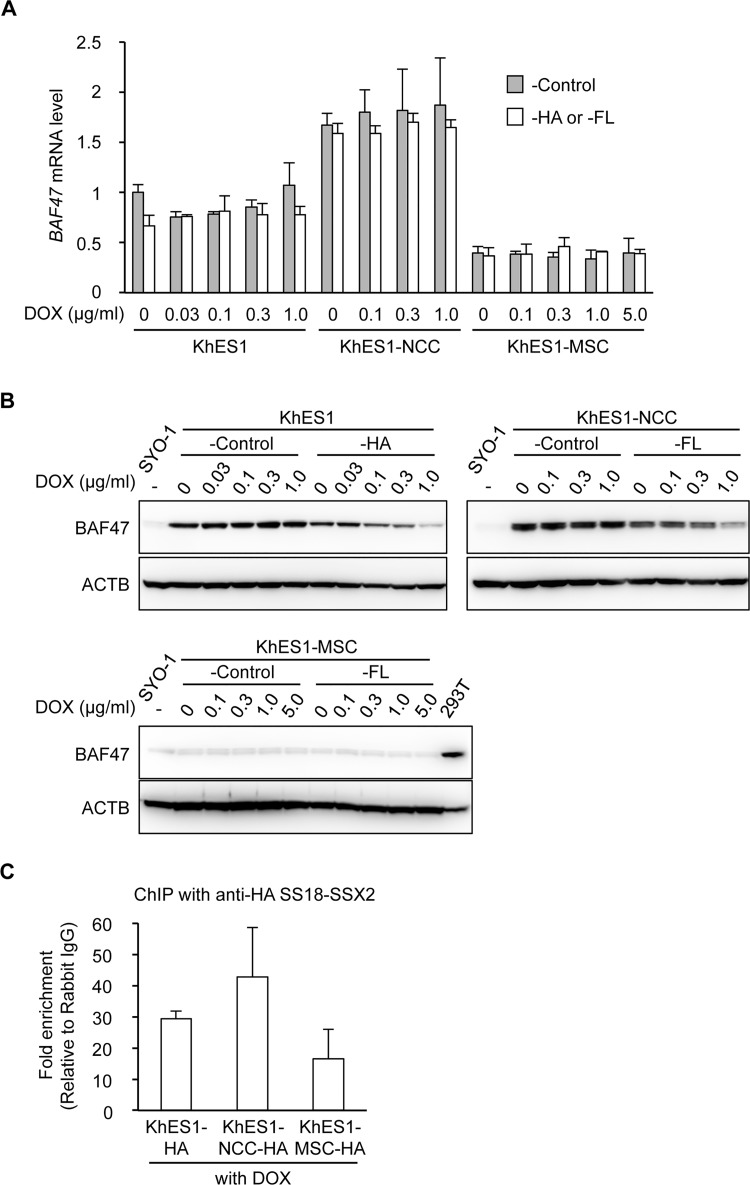
The prevalence and specificity of unique fusion oncogenes are high in a number of soft tissue sarcomas (STSs). The close relationship between fusion genes and clinicopathological features suggests that a correlation may exist between the function of fusion proteins and cellular context of the cell-of-origin of each tumor. However, most STSs are origin-unknown tumors and this issue has not yet been investigated in detail. In the present study, we examined the effects of the cellular context on the function of the synovial sarcoma (SS)-specific fusion protein, SS18-SSX, using human pluripotent stem cells (hPSCs) containing the drug-inducible SS18-SSX gene. We selected the neural crest cell (NCC) lineage for the first trial of this system, induced SS18-SSX at various differentiation stages from PSCs to NCC-derived mesenchymal stromal cells (MSCs), and compared its biological effects on each cell type. We found that the expression of FZD10, identified as an SS-specific gene, was induced by SS18-SSX at the PSC and NCC stages, but not at the MSC stage. This stage-specific induction of FZD10 correlated with stage-specific changes in histone marks associated with the FZD10 locus and also with the loss of the BAF47 protein, a member of the SWI/SNF chromatin-remodeling complex. Furthermore, the global gene expression profile of hPSC-derived NCCs was the closest to that of SS cell lines after the induction of SS18-SSX. These results clearly demonstrated that the cellular context is an important factor in the function of SS18-SSX as an epigenetic modifier.


Conflict of interest statement

Competing Interests: One of co-authors, Makoto Watanabe is an employee of Shimadzu Cooperation. The Life Science Research Center, Shimadzu Cooperation provided support in the form of his salary, but did not have any additional role in the study design, data collection and analysis, decision to publish, or preparation of the manuscript. The specific roles of Makoto Watanabe (analyzed the data) are articulated in the “Authors’ contributions” section. His affiliation to the company does not alter all authors' adherence to PLOS ONE policies on sharing data and materials.

Figures

Fig 1
Fig 1. Direct regulation of the FZD10 gene by the SS18-SSX2 fusion protein.
A) Promoter activity in the regulatory region of the FZD10 gene. The upper panel indicated the 5’ region of the FZD 10 gene with amplified regions in the ChIP-qPCR analysis. The number indicates relative to the transcription start site, and the positions of the amplified region are: -1206 to -955 bp; -825 to -569 bp; -93 to +47 bp; +621 to +869 bp. The lower panel showed the promoter activities of fragments derived from this 5’ region in SYO-1. Error bars reflect SD in 3 experiments. B) Induction of SS18-SSX2 in U2OS. U2OS was transfected with an empty vector (EV) or 3xHA-tagged SS18-SSX2, and 48 h after the transfection, the expression of SS18-SSX2 was analyzed by Western blotting. The SS18-SSX2 and SS18 proteins were detected by an anti-SS18 antibody (top panel), and the 3xHA-SS18-SSX2 protein was detected by an anti-HA antibody (middle panel). C) Induction of FZD10 expression by SS18-SSX2 in U2OS. The expression of FZD10 was analyzed by RT-qPCR. Expression levels were normalized to those of human ACTB and calculated as fold changes relative to SYO-1. Error bars reflect SD in 3 experiments. **, p<0.01 by the t-test. D) Binding of SS18-SSX2 to the FZD10 locus. A ChIP assay with an anti-HA antibody and RT-qPCR were performed. The values indicate relative to rabbit IgG. Error bars reflect SD in 3 experiments.
Fig 2
Fig 2. Induction of SS18-SSX2 in hESCs, hNCCs, and hNCC-derived MSCs.
A-C) DOX dose-dependently induced the SS18-SSX2 protein in KhES1-HA (A), KhES1-NCC-FL (B), and KhES1-MSC-FL (C) cells. Cells with Stuffer (-Control) and SS18-SSX2 were treated with the indicated concentrations of DOX for 24 h, and the expression of SS18-SSX2 was analyzed by Western blotting. The SS18-SSX2 and SS18 proteins were detected using an anti-SS18 antibody (top panel), and the 3xHA-SS18-SSX2 or FLAG-SS18-SSX2 protein was detected by an anti-HA or anti-FLAG antibody (middle panel). D and E) Morphology (left panels) and expression of mCherry (right panels) in KhES1-HA cells treated with 0 (D) or 0.3 (E) μg/ml of DOX for 24 and 96 h. Scale bar, 200 μm. F) Effects of SS18-SSX2 on the cell viability of KhES1-NCC-FL and KhES1-MSC-FL cells. Cells with Stuffer (-Control) or SS18-SSX2 were treated with the indicated concentrations of DOX for 48 h, and cell viability was measured using the AlamarBlue assay. n.s. means not significant. Error bars reflect SD in 4 experiments. G) Induction of FZD10 expression by SS18-SSX2 in KhES1-HA, KhES1-NCC-FL, and KhES1-MSC-FL cells. Cells with Stuffer (-Control) or SS18-SSX2 were treated with the indicated concentrations of DOX for 24 h, and the expression of FZD10 was analyzed by RT-qPCR. Expression levels were normalized to those of human ACTB and calculated as fold changes relative to SYO-1. Error bars reflect SD in 3 experiments. **, p<0.01 by the t-test.
Fig 3
Fig 3. Global gene expression profiles of hPSCs, hPSC-NCCs, and hPSC-MSCs with or without the induction of SS18-SSX2.
A and B) Venn-diagram showing up- (A) and down-regulated (B) genes in hPSCs, hPSC-NCCs, and hPSC-MSCs with the induction of SS18-SSX2. mRNA was extracted from each cell line 24 h after the DOX treatment. Each gene showed a fold change > 2.0 with or without the induction of SS18-SSX2. The numbers of up- and down-regulated genes are shown in the diagram. C) PCA of hPSCs, hPSC-NCCs, and hPSC-MSCs with or without the induction of SS18-SSX2, and SS cell lines. The cell lines and conditions used in this experiment are described in S3 Table. mRNA was extracted from each cell line 12 h after the DOX treatment.
Fig 4
Fig 4. Cell type-dependent effects of SS18-SSX2 on histone modifications at the FZD10 locus.
A-C) Changes in histone modifications at the FZD10 locus by SS18-SSX2 in KhES1-HA, KhES1-NCC-FL, and KhES1-MSC-FL cells. Cells with Stuffer (-Control) and SS18-SSX2 were treated for 24 h with DOX (0.1, 0.3, and 1.0 μg/ml for KhES1-HA, KhES1-NCC-FL, and KhES1-MSC-FL cells, respectively). The levels of H3Ac (A), H3K4me3 (B), and H3K27me3 (C) were analyzed by ChIP-qPCR. The values indicate relative to the input. Error bars reflect SD in 3 experiments. **, p<0.01 by the t-test.
Fig 5
Fig 5. Cell type-dependent differences in BAF complexes.
A and B) The mRNA (A) and protein (B) expression of BAF47 in KhES1-HA, KhES1-NCC-FL, and KhES1-MSC-FL cells. Cells with Stuffer (-Control) and SS18-SSX2 cells were treated with the indicated concentrations of DOX for 24 h, and the expression of BAF47 was analyzed. A) mRNA expression analysis using RT-qPCR. Expression levels were normalized to those of human ACTB. Error bars reflect SD in 3 experiments. B) Protein expression analysis by Western blotting. The BAF47 protein was detected using an anti-BAF47 antibody. 293T cells were used as a positive control. C) Recruitment of SS18-SSX2 to the FZD10 core promoter region (from -93 to +47 bp) in KhES1-HA, KhES1-NCC-HA, and KhES1-MSC-HA cells. Cells were treated with DOX (0.1, 1.0, and 3.0 μg/ml for KhES1-HA, KhES1-NCC-HA, and KhES1-MSC-HA cells, respectively) for 24 h. A ChIP assay with an anti-HA antibody and RT-qPCR were performed. The values indicate relative to rabbit IgG. Error bars reflect SD in 3 experiments.

References

    1. Eifert C, Powers RS (2012) From cancer genomes to oncogenic drivers, tumour dependencies and therapeutic targets. Nat Rev Cancer 12: 572–578. - PubMed
    1. Aryee MJ, Liu W, Engelmann JC, Nuhn P, Gurel M, Haffner MC, et al. (2013) DNA methylation alterations exhibit intraindividual stability and interindividual heterogeneity in prostate cancer metastases. Sci Transl Med 5: 169ra110 10.1126/scitranslmed.3005211 - DOI - PMC - PubMed
    1. Sreekantaiah C, Ladanyi M, Rodriguez E, Chaganti RS (1994) Chromosomal aberrations in soft tissue tumors. Relevance to diagnosis, classification, and molecular mechanisms. Am J Pathol 144: 1121–1134. - PMC - PubMed
    1. Jain S, Xu R, Prieto VG, Lee P (2010) Molecular classification of soft tissue sarcomas and its clinical applications. Int J Clin Exp Pathol 3: 416–428. - PMC - PubMed
    1. Zucman J, Delattre O, Desmaze C, Epstein AL, Stenman G, Speleman F, et al. (1993) EWS and ATF-1 gene fusion induced by t(12;22) translocation in malignant melanoma of soft parts. Nat Genet 4: 341–345. - PubMed

Publication types

MeSH terms