. 2015 Nov 23;211(4):775-84.
doi: 10.1083/jcb.201504109. Epub 2015 Nov 16.

Forcible destruction of severely misfolded mammalian glycoproteins by the non-glycoprotein ERAD pathway

Affiliations

Forcible destruction of severely misfolded mammalian glycoproteins by the non-glycoprotein ERAD pathway

Satoshi Ninagawa et al. J Cell Biol. .

Abstract

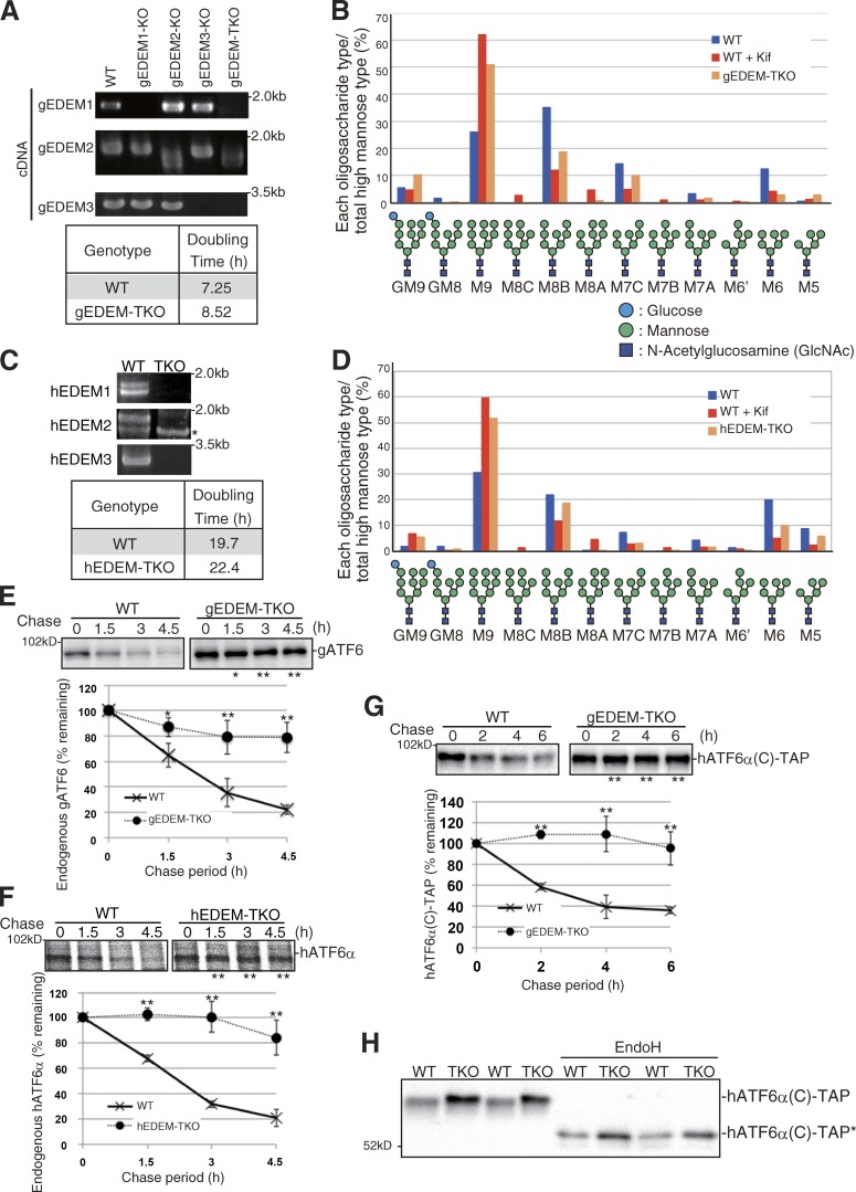
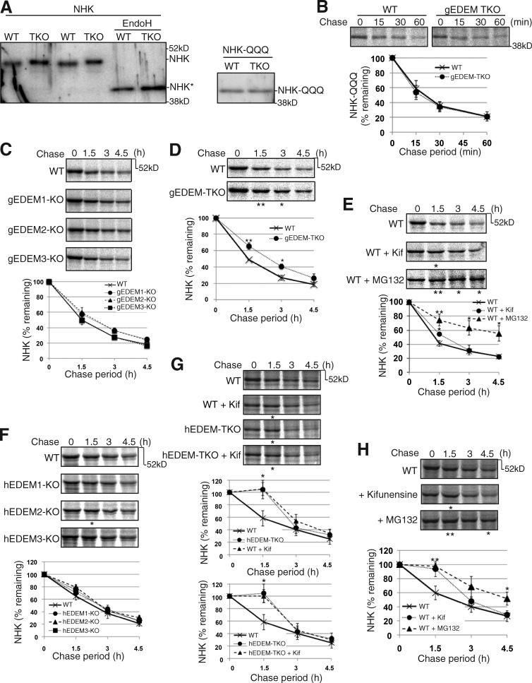
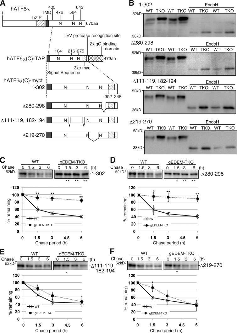
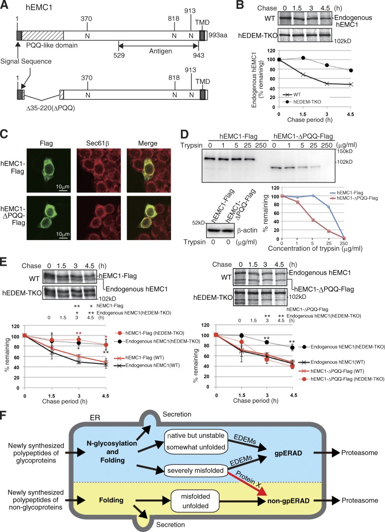
Glycoproteins and non-glycoproteins possessing unfolded/misfolded parts in their luminal regions are cleared from the endoplasmic reticulum (ER) by ER-associated degradation (ERAD)-L with distinct mechanisms. Two-step mannose trimming from Man9GlcNAc2 is crucial in the ERAD-L of glycoproteins. We recently showed that this process is initiated by EDEM2 and completed by EDEM3/EDEM1. Here, we constructed chicken and human cells simultaneously deficient in EDEM1/2/3 and analyzed the fates of four ERAD-L substrates containing three potential N-glycosylation sites. We found that native but unstable or somewhat unfolded glycoproteins, such as ATF6α, ATF6α(C), CD3-δ-ΔTM, and EMC1, were stabilized in EDEM1/2/3 triple knockout cells. In marked contrast, degradation of severely misfolded glycoproteins, such as null Hong Kong (NHK) and deletion or insertion mutants of ATF6α(C), CD3-δ-ΔTM, and EMC1, was delayed only at early chase periods, but they were eventually degraded as in wild-type cells. Thus, higher eukaryotes are able to extract severely misfolded glycoproteins from glycoprotein ERAD and target them to the non-glycoprotein ERAD pathway to maintain the homeostasis of the ER.


Figures

Figure 1.
Figure 1.
Effect of EDEM-TKO on N-glycan profiles and ATF6α degradation. (A) RT-PCR to amplify cDNA corresponding to gEDEM1/2/3 mRNA in DT40 cells of various genotypes and doubling times of WT and gEDEM-TKO cells. (B) Isomer composition of N-glycans prepared from total cellular glycoproteins of WT, WT treated with kifunensine (Kif, 10 µg/ml, 6 h), and gEDEM-TKO cells. This experiment was completed once. (C) RT-PCR to amplify cDNA corresponding to hEDEM1/2/3 mRNA in WT and hEDEM-TKO cells and their doubling times. The asterisk denotes a nonspecific band. (D) Isomer composition of N-glycans prepared from total cellular glycoproteins of WT, WT treated with kifunensine (10 µg/ml, 12 h), and hEDEM-TKO cells. This experiment was completed once. (E) Cycloheximide chase to determine the degradation rate of endogenous gATF6 (only one ATF6 gene in chicken genome) in WT and gEDEM-TKO cells using anti-gATF6 (n = 3). (F) Pulse-chase to determine the degradation rate of endogenous hATF6α in WT and hEDEM-TKO cells using anti-hATF6α (n = 3). (G) Cycloheximide chase to determine the degradation rate of transfected hATF6α(C)-TAP in WT and gEDEM-TKO cells using anti–c-myc (n = 3). (E–G) Means ± SD are shown. *, P < 0.05; **, P < 0.01. (H) Immunoblotting of cell lysates prepared from WT and gEDEM-TKO cells expressing transfected hATF6α(C)-TAP with or without EndoH treatment using anti–c-myc. hATF6α(C)-TAP* denotes the nonglycosylated hATF6α(C)-TAP.
Figure 2.
Figure 2.
Effect of EDEM-TKO on NHK degradation. (A) Immunoblotting of cell lysates prepared from WT and gEDEM-TKO cells expressing transfected NHK with or without EndoH treatment and those expressing transfected NHK-QQQ, using anti-α1PI. NHK* denotes the nonglycosylated NHK. (B) Pulse-chase to determine the degradation rate of transfected NHK-QQQ in WT and gEDEM-TKO cells using anti-α1PI (n = 3). (C) Pulse-chase to determine the degradation rate of transfected NHK in WT and gEDEM1/2/3 single KO cells using anti-α1PI (n = 3). (D) Pulse-chase to determine the degradation rate of transfected NHK in WT and gEDEM-TKO cells using anti-α1PI (n = 3). (E) Pulse-chase to determine the degradation rate of transfected NHK in WT DT40 cells and those treated with kifunensine (10 µg/ml) or MG132 (30 µM) using anti-α1PI (n = 3). (F) Pulse-chase to determine the degradation rate of transfected NHK in WT and hEDEM1/2/3 single KO cells using anti-α1PI (n = 3). (G) Pulse-chase to determine the degradation rate of transfected NHK in WT and hEDEM-TKO cells in the presence or absence of kifunensine (10 µg/ml) using anti-α1PI (n = 3). (H) Pulse-chase to determine the degradation rate of transfected NHK in WT HCT116 cells and those treated with kifunensine (10 µg/ml) or MG132 (20 µM) using anti-α1PI (n = 3). (B–H) Means ± SD are shown. (D–H) *, P < 0.05; **, P < 0.01.
Figure 3.
Figure 3.
Effect of EDEM-TKO on hATF6α(C)-myct degradation. (A) Schematic structures of hATF6α, hATF6α(C)-TAP, hATF6α(C)-myct, and its three deletion mutants. bZIP and TMD denote basic leucine zipper and transmembrane domains, respectively. N indicates a potential glycosylation site. Both the amino acids 111–119 and 182–194 are expected to form an α-helix. (B) Immunoblotting of cell lysates prepared from WT and gEDEM-TKO cells expressing one of the four hATF6α(C) derivatives by transfection with or without EndoH treatment using anti–c-myc. (C–F) Cycloheximide chase of WT and gEDEM-TKO cells expressing one of the four hATF6α(C) derivatives by transfection using anti–c-myc (n = 3). Means ± SD are shown. *, P < 0.05; **, P < 0.01.
Figure 4.
Figure 4.
Effect of EDEM-TKO on mCD3-δ–ΔTM degradation. (A) Schematic structures of WT and two insertion mutants of mCD3-δ–ΔTM. (B) Immunofluorescence of HCT116 cells expressing transfected WT or insertion mutant of mCD3-δ–ΔTM using anti-HA. (C) PNGase sensitivity assay of WT and two insertion mutants of mCD3-δ–ΔTM expressed in HCT116 cells by transfection (immunoblotting with anti-HA). The asterisk denotes partially deglycosylated mCD3-δ–ΔTM. (D) Pulse-chase to determine the degradation rate of transfected WT mCD3-δ–ΔTM in WT and hEDEM-TKO cells using anti-HA (n = 3). (E) Pulse-chase to determine the degradation rate of transfected WT mCD3-δ–ΔTM in WT cells and those treated with 40 µM MG132 using anti-HA. This experiment was completed once. (F) Pulse-chase to determine the degradation rate of transfected insertion mutants of mCD3-δ–ΔTM in WT and hEDEM-TKO cells using anti-HA (n = 3). (D–F) Means ± SD are shown. (D and F) *, P < 0.05; **, P < 0.01.
Figure 5.
Figure 5.
Effect of EDEM-TKO on hEMC1 degradation. (A) Schematic structures of WT and deletion mutant of hEMC1. (B) Pulse-chase to determine the degradation rate of endogenous hEMC1 in WT and hEDEM-TKO cells using anti-hEMC1. This experiment was completed once. (C) Immunofluorescence of HCT116 cells expressing transfected hEMC1-Flag or hEMC1-ΔPQQ-Flag using anti-Flag. (D) Trypsin sensitivity assay of hEMC1-Flag and hEMC1-ΔPQQ-Flag expressed in HCT116 cells by transfection (immunoblotting with anti-Flag). The data shown are from a single representative experiment out of two repeats. (E) Pulse-chase to determine the degradation rate of transfected hEMC1-Flag and hEMC1-ΔPQQ-Flag, as well as endogenous hEMC1 in WT and hEDEM-TKO cells using anti-hEMC1 (n = 3). Means ± SD are shown. *, P < 0.05; **, P < 0.01. (F) Model. Newly synthesized polypeptides of non-glycoproteins are subjected to productive folding. Folded non-glycoproteins are secreted, whereas unfolded or misfolded proteins are targeted to non-gpERAD for proteasomal degradation. Newly synthesized polypeptides of glycoproteins are subjected to N-glycosylation and productive folding. Folded glycoproteins are secreted, whereas native but unstable glycoproteins, somewhat unfolded glycoproteins, and severely misfolded glycoproteins are subjected to EDEM-mediated mannose trimming, followed by proteasomal degradation via gpERAD. In addition, severely misfolded glycoproteins are subjected to non-gpERAD via protein X.

References

    1. Aikawa J., Matsuo I., and Ito Y.. 2012. In vitro mannose trimming property of human ER α-1,2 mannosidase I. Glycoconj. J. 29:35–45. 10.1007/s10719-011-9362-1 - DOI - PubMed
    1. Bernasconi R., Galli C., Calanca V., Nakajima T., and Molinari M.. 2010. Stringent requirement for HRD1, SEL1L, and OS-9/XTP3-B for disposal of ERAD-LS substrates. J. Cell Biol. 188:223–235. 10.1083/jcb.200910042 - DOI - PMC - PubMed
    1. Brodsky J.L. 2012. Cleaning up: ER-associated degradation to the rescue. Cell. 151:1163–1167. 10.1016/j.cell.2012.11.012 - DOI - PMC - PubMed
    1. Fujimori T., Kamiya Y., Nagata K., Kato K., and Hosokawa N.. 2013. Endoplasmic reticulum lectin XTP3-B inhibits endoplasmic reticulum-associated degradation of a misfolded α1-antitrypsin variant. FEBS J. 280:1563–1575. 10.1111/febs.12157 - DOI - PubMed
    1. Haze K., Yoshida H., Yanagi H., Yura T., and Mori K.. 1999. Mammalian transcription factor ATF6 is synthesized as a transmembrane protein and activated by proteolysis in response to endoplasmic reticulum stress. Mol. Biol. Cell. 10:3787–3799. 10.1091/mbc.10.11.3787 - DOI - PMC - PubMed

Publication types

MeSH terms