Review
. 2016 Jun;43(6):1139-50.
doi: 10.1007/s00259-015-3231-2. Epub 2015 Nov 16.

Tau imaging in neurodegenerative diseases

Affiliations
Review

Tau imaging in neurodegenerative diseases

M Dani et al. Eur J Nucl Med Mol Imaging. 2016 Jun.

Abstract

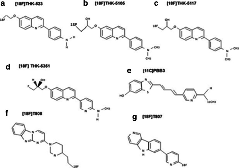
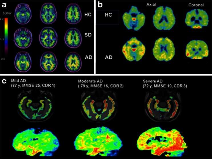
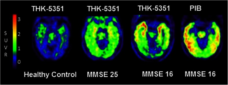
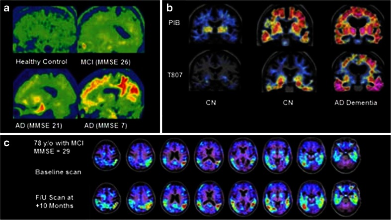
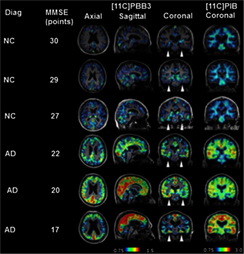
Aggregated tau protein is a major neuropathological substrate central to the pathophysiology of neurodegenerative diseases such as Alzheimer's disease (AD), frontotemporal dementia, progressive supranuclear palsy, corticobasal degeneration and chronic traumatic encephalopathy. In AD, it has been shown that the density of hyperphosphorylated tau tangles correlates closely with neuronal dysfunction and cell death, unlike β-amyloid. Until now, diagnostic and pathologic information about tau deposition has only been available from invasive techniques such as brain biopsy or autopsy. The recent development of selective in-vivo tau PET imaging ligands including [(18)F]THK523, [(18)F]THK5117, [(18)F]THK5105 and [(18)F]THK5351, [(18)F]AV1451(T807) and [(11)C]PBB3 has provided information about the role of tau in the early phases of neurodegenerative diseases, and provided support for diagnosis, prognosis, and imaging biomarkers to track disease progression. Moreover, the spatial and longitudinal relationship of tau distribution compared with β - amyloid and other pathologies in these diseases can be mapped. In this review, we discuss the role of aggregated tau in tauopathies, the challenges posed in developing selective tau ligands as biomarkers, the state of development in tau tracers, and the new clinical information that has been uncovered, as well as the opportunities for improving diagnosis and designing clinical trials in the future.

Keywords: Dementia; Neurodegenerative diseases; Tau imaging.


Figures

Fig. 1
Fig. 1
Chemical structures of current tau tracers. The chemical structures of: a [18F] THK-523, b [18F]THK-5105, c [18F] THK-5117, d [18F] THK-5351, e [11C] PBB3, f [18F]T808, and g [18F]-T807
Fig. 2
Fig. 2
PET images using the [18F] THK family of tracers. a The first tau tracer, [18F] THK 523 in a healthy control, a subject with semantic dementia and an AD subject. There is increased tracer retention in the AD subject, but no difference between the control and SD Reproduced from Villemagne 2014 [53]. b [18F] THK5105 PET images in a 72-year-old healthy control (MMSE 29) and a 68-year-old AD subject (MMSE 20). Reproduced from Okamura 2014 [54]. c Tau tracer [18F]THK 5117 in a subject with mild, moderate, and severe AD, showing increasing retention of tracer as disease progresses from the medial, anterior, and inferior temporal cortex in mild AD, spreading to association areas in moderate AD, and throughout the neocortex in severe AD. Reproduced from Okamura 2014 [44]
Fig. 3
Fig. 3
Novel tau tracer [18F]THK-5351 in different stages of cognitive impairment. PET images of [18F]THK-5351 in a healthy control, an MCI subject (MMSE 25), and an AD subject (MMSE 16). There is increasing tracer retention as disease progresses. In the AD patient, a [11C]PIB PET scan shows amyloid deposition in discrete separate areas of cortex. Courtesy of Nobayaki Okamura, unpublished work
Fig. 4
Fig. 4
Novel tau tracer [18F]AV-1451 (previously [18F]-T807. a [18F]T807 in a healthy control (top left), through increasing severity of cognitive impairment to severe AD (bottom right). Increased tracer retention is seen as disease progresses, with widespread neocortical deposition in severe disease. Reproduced from Chien 2013 [55]. b PET images from two cognitively normal individuals, and one with AD dementia, with amyloid PET images on the top row ([11C]PIB) and tau PET images on the bottom row ([18F] T807). From left to right, increasing amyloid deposition is seen in the neocortex, as well as increasing tau in the inferior temporal cortices. Reproduced from Sperling 2014) [56]. c [18F] T807 in a subject with MCI at baseline, and after 10 months, showing a significant increase in tracer deposition in the temporal and parietal lobes. This indicates the clinical utility of tau imaging in detecting disease progression over relatively short time periods. Reproduced from Mark Mintun, 2015 [57]
Fig. 5
Fig. 5
Tau tracer [11C]-PBB in differing stages of cognitive impairment. [11C]PBB3 and [11C]PIB in normal controls and AD patients with increasing severity of disease. The arrowheads indicate the hippocampi. While there is minimal tracer retention in the hippocampi of normal controls, there is increasing retention in the AD patients, especially as MMSE declines, with spread from the hippocampus to the neocortex, consistent with Braak staging. Reproduced from Maruyama 2013 [58]

References

    1. Spillantini MG, Goedert M. Tau pathology and neurodegeneration. Lancet Neurol. 2013;12:609–22. doi: 10.1016/S1474-4422(13)70090-5. - DOI - PubMed
    1. Gomez-Isla Neuronal loss correlates with but exceeds neurofibrillary tangles in Alzheimer’s disease. Ann Neurol. 1997;41:17–24. doi: 10.1002/ana.410410106. - DOI - PubMed
    1. Ingelsson M, Fukumoto H, Newell K, Growdon W, Hedley-White E, Frosch M, et al. Early amyloid accumulation and progressive synaptic loss, gliosis and tangle formation in the Alzheimer’s disease brain. Neurology. 2004;62:925–31. doi: 10.1212/01.WNL.0000115115.98960.37. - DOI - PubMed
    1. Lee HG, Perry G, Moreira PI, Garrett MR, Liu Q, Zhu X, et al. Tau phosphorylation in Alzheimer’s disease: pathogen or protector? Trends Mol Med. 2005;11:164–9. doi: 10.1016/j.molmed.2005.02.008. - DOI - PubMed
    1. Giacobini E, Gold G. Alzheimer disease therapy—moving from amyloid-beta to tau. Nat Rev Neurol. 2013;9:677–86. doi: 10.1038/nrneurol.2013.223. - DOI - PubMed

MeSH terms