

AXL is a potential therapeutic target in dedifferentiated and pleomorphic liposarcomas
- PMID: 26573603
- PMCID: PMC4647521
- DOI: 10.1186/s12885-015-1916-3
AXL is a potential therapeutic target in dedifferentiated and pleomorphic liposarcomas
Abstract
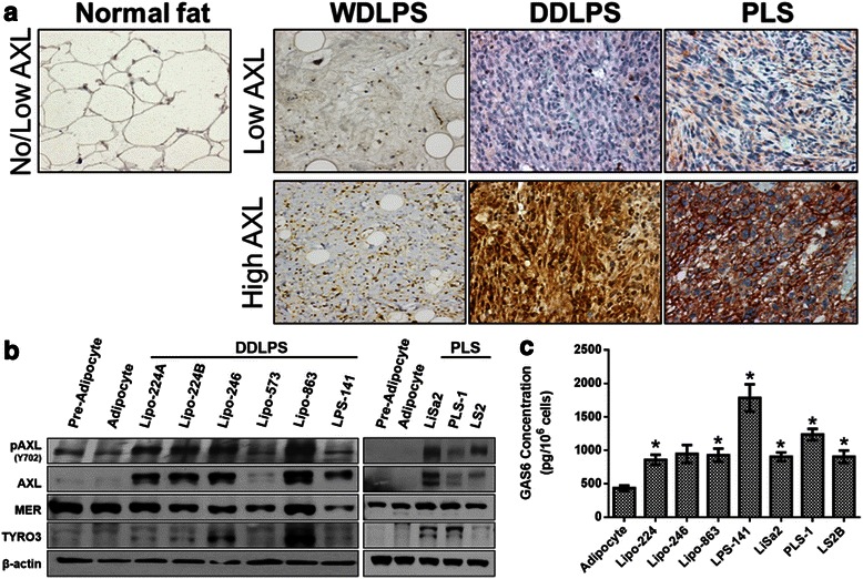
Background: AXL is a well-characterized, protumorigenic receptor tyrosine kinase that is highly expressed and activated in numerous human carcinomas and sarcomas, including aggressive subtypes of liposarcoma. However, the role of AXL in the pathogenesis of well-differentiated (WDLPS), dedifferentiated (DDLPS), and pleomorphic liposarcoma (PLS) has not yet been determined.
Methods: Immunohistochemical analysis of AXL expression was conducted on two tissue microarrays containing patient WDLPS, DDLPS, and PLS samples. A panel of DDLPS and PLS cell lines were interrogated via western blot for AXL expression and activity and by ELISA for growth arrest-specific 6 (GAS6) production. AXL knockdown was achieved by siRNA or shRNA. The effects of AXL knockdown on cell proliferation, migration, and invasion were measured in vitro. In addition, AXL shRNA-containing DDLPS cells were assessed for their tumor-forming capacity in vivo.
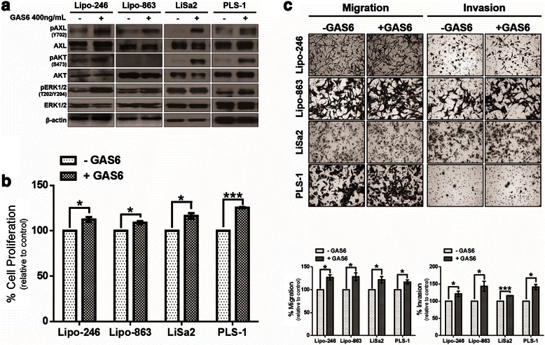
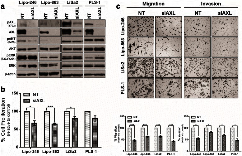
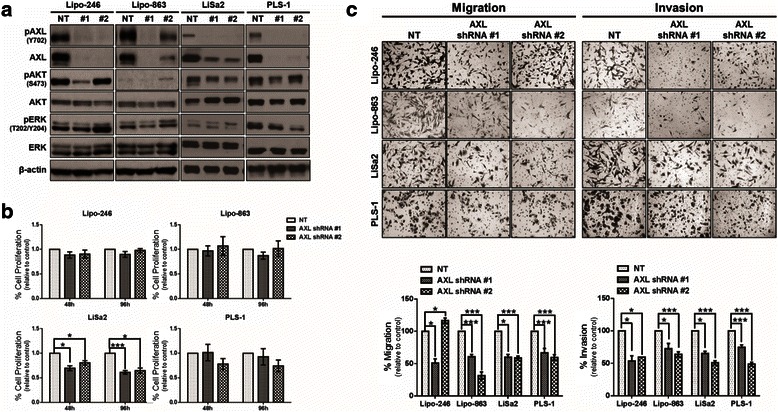
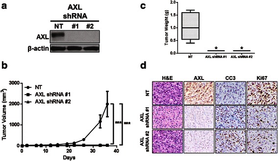
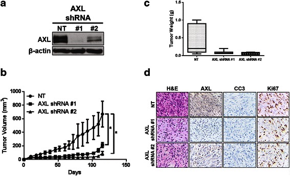
Results: In this study, we determined that AXL is expressed in a subset of WDLPS, DDLPS, and PLS patient tumor samples. In addition, AXL and its ligand GAS6 are expressed in a panel of DDLPS and PLS cell lines. We show that the in vitro activation of AXL via stimulation with exogenous GAS6 resulted in a significant increase in cell proliferation, migration, and invasion in DDLPS and PLS cell lines. Transient knockdown of AXL resulted in attenuation of these protumorigenic phenotypes in vitro. Stable AXL knockdown not only decreased migratory and invasive characteristics of DDLPS and PLS cells in vitro but also significantly diminished tumorigenicity of two dedifferentiated liposarcoma xenograft models in vivo.
Conclusions: Our results suggest that AXL signaling contributes to the aggressiveness of DDLPS and PLS, and that AXL is therefore a potential therapeutic target for treatment of these rare, yet devastating tumors.
Figures
Similar articles
-
An experimental model for the study of well-differentiated and dedifferentiated liposarcoma; deregulation of targetable tyrosine kinase receptors.Lab Invest. 2011 Mar;91(3):392-403. doi: 10.1038/labinvest.2010.185. Epub 2010 Nov 8. Lab Invest. 2011. PMID: 21060307 Free PMC article.
-
Expression of the preadipocyte marker ZFP423 is dysregulated between well-differentiated and dedifferentiated liposarcoma.BMC Cancer. 2022 Mar 21;22(1):300. doi: 10.1186/s12885-022-09379-6. BMC Cancer. 2022. PMID: 35313831 Free PMC article.
-
The hepatocyte growth factor receptor as a potential therapeutic target for dedifferentiated liposarcoma.Lab Invest. 2015 Aug;95(8):951-61. doi: 10.1038/labinvest.2015.62. Epub 2015 Jun 1. Lab Invest. 2015. PMID: 26006023 Free PMC article.
-
Update on genomic and molecular landscapes of well-differentiated liposarcoma and dedifferentiated liposarcoma.Mol Biol Rep. 2021 Apr;48(4):3637-3647. doi: 10.1007/s11033-021-06362-5. Epub 2021 Apr 24. Mol Biol Rep. 2021. PMID: 33893924 Review.
-
Therapeutic Targeting of the Gas6/Axl Signaling Pathway in Cancer.Int J Mol Sci. 2021 Sep 15;22(18):9953. doi: 10.3390/ijms22189953. Int J Mol Sci. 2021. PMID: 34576116 Free PMC article. Review.
Cited by
-
PDE3A Is a Highly Expressed Therapy Target in Myxoid Liposarcoma.Cancers (Basel). 2023 Nov 7;15(22):5308. doi: 10.3390/cancers15225308. Cancers (Basel). 2023. PMID: 38001568 Free PMC article.
-
The First-In-Class Anti-AXL×CD3ε Pronectin™-Based Bispecific T-Cell Engager Is Active in Preclinical Models of Human Soft Tissue and Bone Sarcomas.Cancers (Basel). 2023 Mar 8;15(6):1647. doi: 10.3390/cancers15061647. Cancers (Basel). 2023. PMID: 36980534 Free PMC article.
-
Role of the Gas6/TAM System as a Disease Marker and Potential Drug Target.Dis Markers. 2021 Jan 13;2021:2854925. doi: 10.1155/2021/2854925. eCollection 2021. Dis Markers. 2021. PMID: 33532004 Free PMC article. No abstract available.
-
Potential of Stem Cells and CART as a Potential Polytherapy for Small Cell Lung Cancer.Front Cell Dev Biol. 2021 Dec 3;9:778020. doi: 10.3389/fcell.2021.778020. eCollection 2021. Front Cell Dev Biol. 2021. PMID: 34926461 Free PMC article. Review.
-
The Gas6/TAM System and Multiple Sclerosis.Int J Mol Sci. 2016 Oct 28;17(11):1807. doi: 10.3390/ijms17111807. Int J Mol Sci. 2016. PMID: 27801848 Free PMC article. Review.
References
-
- Fletcher CDM, Bridge JA, Hogendoom PCW, Mertens F. World health organization classification of tumours of soft tissue and bone. 4. Lyon: IARC press; 2013.
-
- Fabre-Guillevin E, Coindre JM, Somerhausen Nde S, Bonichon F, Stoeckle E, Bui NB. Retroperitoneal liposarcomas: follow-up analysis of dedifferentiation after clinicopathologic reexamination of 86 liposarcomas and malignant fibrous histiocytomas. Cancer. 2006;106:2725–33. doi: 10.1002/cncr.21933. - DOI - PubMed
Publication types
MeSH terms
Substances
Grants and funding
LinkOut - more resources
Full Text Sources
Other Literature Sources
Research Materials
Miscellaneous
