

Inhibition of Brain Mitogen-Activated Protein Kinase Signaling Reduces Central Endoplasmic Reticulum Stress and Inflammation and Sympathetic Nerve Activity in Heart Failure Rats
- PMID: 26573710
- PMCID: PMC4679490
- DOI: 10.1161/HYPERTENSIONAHA.115.06329
Inhibition of Brain Mitogen-Activated Protein Kinase Signaling Reduces Central Endoplasmic Reticulum Stress and Inflammation and Sympathetic Nerve Activity in Heart Failure Rats
Erratum in
-
Correction to: Inhibition of Brain Mitogen-Activated Protein Kinase Signaling Reduces Central Endoplasmic Reticulum Stress and Inflammation and Sympathetic Nerve Activity in Heart Failure Rats.Hypertension. 2018 Sep;72(3):e32. doi: 10.1161/HYP.0000000000000078. Hypertension. 2018. PMID: 30354764 No abstract available.
Abstract
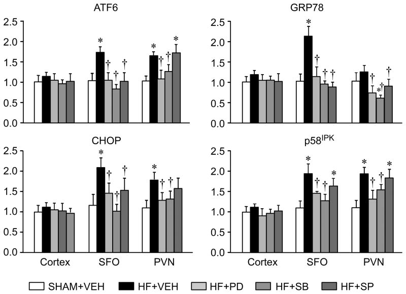
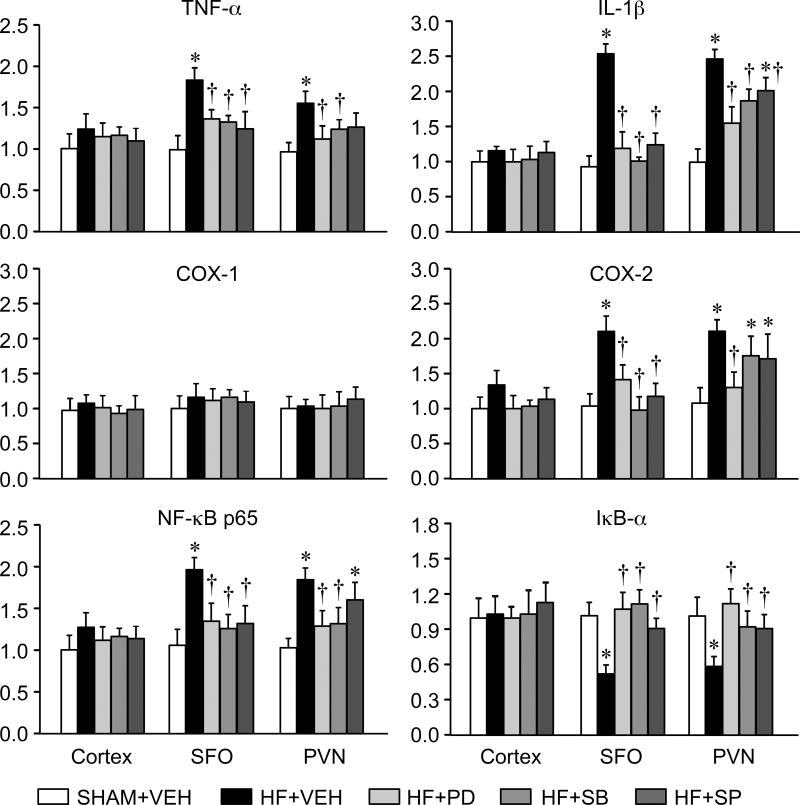
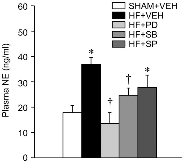
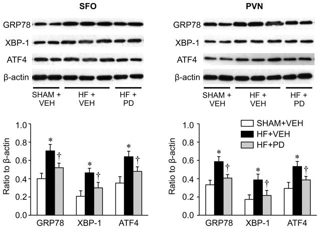
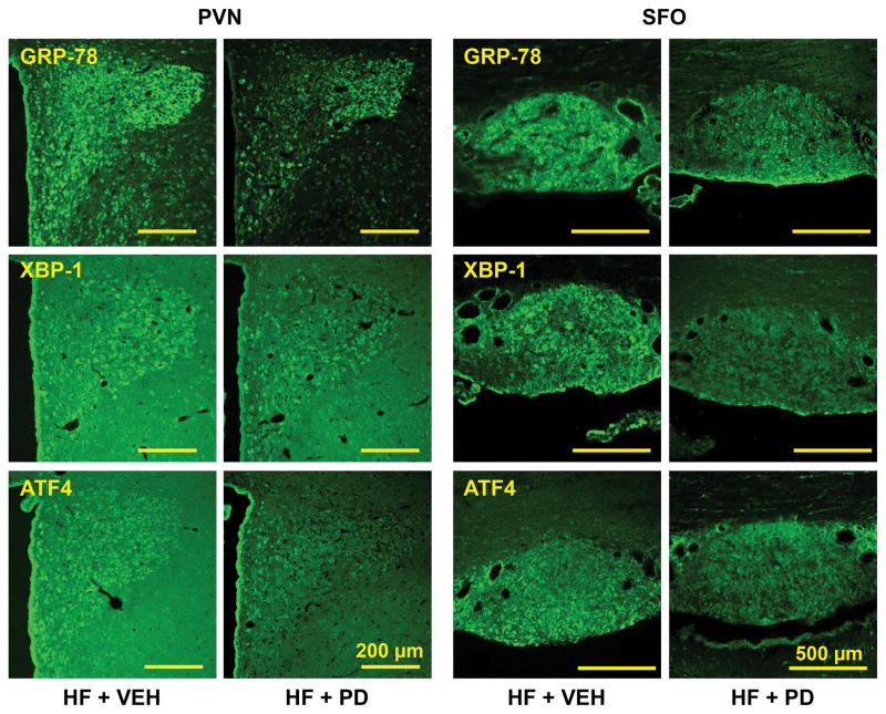
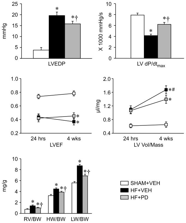
Mitogen-activated protein kinase (MAPK) signaling and endoplasmic reticulum (ER) stress in the brain have been implicated in the pathophysiology of hypertension. This study determined whether ER stress occurs in subfornical organ and hypothalamic paraventricular nucleus in heart failure (HF) and how MAPK signaling interacts with ER stress and other inflammatory mediators. HF rats had significantly higher levels of the ER stress biomarkers (glucose-regulated protein 78, activating transcription factor 6, activating transcription factor 4, X-box binding protein 1, P58(IPK), and C/EBP homologous protein) in subfornical organ and paraventricular nucleus, which were attenuated by a 4-week intracerebroventricular infusion of inhibitors selective for p44/42 MAPK (PD98059), p38 MAPK (SB203580), or c-Jun N-terminal kinase (SP600125). HF rats also had higher mRNA levels of tumor necrosis factor-α, interleukin-1β, cyclooxygenase-2, and nuclear factor-κB p65, and a lower mRNA level of IκB-α, in subfornical organ and paraventricular nucleus, compared with SHAM rats, and these indicators of increased inflammation were attenuated in the HF rats treated with the MAPK inhibitors. Plasma norepinephrine level was higher in HF rats than in SHAM rats but was reduced in the HF rats treated with PD98059 and SB203580. A 4-week intracerebroventricular infusion of PD98059 also improved some hemodynamic and anatomic indicators of left ventricular function in HF rats. These data demonstrate that ER stress increases in the subfornical organ and paraventricular nucleus of rats with ischemia-induced HF and that inhibition of brain MAPK signaling reduces brain ER stress and inflammation and decreases sympathetic excitation in HF. An interaction between MAPK signaling and ER stress in cardiovascular regions of the brain may contribute to the development of HF.
Keywords: brain; endoplasmic reticulum stress; heart failure; hypothalamic paraventricular nucleus; subfornical organ; sympathetic nerve activity.
© 2015 American Heart Association, Inc.
Figures
Similar articles
-
Endoplasmic reticulum stress increases brain MAPK signaling, inflammation and renin-angiotensin system activity and sympathetic nerve activity in heart failure.Am J Physiol Heart Circ Physiol. 2016 Oct 1;311(4):H871-H880. doi: 10.1152/ajpheart.00362.2016. Epub 2016 Aug 5. Am J Physiol Heart Circ Physiol. 2016. PMID: 27496879 Free PMC article.
-
Mitogen-activated protein kinases mediate upregulation of hypothalamic angiotensin II type 1 receptors in heart failure rats.Hypertension. 2008 Oct;52(4):679-86. doi: 10.1161/HYPERTENSIONAHA.108.113639. Epub 2008 Sep 2. Hypertension. 2008. PMID: 18768402 Free PMC article.
-
Angiotensin II-triggered p44/42 mitogen-activated protein kinase mediates sympathetic excitation in heart failure rats.Hypertension. 2008 Aug;52(2):342-50. doi: 10.1161/HYPERTENSIONAHA.108.110445. Epub 2008 Jun 23. Hypertension. 2008. PMID: 18574076 Free PMC article.
-
Chronic heart failure: a disease of the brain.Heart Fail Rev. 2019 Mar;24(2):301-307. doi: 10.1007/s10741-018-9747-3. Heart Fail Rev. 2019. PMID: 30341700 Review.
-
Neurohumoral activation in heart failure: role of paraventricular nucleus.Clin Exp Pharmacol Physiol. 1996 Aug;23(8):722-6. doi: 10.1111/j.1440-1681.1996.tb01765.x. Clin Exp Pharmacol Physiol. 1996. PMID: 8886497 Review.
Cited by
-
Inhibition of endoplasmic reticulum stress signaling pathway: A new mechanism of statins to suppress the development of abdominal aortic aneurysm.PLoS One. 2017 Apr 3;12(4):e0174821. doi: 10.1371/journal.pone.0174821. eCollection 2017. PLoS One. 2017. PMID: 28369137 Free PMC article.
-
Hypothalamic dysfunction in heart failure: pathogenetic mechanisms and therapeutic implications.Heart Fail Rev. 2018 Jan;23(1):55-61. doi: 10.1007/s10741-017-9659-7. Heart Fail Rev. 2018. PMID: 29052045 Review.
-
The biochemical alterations underlying post-burn hypermetabolism.Biochim Biophys Acta Mol Basis Dis. 2017 Oct;1863(10 Pt B):2633-2644. doi: 10.1016/j.bbadis.2017.02.019. Epub 2017 Feb 20. Biochim Biophys Acta Mol Basis Dis. 2017. PMID: 28219767 Free PMC article. Review.
-
Transforming Growth Factor-α Acts in Hypothalamic Paraventricular Nucleus to Upregulate ERK1/2 Signaling and Expression of Sympathoexcitatory Mediators in Heart Failure Rats.Neuroscience. 2022 Feb 10;483:13-23. doi: 10.1016/j.neuroscience.2021.12.030. Epub 2021 Dec 27. Neuroscience. 2022. PMID: 34968668 Free PMC article.
-
ERK1/2 MAPK signaling in hypothalamic paraventricular nucleus contributes to sympathetic excitation in rats with heart failure after myocardial infarction.Am J Physiol Heart Circ Physiol. 2016 Mar 15;310(6):H732-9. doi: 10.1152/ajpheart.00703.2015. Epub 2016 Jan 22. Am J Physiol Heart Circ Physiol. 2016. PMID: 26801309 Free PMC article.
References
-
- Pearson G, Robinson F, Beers Gibson T, Xu BE, Karandikar M, Berman K, Cobb MH. Mitogen-activated protein (MAP) kinase pathways: Regulation and physiological functions. Endocr Rev. 2001;22:153–183. - PubMed
-
- Ron D, Walter P. Signal integration in the endoplasmic reticulum unfolded protein response. Nat Rev Mol Cell Biol. 2007;8:519–529. - PubMed
-
- Darling NJ, Cook SJ. The role of MAPK signalling pathways in the response to endoplasmic reticulum stress. Biochim Biophys Acta. 2014;1843:2150–2163. - PubMed
Publication types
MeSH terms
Substances
Grants and funding
LinkOut - more resources
Full Text Sources
Other Literature Sources
Medical
Research Materials
Miscellaneous
