. 2015 Nov 17:5:16865.
doi: 10.1038/srep16865.

Cervical intraepithelial neoplasia disease progression is associated with increased vaginal microbiome diversity

Affiliations

Cervical intraepithelial neoplasia disease progression is associated with increased vaginal microbiome diversity

A Mitra et al. Sci Rep. .

Abstract

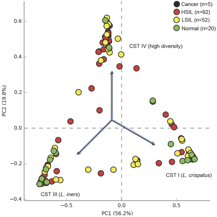
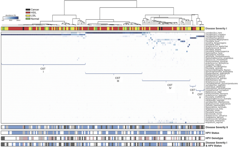
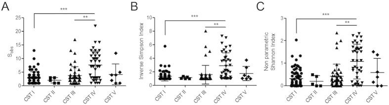
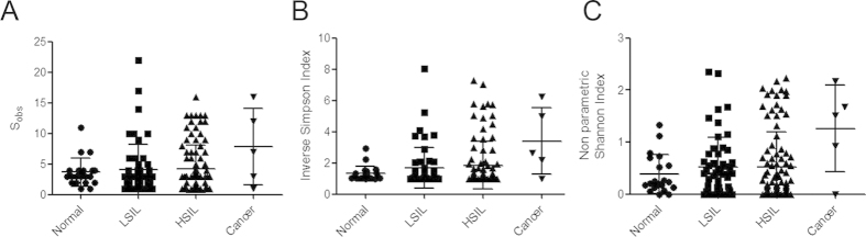
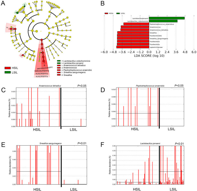
Persistent infection with oncogenic Human Papillomavirus (HPV) is necessary for cervical carcinogenesis. Although evidence suggests that the vaginal microbiome plays a functional role in the persistence or regression of HPV infections, this has yet to be described in women with cervical intra-epithelial neoplasia (CIN). We hypothesised that increasing microbiome diversity is associated with increasing CIN severity. llumina MiSeq sequencing of 16S rRNA gene amplicons was used to characterise the vaginal microbiota of women with low-grade squamous intra-epithelial lesions (LSIL; n = 52), high-grade (HSIL; n = 92), invasive cervical cancer (ICC; n = 5) and healthy controls (n = 20). Hierarchical clustering analysis revealed an increased prevalence of microbiomes characterised by high-diversity and low levels of Lactobacillus spp. (community state type-CST IV) with increasing disease severity, irrespective of HPV status (Normal = 2/20,10%; LSIL = 11/52,21%; HSIL = 25/92,27%; ICC = 2/5,40%). Increasing disease severity was associated with decreasing relative abundance of Lactobacillus spp. The vaginal microbiome in HSIL was characterised by higher levels of Sneathia sanguinegens (P < 0.01), Anaerococcus tetradius (P < 0.05) and Peptostreptococcus anaerobius (P < 0.05) and lower levels of Lactobacillus jensenii (P < 0.01) compared to LSIL. Our results suggest advancing CIN disease severity is associated with increasing vaginal microbiota diversity and may be involved in regulating viral persistence and disease progression.


Figures

Figure 1
Figure 1. Bacterial species diversity in study cohort and controls.
Principle component analysis (PCA) of vaginal bacterial species data identified 3 major clusters corresponding to samples dominated by three community state types (CSTs): L. iners (CST III), L. crispatus (CST I) and high diversity samples (CST IV). KEY - Cancer: Black; High-grade squamous intra-epithelial lesions (HSIL): Red; Low-grade squamous intra-epithelial lesions (LSIL): Yellow; Normal: Green.
Figure 2
Figure 2. Vaginal microbiome composition according to disease and HPV status.
Hierarchical clustering analysis (HCA) using centroid clustering showed the distribution of CSTs differs in healthy, normal control women (green), compared to those with disease (Disease Severity I). Frequency of CST IV was 2-fold greater in women with LSIL (yellow), 3-fold greater in HSIL (red) and 4-fold greater in women with ICC (black), compared to controls, with a reciprocal decrease in frequency of CST I with increasing disease severity. When HPV/ASCUS (light grey) and LSIL (dark grey) were examined as two separate groups, the stepwise increase in CST IV prevalence was maintained (Disease Severity II). HPV positivity (blue) was associated with increased rates of CST IV, compared to HPV negative women (light grey) who were most likely to have CST I (HPV status). HPV-16 (pink) was most frequently associated with CST IV (HPV genotype). Rates of CST IV were similar in women with normal or LSIL regardless of HPV status (negative = light grey, positive = dark grey), and were substantially higher in women with HSIL and positive for HPV (blue) (Disease severity I & HPV status). DISEASE SEVERITY I - Normal: Green; Low-grade squamous intra-epithelial lesions (LSIL): Yellow; High-grade squamous intra-epithelial lesions (HSIL): Red; Cancer: Black. DISEASE SEVERITY II - Normal: white; ASCUS: light grey; LSIL: dark grey; HSIL: blue; Cancer: pink. HPV STATUS - HPV negative: light grey; HPV positive: blue; Unknown: white. HPV GENOTYPE - HPV negative: light grey; HPV16: pink; HPV18: blue; Other HR-HPV: dark grey; Unknown: white. DISEASE SEVERITY I & HPV STATUS - LSIL/Normal HPV negative: light grey; LSIL/Normal HPV positive: dark grey; HSIL/HPV positive: blue; Cancer: pink; unknown/other: white. KEY - ASCUS: atypical squamous cells of undetermined significance; CST: community state type; HPV: Human Papillomavirus; HR-HPV: high-risk HPV; HSIL: High-grade squamous intra-epithelial lesion; ICC: invasive cervical cancer; LSIL: Low-grade squamous intra-epithelial lesion.
Figure 3
Figure 3. Analysis of richness (A) and diversity indices (B&C) with attributed CSTs for the patient cohort.
A significantly higher number of species observed in samples classified as CST IV (A) compared to CST I (P < 0.001) and CST III (P < 0.01). Diversity was also significantly higher in CST IV classified samples as assessed by the Inverse Simpson (B) and non-parametric Shannon (C) indices compared to CST I (P < 0.001) and CST III (P < 0.01). Kruskall-Wallis test (Dunn’s post hoc). KEY - CST: Community state type; Sobs: Species observed; ** = P < 0.01; *** = P < 0.001.
Figure 4
Figure 4. Vaginal microbiome richness and diversity indices associated with disease status (normal, LSIL, HSIL and cervical cancer patients).
The number of species observed increased with disease severity with lowest richness observed in healthy controls and highest in HSIL and ICC (A). Diversity, as assessed by Inverse Simpson (B) and non-parametric Shannon (C) indices followed the same pattern. KEY - HSIL: High-grade squamous intra-epithelial lesion; ICC: invasive cervical cancer; LSIL: Low-grade squamous intra-epithelial lesion; Sobs: Species observed; ** = P < 0.01; *** = P < 0.001.
Figure 5
Figure 5. Identification of vaginal microbiota biomarkers of LSIL vs. HSIL by LEfSe analysis.
(A) Cladogram representing taxa with different abundance according to disease severity. Size of circle is proportionate to abundance of taxon. (B) Histogram of the LDA scores computed for features differentially abundant between LSIL and HSIL disease states. Relative abundance counts of Anaerococcus tetradius (C), Peptostreptococcus anaerobis (D) and Sneathia sanguinegens (E), which were found to be significantly over-represented in HSIL whereas Lactobacillus jensenii (F) was enriched in LSIL samples (Welch’s t-test). KEY - HSIL: High-grade squamous intra-epithelial lesion; LDA score: Linear discriminant analysis score; LSIL: Low-grade squamous intra-epithelial lesion.

References

    1. Munoz N. Human papillomavirus and cancer: the epidemiological evidence. J. Clin. Virol. 19, 1–5 (2000). - PubMed
    1. Myers E. R., McCrory D. C., Nanda K., Bastian L. & Matchar D. B. Mathematical model for the natural history of human papillomavirus infection and cervical carcinogenesis. Am. J. Epidemiol. 151, 1158–1171 (2000). - PubMed
    1. Richardson H. et al.. The natural history of type-specific human papillomavirus infections in female university students. Cancer Epidemiol. Biomarkers Prev. 12, 485–490 (2003). - PubMed
    1. Martin D. H. The microbiota of the vagina and its influence on women’s health and disease. Am. J. Med. Sci. 343, 2–9, doi: 10.1097/MAJ.0b013e31823ea228 (2012). - DOI - PMC - PubMed
    1. Boskey E. R., Cone R. A., Whaley K. J. & Moench T. R. Origins of vaginal acidity: high D/L lactate ratio is consistent with bacteria being the primary source. Hum. Reprod. 16, 1809–1813 (2001). - PubMed

Publication types

MeSH terms