. 2016 Jan 8;291(2):582-92.
doi: 10.1074/jbc.M115.667436. Epub 2015 Nov 16.

The Non-structural Protein of Crimean-Congo Hemorrhagic Fever Virus Disrupts the Mitochondrial Membrane Potential and Induces Apoptosis

Affiliations

The Non-structural Protein of Crimean-Congo Hemorrhagic Fever Virus Disrupts the Mitochondrial Membrane Potential and Induces Apoptosis

Bhaskar Barnwal et al. J Biol Chem. .

Abstract

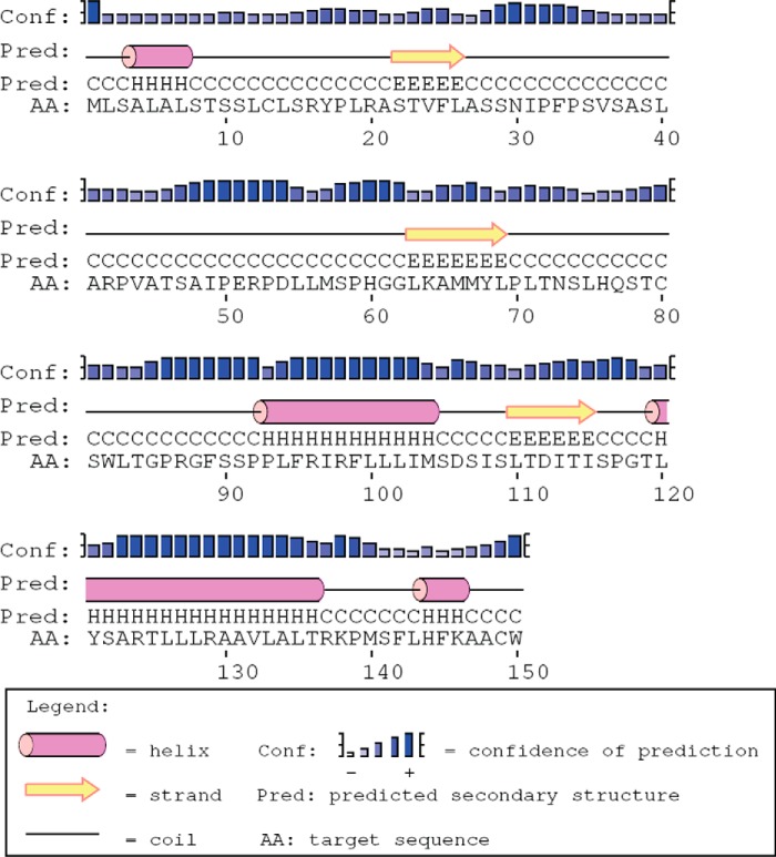
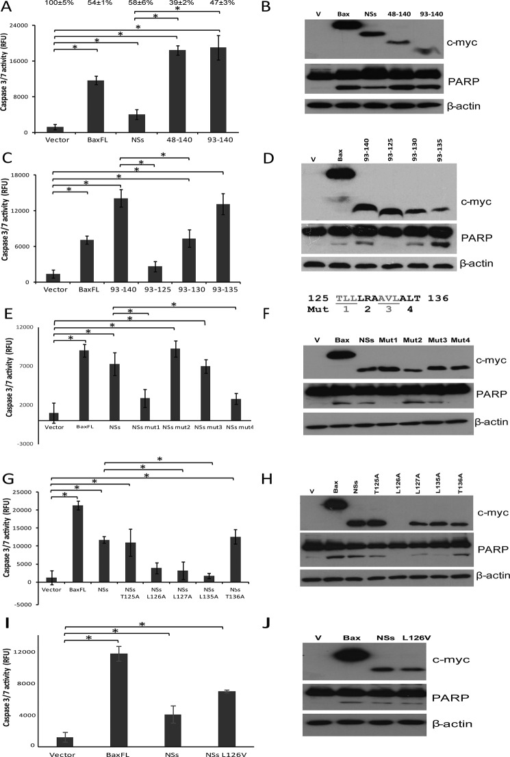
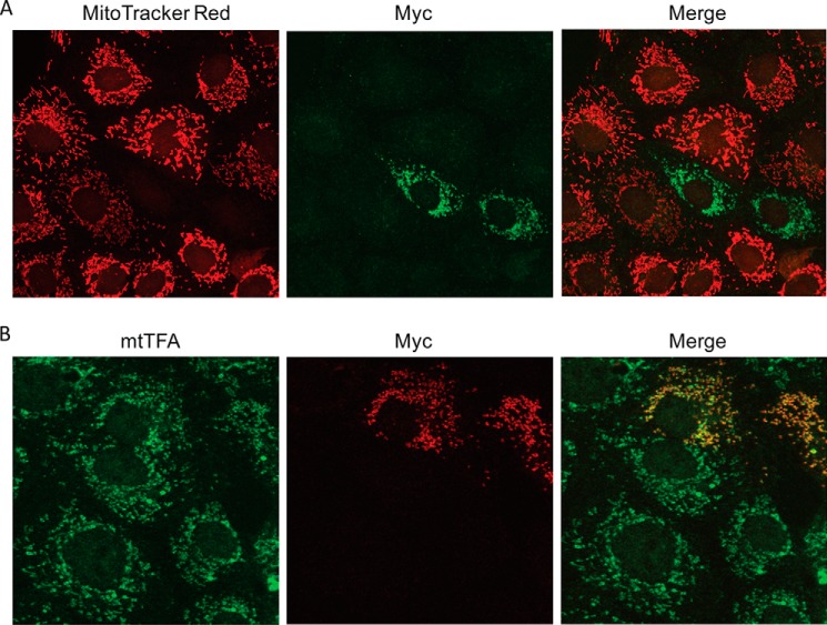
Viruses have developed distinct strategies to overcome the host defense system. Regulation of apoptosis in response to viral infection is important for virus survival and dissemination. Like other viruses, Crimean-Congo hemorrhagic fever virus (CCHFV) is known to regulate apoptosis. This study, for the first time, suggests that the non-structural protein NSs of CCHFV, a member of the genus Nairovirus, induces apoptosis. In this report, we demonstrated the expression of CCHFV NSs, which contains 150 amino acid residues, in CCHFV-infected cells. CCHFV NSs undergoes active degradation during infection. We further demonstrated that ectopic expression of CCHFV NSs induces apoptosis, as reflected by caspase-3/7 activity and cleaved poly(ADP-ribose) polymerase, in different cell lines that support CCHFV replication. Using specific inhibitors, we showed that CCHFV NSs induces apoptosis via both intrinsic and extrinsic pathways. The minimal active region of the CCHFV NSs protein was determined to be 93-140 amino acid residues. Using alanine scanning, we demonstrated that Leu-127 and Leu-135 are the key residues for NSs-induced apoptosis. Interestingly, CCHFV NSs co-localizes in mitochondria and also disrupts the mitochondrial membrane potential. We also demonstrated that Leu-127 and Leu-135 are important residues for disruption of the mitochondrial membrane potential by NSs. Therefore, these results indicate that the C terminus of CCHFV NSs triggers mitochondrial membrane permeabilization, leading to activation of caspases, which, ultimately, leads to apoptosis. Given that multiple factors contribute to apoptosis during CCHFV infection, further studies are needed to define the involvement of CCHFV NSs in regulating apoptosis in infected cells.

Keywords: Crimean-Congo hemorrhagic fever virus; apoptosis; caspase; mitochondria; mitochondrial membrane potential; non-structural protein; proteasome.


Figures

FIGURE 1.
FIGURE 1.
Predicted secondary structure of CCHFV NSs. The secondary structure of NSs containing 150 amino acid residues was predicted by PSIPRED.
FIGURE 2.
FIGURE 2.
NSs expression in CCHFV-infected or NSs-transfected cells. A, SW13 cells were mock-infected or infected with CCHFV and either left untreated or treated with MG132, followed by cell harvesting at 48 and 72 h.p.i. Alternatively, SW13 cells were transfected with empty vector or myc-NSs and either left untreated or treated with MG132, after which cells were harvested 24 h post-transfection. Western blot analysis was performed to determine the expression levels of CCHFV NSs (bottom row) and calnexin as a loading control (top row). The Western blots shown represent one of three independent experiments. B, densitometric analysis of NSs band intensities normalized to calnexin. C, immunofluorescence analysis was performed on CCHFV-infected cells. NSs (green) and nucleocapsid protein (NP, red) were analyzed by fluorescence microscopy. Nuclei were counterstained with DAPI (blue). Three independent experiments were performed, with a representative set of images shown here.
FIGURE 3.
FIGURE 3.
Induction of apoptosis by NSs in different cell lines that support CCHFV replication. The cell lines used were Vero E6 (A and B), HeLa (C and D), and 293FT (E and F). A, C, and E, the apo-ONE homogeneous caspase-3/7 assay was used to measure the activation of caspase-3/7 in three different cell lines that were transiently transfected with empty vector, myc-NSs, or myc-Bax. All experiments were performed in triplicate, and the results are expressed as mean ± S.D. Three independent experiments were performed, and a representative dataset is shown. *, p < 0.05. B, D, and F, Western blot analysis was performed to determine the expression levels of the myc-tagged proteins (top panels), the cleavage of endogenous PARP (center panels), and the levels of endogenous actin as a loading control (bottom panels). The Western blots shown represent one of three independent experiments. RFU, relative fluorescence units; V, vector.
FIGURE 4.
FIGURE 4.
NSs induces apoptosis via both the intrinsic and extrinsic pathways. A, the apo-ONE homogeneous caspase-3/7 assay was used to measure the activation of caspase-3/7 in 293FT cells that were transiently transfected with empty vector or myc-NSs. The cells were treated with an irrelevant peptide (Z-FA-fmk) or caspase-3, caspase-8, and caspase-9 inhibitors (Z-DEVD-fmk, Z-IETD-fmk, and Z-LEHD-fmk, respectively). All experiments were performed in triplicate, and the results are expressed as mean ± S.D. Three independent experiments were performed, and a representative dataset is shown. *, p < 0.05. B, Western blot analysis was performed to determine the expression levels of myc-NSs (top panel), the cleavage of endogenous PARP (center panel), and the levels of endogenous actin as a loading control (bottom panel). The Western blots shown represent one of three independent experiments. C, the caspase-Glo 8 assay was used to measure the activation of caspase-8 in 293FT cells that were transiently transfected with empty vector or NSs. Cells treated with TNF-α + CHX were used as a positive control, and those treated with 0.1% BSA in PBS + dimethyl sulfoxide (DMSO) were used as a negative control. All experiments were performed in triplicate, and the results are expressed as mean ± S.D. Four independent experiments were performed, and a representative dataset is shown. *, p < 0.05. D, Western blot analysis was performed to determine the expression levels of myc-NSs (top panel), the cleavage of endogenous PARP (center panel), and the levels of endogenous actin as a loading control (bottom panel). The Western blots shown represent one of three independent experiments. RFU, relative fluorescence units; V, vector.
FIGURE 5.
FIGURE 5.
Leu-127 and Leu-135 are key residues for the apoptotic activity of NSs protein. A, C, E, G, and I, the apo-one homogeneous caspase-3/7 assay was used to measure the activation of caspase-3/7 in 293FT cells that were transiently transfected with empty vector, Bax, NSs, NSs(48–140), or NSs(93–140) (A); empty vector, Bax, NSs(93–140), NSs(93–135), NSs(93–130), or NSs(93–125) (C); empty vector, Bax, NSs, or triple alanine substitution mutants such as NSs mut1 (TLL(125–127)AAA), NSs mut2 (LRA(128–130)AAA), NSs mut3 (AVL(131–133)AAA), or NSs mut4 (ALT(134–136)AAA) (E); empty vector, Bax, NSs, or single alanine substitution mutants like NSs T125A, L126A, L127A, L135A, or T136A (G); and empty vector, Bax, NSs, or NSs L126V (I). The percentages of live cells compared with that of the empty vector, which was normalized to 100%, are shown above each column. All experiments were performed in triplicate, and the results are expressed as mean ± S.D. At least three independent experiments were performed, and a representative dataset is shown.*, p < 0.05. B, D, F, H, and J, Western blot analysis was performed for the samples to determine the expression levels of myc-tagged proteins (top panels), the cleavage of endogenous PARP (center panels), and the levels of endogenous actin as a loading control (bottom panels). The Western blots shown represent one of three independent experiments. RFU, relative fluorescence units; Mut, NSs triple alanine substitution mutant; V, vector.
FIGURE 6.
FIGURE 6.
NSs co-localizes in mitochondria and disrupts the mitochondrial membrane potential. A, myc-NSs was transiently transfected into Vero E6 cells. Subcellular localization of NSs was detected by anti-myc antibody and Alexa Fluor 488-conjugated secondary antibody (green). Mitochondria were labeled with MitoTracker Red CMXRos (red). B, the subcellular localization of NSs in Vero E6 cells was detected by anti-myc antibody and Rhodamine-conjugated secondary antibody (red). Endogenous mtTFA was detected by anti-mtTFA antibody and Alexa Fluor 488-conjugated secondary antibody (green). Three independent experiments were performed, with a representative set of images shown here.
FIGURE 7.
FIGURE 7.
NSs disrupts the mitochondrial membrane potential in HeLa cells. A, the subcellular localization of transiently transfected DN-GRIM19-HA was detected by anti-HA antibody and Alexa Fluor 488-conjugated secondary antibody (green). The subcellular localization of transiently transfected myc-NSs (B) and myc-NSs L127A and L135A (C) was detected by anti-myc antibody and Alexa Fluor 488-conjugated secondary antibody (green). Mitochondria were labeled with MitoTracker Red CMXRos (red), and nuclei were counterstained with DAPI (blue). At least three independent experiments were performed, with a representative set of images shown here.

References

    1. Whitehouse C. A. (2004) Crimean-Congo hemorrhagic fever. Antiviral Res. 64, 145–160 - PubMed
    1. Keshtkar-Jahromi M., Kuhn J. H., Christova I., Bradfute S. B., Jahrling P. B., and Bavari S. (2011) Crimean-Congo hemorrhagic fever: current and future prospects of vaccines and therapies. Antiviral Res. 90, 85–92 - PubMed
    1. Bente D. A., Forrester N. L., Watts D. M., McAuley A. J., Whitehouse C. A., and Bray M. (2013) Crimean-Congo hemorrhagic fever: history, epidemiology, pathogenesis, clinical syndrome and genetic diversity. Antiviral Res. 100, 159–189 - PubMed
    1. Ergönül O. (2006) Crimean-Congo haemorrhagic fever. Lancet Infect. Dis. 6, 203–214 - PMC - PubMed
    1. Elliott R. M. (1990) Molecular biology of the Bunyaviridae. J. Gen. Virol. 71, 501–522 - PubMed

Publication types

MeSH terms