. 2016 Jan;5(1):45-55.
doi: 10.5966/sctm.2015-0076. Epub 2015 Nov 16.

Propranolol Targets Hemangioma Stem Cells via cAMP and Mitogen-Activated Protein Kinase Regulation

Affiliations

Propranolol Targets Hemangioma Stem Cells via cAMP and Mitogen-Activated Protein Kinase Regulation

Naikhoba C O Munabi et al. Stem Cells Transl Med. 2016 Jan.

Abstract

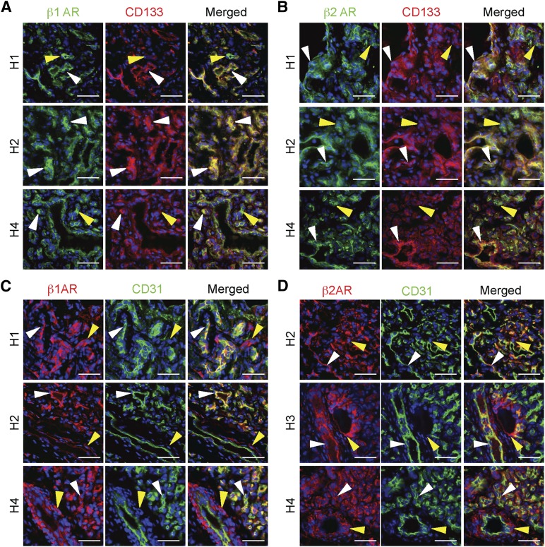
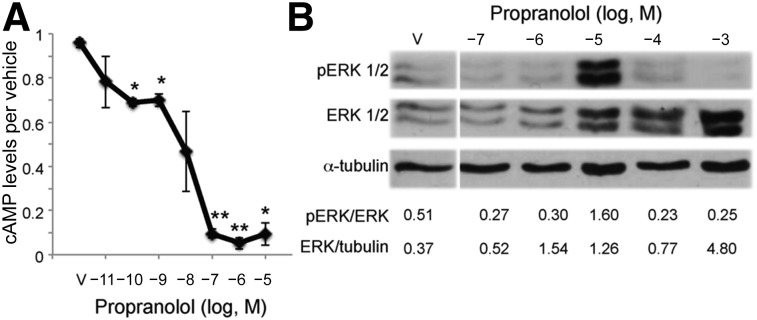
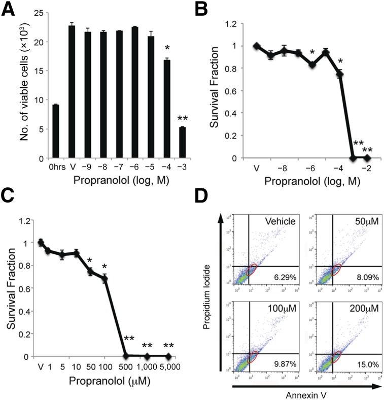
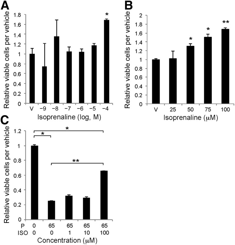
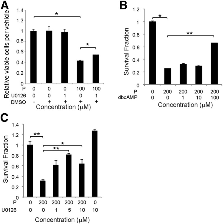
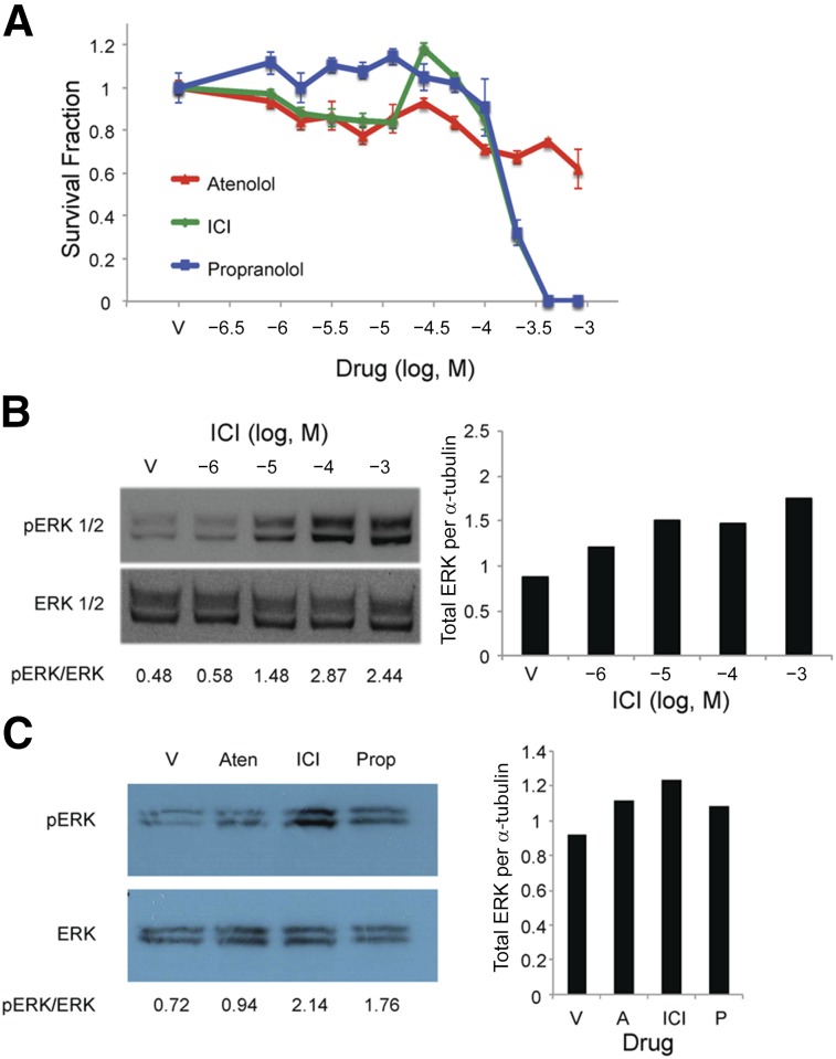
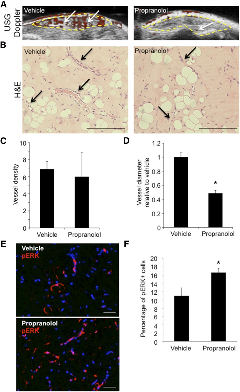
Infantile hemangiomas (IHs) are the most common vascular tumor and arise from a hemangioma stem cell (HemSC). Propranolol has proved efficacious for problematic IHs. Propranolol is a nonselective β-adrenergic receptor (βAR) antagonist that can lower cAMP levels and activate the mitogen-activated protein kinase (MAPK) pathway downstream of βARs. We found that HemSCs express β1AR and β2AR in proliferating IHs and determined the role of these βARs and the downstream pathways in mediating propranolol's effects. In isolated HemSCs, propranolol suppressed cAMP levels and activated extracellular signal-regulated kinase (ERK)1/2 in a dose-dependent fashion. Propranolol, used at doses of <10(-4) M, reduced cAMP levels and decreased HemSC proliferation and viability. Propranolol at ≥10(-5) M reduced cAMP levels and activated ERK1/2, and this correlated with HemSC apoptosis and cytotoxicity at ≥10(-4) M. Stimulation with a βAR agonist, isoprenaline, promoted HemSC proliferation and rescued the antiproliferative effects of propranolol, suggesting that propranolol inhibits βAR signaling in HemSCs. Treatment with a cAMP analog or a MAPK inhibitor partially rescued the HemSC cell viability suppressed by propranolol. A selective β2AR antagonist mirrored propranolol's effects on HemSCs in a dose-dependent fashion, and a selective β1AR antagonist had no effect, supporting a role for β2AR signaling in IH pathobiology. In a mouse model of IH, propranolol reduced the vessel caliber and blood flow assessed by ultrasound Doppler and increased activation of ERK1/2 in IH cells. We have thus demonstrated that propranolol acts on HemSCs in IH to suppress proliferation and promote apoptosis in a dose-dependent fashion via β2AR perturbation, resulting in reduced cAMP and MAPK activation.

Significance: The present study investigated the action of propranolol in infantile hemangiomas (IHs). IHs are the most common vascular tumor in children and have been proposed to arise from a hemangioma stem cell (HemSC). Propranolol, a nonselective β-adrenergic receptor (βAR) antagonist, has proven efficacy; however, understanding of its mechanism of action on HemSCs is limited. The presented data demonstrate that propranolol, via βAR perturbation, dose dependently suppresses cAMP levels and activated extracellular signal-regulated kinase 1/2. Furthermore, propranolol acts via perturbation of β2AR, and not β1AR, although both receptors are expressed in HemSCs. These results provide important insight into propranolol's action in IHs and can be used to guide the development of more targeted therapy.

Keywords: Cell death; Cell proliferation; Hemangioma; Mitogen-activated protein kinase; Propranolol; Stem cell; cAMP; β-Adrenergic receptor.


Figures

Figure 1.
Figure 1.
Hemangioma stem cells (HemSCs) and hemangioma endothelial cells (HemECs) in proliferating infantile hemangiomas (IHs) expressed β1AR and β2AR. IH tissue sections were costained for β1AR or β2AR and either a stem cell marker (CD133) or an endothelial cell marker (CD31) (n = 7; representative images from three specimens shown). (A): β1AR and CD133 staining. White arrowheads mark β1AR-expressing CD133+ HemSCs. Yellow arrowheads mark cells that only express β1AR. (B): β2AR and CD133 staining. White arrowheads mark β2AR-expressing CD133+ HemSCs. Yellow arrowheads mark CD133− and β2AR+ cells. (C): β1AR and CD31 staining. White arrowheads mark β1AR-expressing CD31+ HemECs. Yellow arrowheads mark cells that only express β1AR localized to the perivascular region. (D): β2AR and CD31 staining. White arrowheads mark β2AR-expressing CD31+ HemECs. Yellow arrowheads mark CD31− and β2AR+ cells localized to perivascular regions. Scale bars = 50 μm. Abbreviations: β1AR, β1-adrenergic receptor; β2-adrenergic receptor.
Figure 2.
Figure 2.
Propranolol dose dependently decreased cAMP levels and activated ERK1/2 in hemangioma stem cells (HemSCs). (A): HemSCs were treated with increasing doses of propranolol over a 7-log dose range (10−11 M to 10−5 M) and cAMP levels determined. Data presented as the fold-difference between propranolol-treated HemSCs relative to vehicle-treated HemSCs ± SEM; ∗, p < .005; ∗∗, p < .00002. (B): HemSCs were treated with increasing doses of propranolol over a 5-log dose range (10−7 M to 10−3 M), and ERK1/2 activation was determined at 30 minutes. Total and pERK1/2 expression was assessed by Western blot. Blots were serially stained for α-tubulin as a protein-loading control. Ratios of total ERK1/2 to α-tubulin and pERK1/2 to total ERK1/2 as determined by densitometry are presented below the blots. Abbreviations: ERK, extracellular signal-regulated kinase; pERK, phosphorylated ERK; V, vehicle.
Figure 3.
Figure 3.
Propranolol inhibited proliferation and induced apoptosis of hemangioma stem cells (HemSCs). (A): HemSCs were treated with increasing doses of propranolol over a 10-log dose range and the number of viable HemSCs determined at time 0 before treatment or 24 hours after treatment. ∗, p < .002 and ∗∗, p < .001 compared with vehicle. (B): HemSCs were treated with increasing doses of propranolol over an 8-log dose range (10−9 M to 10−2 M), and viability was assessed by Digital Imaging Microscopy System (DIMSCAN) assay at 24 hours. ∗, p < .0005; ∗∗, p < .000001. (C): HemSCs were treated with increasing doses of propranolol from 1 μM to 5 mM, and viability was assessed by DIMSCAN assay at 24 hours. ∗, p < .001; ∗∗, p < .00001. (B, C): Data presented as survival fraction of propranolol-treated HemSCs relative to vehicle controls ± SEM. (D): HemSCs were treated with 50 μM, 200 μM, and 400 μM propranolol (corresponding to LD10, LD50, and LD90), and Annexin V assay was performed at 24 hours. Annexin V detects actively apoptotic cells (x-axis), and propidium iodide (y-axis) detects necrotic cells. Annexin V-positive and propidium iodide-negative apoptotic HemSCs are circled in red and their percentages shown in the lower right corner. Abbreviation: V, vehicle.
Figure 4.
Figure 4.
Isoprenaline induced hemangioma stem cell (HemSC) proliferation and blocked propranolol’s antiproliferative effects on HemSCs. (A): HemSCs were treated with increasing doses of isoprenaline over a 6-log dose range (10−9 M to 10−4 M), and proliferation was determined at 48 hours. ∗, p < .05. (B): HemSCs were treated with a narrower dose range of isoprenaline from 25 μM to 100 μM (10−4.6 M to 10−4 M), and proliferation was determined at 48 hours. ∗, p < .05; ∗∗, p < .005. (C): HemSCs were pretreated with increasing doses of isoprenaline for 1 hour. Next, 65 μM propranolol (antiproliferative dose) was added and proliferation determined at 24 hours. ∗, p < .05; ∗∗, p < .0005. (A–C): Data presented as fold-difference to vehicle-treated HemSCs ± SEM. Abbreviations: ISO, isoprenaline; P, propranolol; V, vehicle.
Figure 5.
Figure 5.
Ectopic cAMP or mitogen-activated protein kinase inhibition partially rescued the propranolol-induced effects on hemangioma stem cells (HemSCs). (A): HemSCs, in either the presence or absence of 100 µM propranolol were treated with 1 µM U0126 or DMSO as a vehicle control, and the number of viable HemSCs was determined at 48 hours. ∗, p < .05. Data presented as fold-difference between treatment group and control ± SEM. (B): HemSCs, in either the presence or absence of 200 µM propranolol (proapoptotic dose), were treated with increasing doses of dbcAMP, and cell viability was determined by Digital Imaging Microscopy System (DIMSCAN) at 24 hours. ∗, p < .02; ∗∗, p < .0001. (C): HemSCs, either in the presence or absence of 200 µM propranolol (proapoptotic dose), were treated with increasing doses of U0126, and cell viability was determined by DIMSCAN at 24 hours. ∗, p < .05; ∗∗, p < .005. Data presented as survival fraction relative to control ± SEM (B, C). Abbreviations: dbcAMP, dibutyryl cAMP; DMSO, dimethyl sulfoxide; P, propranolol.
Figure 6.
Figure 6.
β2-Adrenergic receptor inhibition mirrored propranolol’s effects on hemangioma stem cell (HemSC) viability and ERK1/2 activation. (A): HemSCs were treated with increasing doses of atenolol, ICI, or propranolol (10−6.5 M to 10−3 M), and HemSC viability was assessed by Digital Imaging Microscopy System (DIMSCAN) assay at 24 hours. Data presented as survival fraction of propranolol-treated HemSCs relative to vehicle controls ± SEM. At doses of 200 μM propranolol or greater, HemSC viability was significantly greater for cells treated with atenolol compared with those treated with either ICI or propranolol (p < .005 at 200 μM, p < .001 at 400 μM, and p < .005 at 800 μM). No significant difference was seen in cell viability between the cells treated with ICI and propranolol. (B): HemSCs were treated with increasing doses of ICI over a 4-log dose range (10−6 M to 10−3 M), and ERK1/2 activation was determined at 30 minutes by Western blot. (C): HemSCs were treated with 100 μM atenolol, ICI, or propranolol, and ERK1/2 activation was determined at 30 minutes by Western blot. (B, C): Blots were serially stained for α-tubulin as a protein-loading control. Ratios of pERK1/2 to total ERK1/2 as determined by densitometry are presented below the blots. Ratio of total ERK1/2 to α-tubulin presented in bar graphs to the right. Abbreviations: A, atenolol; Aten, atenolol; ERK, extracellular signal-regulated kinase; pERK, phosphorylated ERK; ICI, ICI-118,551; P, propranolol; Prop, propranolol; V, vehicle.
Figure 7.
Figure 7.
Propranolol reduced vessel caliber and increased ERK1/2 phosphorylation in an infantile hemangioma (IH) mouse model. Hemangioma stem cell (HemSC) Matrigel implants were xenografted into the flanks of immunocompromised mice, which were split into two treatment groups: vehicle and propranolol for 3 weeks (n = 2 cell populations; four Matrigel implants per treatment group). (A): Ultrasound-guided Doppler of implants at 21 days after implantation demonstrated reduced blood flow (red; white arrows) in implants from the propranolol treatment group compared with vehicle. (B): H&E of implant sections at 21 days after implantation. Black arrows highlight blood vessels. (C): Quantification of average blood vessel number per high-power field (HPF) in implants from vehicle and propranolol treatment. (D): Quantification of average blood vessel diameter in propranolol-treated group compared with vehicle for both H49 and H50. ∗, p < .0001 (C, D); n = 4–5 HPF for each line and treatment. (E): Vehicle- and propranolol-treated implants stained for pERK1/2. (F): Quantification of the number of pERK1/2-positive cells divided by the total number of cells. ∗, p < .05. Scale bars = 50 µm. Abbreviations: ERK, extracellular signal-regulated kinase; pERK, phosphorylated ERK; USG, ultrasonography.

References

    1. Mulliken JB, Fishman SJ, Burrows PE. Vascular anomalies. Curr Probl Surg. 2000;37:517–584. - PubMed
    1. Frieden IJ, Haggstrom AN, Drolet BA, et al. Infantile hemangiomas: Current knowledge, future directions. Proceedings of a research workshop on infantile hemangiomas, April 7–9, 2005, Bethesda, Maryland, USA. Pediatr Dermatol. 2005;22:383–406. - PubMed
    1. Kwon EK, Seefeldt M, Drolet BA. Infantile hemangiomas: An update. Am J Clin Dermatol. 2013;14:111–123. - PubMed
    1. Chamlin SL, Haggstrom AN, Drolet BA, et al. Multicenter prospective study of ulcerated hemangiomas. J Pediatr. 2007;151:684–689. - PubMed
    1. Haggstrom AN, Drolet BA, Baselga E, et al. Prospective study of infantile hemangiomas: Clinical characteristics predicting complications and treatment. Pediatrics. 2006;118:882–887. - PubMed

Publication types

MeSH terms