. 2015 Dec;74(12):1147-57.
doi: 10.1097/NEN.0000000000000261.

Blood-Brain Barrier Disruption Is an Early Event That May Persist for Many Years After Traumatic Brain Injury in Humans

Affiliations

Blood-Brain Barrier Disruption Is an Early Event That May Persist for Many Years After Traumatic Brain Injury in Humans

Jennifer R Hay et al. J Neuropathol Exp Neurol. 2015 Dec.

Abstract

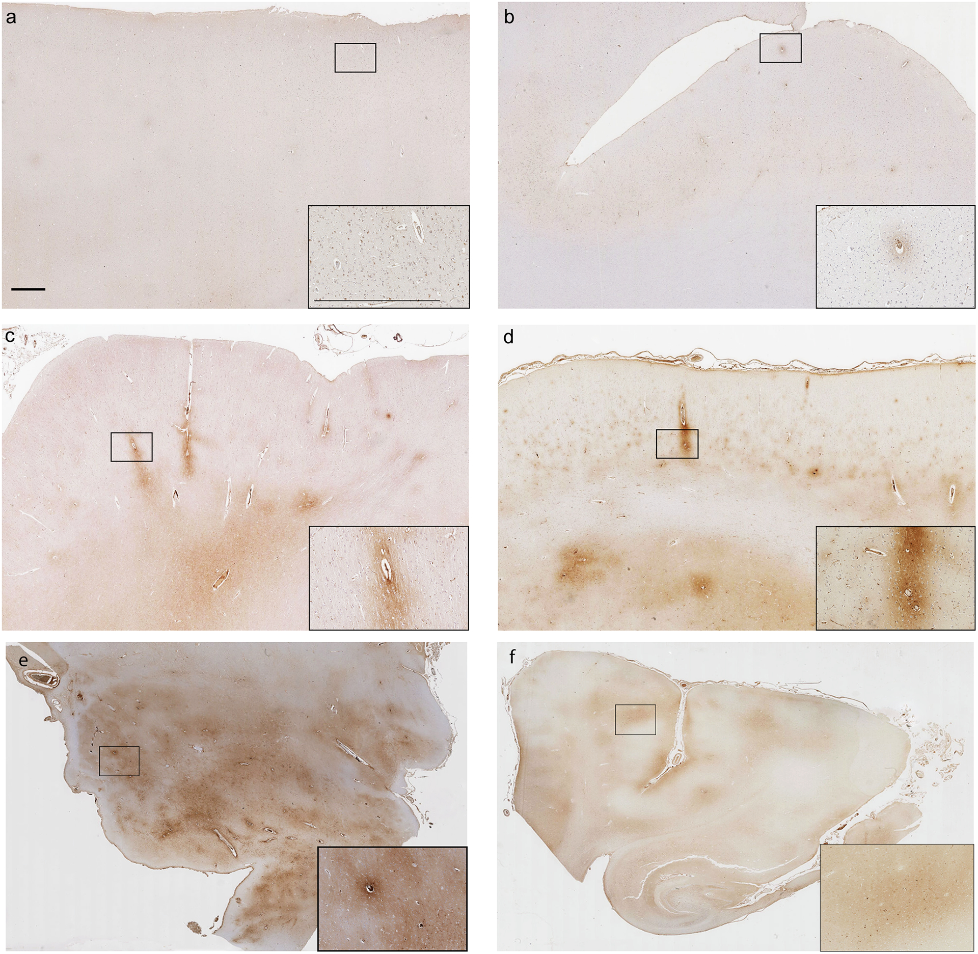
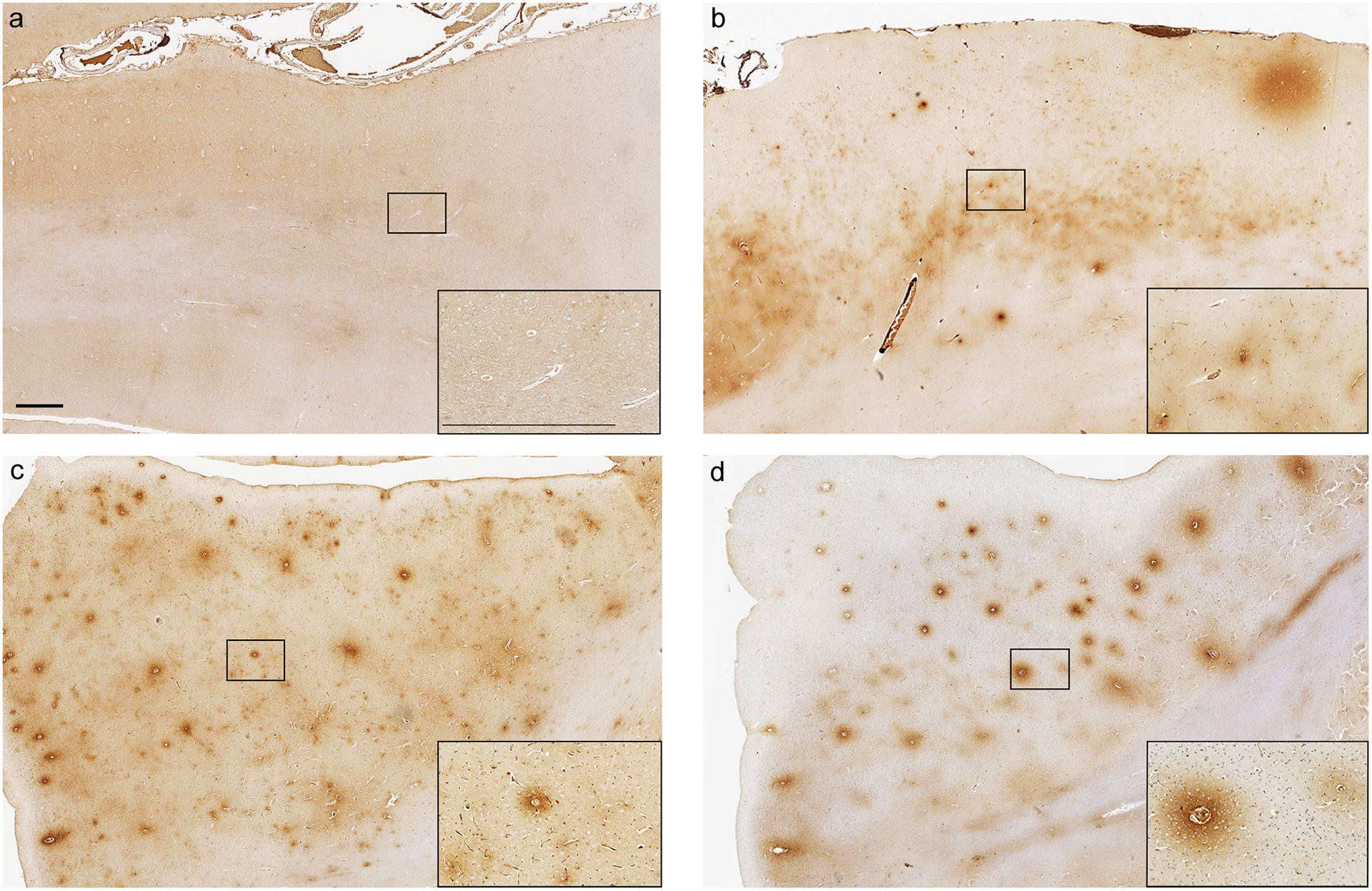
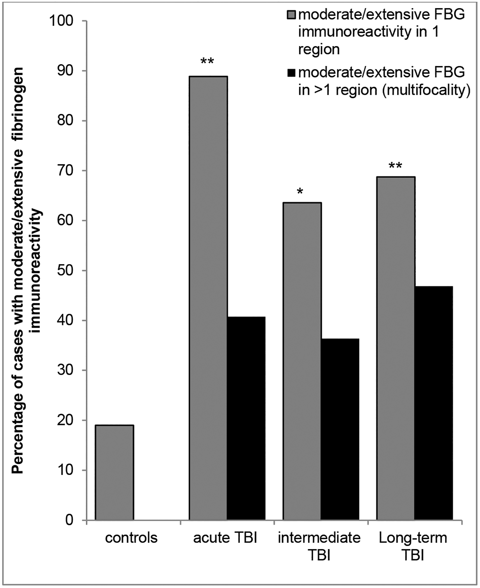
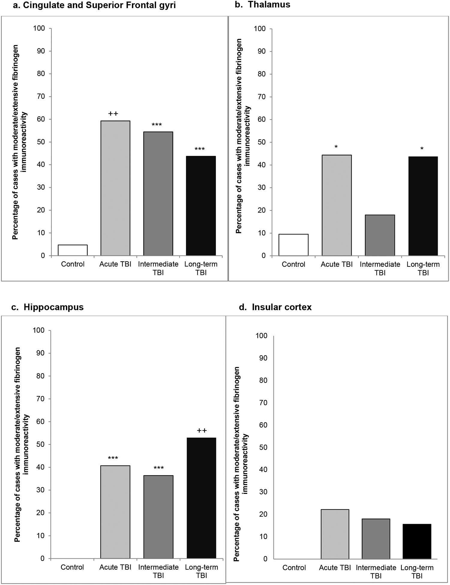
Traumatic brain injury (TBI) is a risk factor for dementia. Mixed neurodegenerative pathologies have been described in late survivors of TBI, but the mechanisms driving post-TBI neurodegeneration remain elusive. Increasingly, blood-brain barrier (BBB) disruption has been recognized in a range of neurologic disorders including dementias, but little is known of the consequences of TBI on the BBB. Autopsy cases of single moderate or severe TBI from the Glasgow TBI Archive (n = 70) were selected to include a range from acute (10 hours-13 days) to long-term (1-47 years) survival, together with age-matched uninjured controls (n = 21). Multiple brain regions were examined using immunohistochemistry for the BBB integrity markers fibrinogen and immunoglobulin G. After TBI, 40% of patients dying in the acute phase and 47% of those surviving a year or more from injury showed multifocal, abnormal, perivascular, and parenchymal fibrinogen and immunoglobulin G immunostaining localized to the gray matter, with preferential distribution toward the crests of gyri and deep neocortical layers. In contrast, when present, controls showed only limited localized immunostaining. These preliminary data demonstrate evidence of widespread BBB disruption in a proportion of TBI patients emerging in the acute phase and, intriguingly, persisting in a high proportion of late survivors.


Figures

Fig. 1
Fig. 1. Representative examples of the patterns of FBG immunoreactivity encountered
(a) Section from the superior frontal gyrus of a 47 year old male TBI patient who died 1 year following a fall. No abnormal FBG immunoreactivity is present (score of 0). (b) Limited, faint perivascular FBG immunoreactivity (score of 1) from the superior frontal gyrus of a 60 year old male TBI patient who died 16 years after a road traffic accident. (c) More widespread, moderate perivascular FBG immunoreactivity (score of 2) in the superior frontal gyrus of a 60 year old male TBI patient who survived 10 hours after a fall. (d) Extensive perivascular and adjacent parenchymal FBG immunoreactivity (score of 3) in the superior frontal gyrus of a 50 year old male TBI patient who survived 1 year after an assault. (e) Extensive perivascular FBG immunoreactivity in thalamic region of a 60 year old male TBI patient who survived 8 days after a fall. (f) Limited perivascular FBG immunoreactivity in parahippocampal region of a 56 year old female TBI patient who survived 24 hours after a road traffic accident. Scale bars = 1mm and apply to all corresponding images.
Fig. 2
Fig. 2. Representative images of FBG and IgG immunoreactivity following TBI and in controls.
Absence of FBG immunoreactivity in the superior frontal gyrus of a 46 year old male with no history of TBI. (b) Extensive FBG immunoreactivity in the superior frontal gyrus, with preferential distribution of staining to the mid and deep cortical layers, in a 20 year old male TBI patient who survived 2 days following an assault. Extensive FBG (c) and IgG (d) immunoreactivity in the adjacent sections from the superior frontal gyrus of a 60 year old male TBI patient who survived 18 years following a fall. Scale bars = 1mm and apply to all corresponding images.
Fig. 3
Fig. 3. Extent of abnormal FBG immunoreactivity following TBI at varying survivals versus controls.
Whilst moderate or extensive FBG immunoreactivity was an uncommon observation in controls, occurring in just 4 of 21 cases (19%), it was a frequent observation following TBI at all survival time points assessed, being present in 88%, 62% and 69% of acute, intermediate and long-term survival cases respectively. Furthermore, in controls abnormal FBG immunostaining was restricted to single anatomical regions, in contrast to the often multifocal pathology in material following TBI. (*p<0.005; **p<0.001; Chi-square TBI cohort v control fibrinogen positivity).
Fig. 4
Fig. 4. Regional distribution of FBG immunoreactivity following TBI versus controls.
At all survival intervals and in each region analyzed there was evidence of BBB disruption following TBI, evidenced by moderate/ extensive FBG immunoreactivity, in a higher proportion of TBI survivors than matched, non-injured controls in material from the (a) cingulate/superior frontal gyri, (b) thalamus (c) hippocampus and (d) insular cortex. (*p<0.05; **p<0.01; ***p<0.005; +p<0.0005; ++p<0.0001; Chi-square TBI cohort v control)
Fig. 5
Fig. 5. Neocortical distribution of FBG following TBI.
(a) Across all survival time points there was a clear preferential distribution of abnormal FBG immunoreactivity to the crests of gyri when compared to the depths of sulci, as illustrated here for the superior frontal gyrus versus the adjacent cingulate sulcus (*p<0.01; **p<0.001; Chi-square sulcus versus gyrus). (b) Further, within the neocortical grey there was preferential distribution of abnormal staining to the mid (layers 3 and 4) and deep (layers 5 and 6) cortical layers when compared to superficial layers (layers 1 and 2). (+p<0.01; ++p<0.005; Chi-square deep versus superficial cortical layers).

References

    1. Coronado VG, McGuire LC, Sarmiento K, Bell J, Lionbarger MR, Jones CD, Geller AI, Khoury N, Xu L. Trends in traumatic brain injury in the U.S. and the public health response: 1995–2009. J Safety Res 2012:43;299–307. - PubMed
    1. Molgaard CA, Stanford EP, Morton DJ, Ryden LA, Schubert KR, Golbeck AL. Epidemiology of head trauma and neurocognitive impairment in a multi-ethnic population. Neuroepidemiology 1990:9;233–42. - PubMed
    1. Mortimer JA, French LR, Hutton JT, Schuman LM. Head injury as a risk factor for Alzheimer’s disease. Neurology 1985:35;264–7. - PubMed
    1. Mortimer JA, van Duijn CM, Chandra V, Fratiglioni L, Graves AB, Heyman A, Jorm AF, Kokmen E, Kondo K, Rocca WA, Head trauma as a risk factor for Alzheimer’s disease: a collaborative re-analysis of case-control studies. EURODEM Risk Factors Research Group. Int J Epidemiol 1991:20 Suppl 2;S28–35. - PubMed
    1. Graves AB, White E, Koepsell TD, Reifler BV, van Belle G, Larson EB, Raskind M. The association between head trauma and Alzheimer’s disease. Am J Epidemiol1990:131;491–501.1990:131;491–501. - PubMed

Publication types