Review
. 2015 Dec;11(12):933-41.
doi: 10.1038/nchembio.1958. Epub 2015 Nov 17.

Progress and challenges for chemical probing of RNA structure inside living cells

Affiliations
Review

Progress and challenges for chemical probing of RNA structure inside living cells

Miles Kubota et al. Nat Chem Biol. 2015 Dec.

Abstract

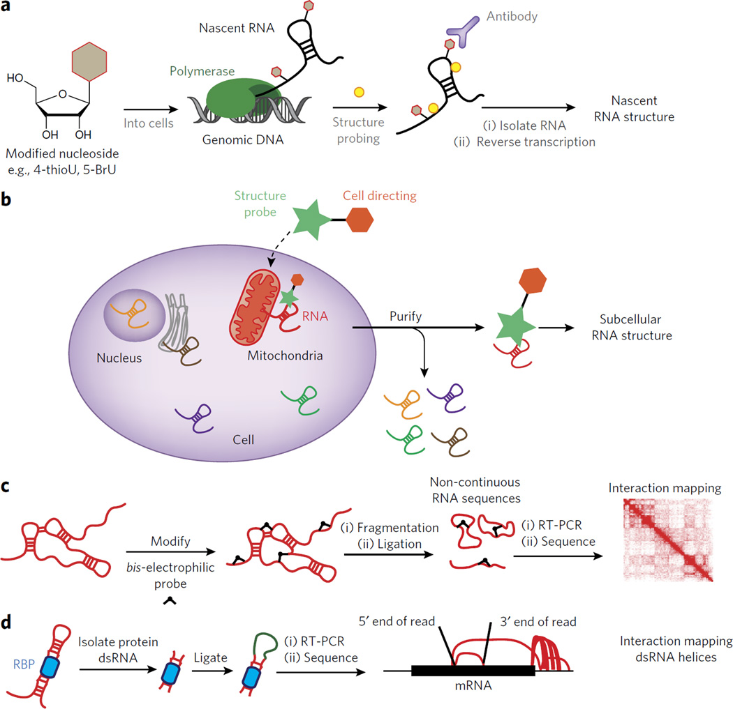
Proper gene expression is essential for the survival of every cell. Once thought to be a passive transporter of genetic information, RNA has recently emerged as a key player in nearly every pathway in the cell. A full description of its structure is critical to understanding RNA function. Decades of research have focused on utilizing chemical tools to interrogate the structures of RNAs, with recent focus shifting to performing experiments inside living cells. This Review will detail the design and utility of chemical reagents used in RNA structure probing. We also outline how these reagents have been used to gain a deeper understanding of RNA structure in vivo. We review the recent merger of chemical probing with deep sequencing. Finally, we outline some of the hurdles that remain in fully characterizing the structure of RNA inside living cells, and how chemical biology can uniquely tackle such challenges.


Figures

Figure 1
Figure 1. The functional and structural complexity of RNA
(a) A cartoon depicting the function of group I introns. (b) A cartoon depicting the function of RNase P in trimming tRNA. (c) A crystal structure of the group I intron (PDB 3BO2). (d) A crystal structure of the catalytic component of RNase P in complex with a tRNA substrate (PDB 3Q1R).
Figure 2
Figure 2. Chemical methods to probe RNA structure
(a) A depiction of a typical chemical probing experiment. The RNA is first folded and then treated with a reagent that reacts covalently. The site of reaction is read out by reverse transcription and synthesis of cDNA that can be resolved by denaturing gel electrophoresis. (b) A depiction of a typical RNA structure probing experiment in which RNA cleavage is used to probe structure. The RNA is first folded and then treated with a reagent that cleaves the RNA backbone. The site of cleavage is read out by reverse transcription and synthesis of cDNA that can be resolved by denaturing gel electrophoresis. (c) Dimethylsulfate can alkylate the N7 of guanosine, the N1 of adenosine and the N3 of cytidine. (d) RNA SHAPE analysis measures the propensity of the 2′-OH to become activated as a nucleophile and undergo an acylation reaction. (e) Hydroxyl radical probing at the C5′ position leads to strand cleavage, resulting in the formation of a 3′-phosphate and 5′-aldehyde. (f) In-line probing results in a 2′,3′-cyclic phosphate and a 5′-hydroxyl.
Figure 3
Figure 3. Methods to measure RNA structure inside living cells
(a) A schematic representation of using 4-thioU (4SU) RNA labeling to enrich for newly transcribed RNAs. DMS chemical probing was merged with 4SU labeling to study the structure of pre-mature rRNA in vivo (see ref. 46). In this method DMS chemical probing is performed in vivo and only the RNA structure pattern for premature ribosomal RNA is obtained. Both the pre-folded and mature RNA are present. However, as a result of 4SU enrichment, only the pre-mature rRNA is probed. (b) A chemical schematic of RNA SHAPE reagents. The site of 2′-OH attack is represented as a red sphere. (c) Demonstration of NAI structure probing of 5S rRNA in living cells. A denaturing gel is shown at right and the B-factors of the 5S rRNA from a corresponding crystal structure at left. Images in c are reproduced from ref. with permission from Nature Publishing Group.
Figure 4
Figure 4. icSHAPE is a novel chemical probing method that permits transcriptome-wide interrogation of RNA structure
(a) Chemical scheme for the preparation of acylated RNA, which can be purified by biotin-streptavidin purification. DIBO, dibenzocyclooctyne. (b) Schematic of icSHAPE modification and purification steps to generate a sequencing library. (c) Schematic of subtractive RNA structure probing, which can be used to study RNA-protein interactions to identify the role of RNA structure in regulating post-transcriptional interactions.
Figure 5
Figure 5. Outstanding challenges for understanding RNA structure inside living cells
(a) Outline of an experiment for interrogating RNA structure formation during transcription. In such a case a modified nucleoside can be introduced into the cell and then used to enrich for co-transcriptionally probed RNA structure. (b) A schematic for the design of a dual-functioning chemical probe to measure RNA structure within unique subcellular compartments. (c) A depiction of how a chemical probe can be used to identify three-dimensional contacts within folded RNAs. The results are mapped to an interaction map, depicting the spatial relationship between two points in the RNA sequence. (d) A schematic for the recently developed method known as hiCLIP. In hiCLIP non-contiguous reads are mapped to genes and represented by rainbow maps to connect primary sequence points through space.

References

    1. Xie X, et al. Systematic discovery of regulatory motifs in human promoters and 3′ UTRs by comparison of several mammals. Nature. 2005;434:338–345. - PMC - PubMed
    1. Badis G, et al. Diversity and complexity in DNA recognition by transcription factors. Science. 2009;324:1720–1723. - PMC - PubMed
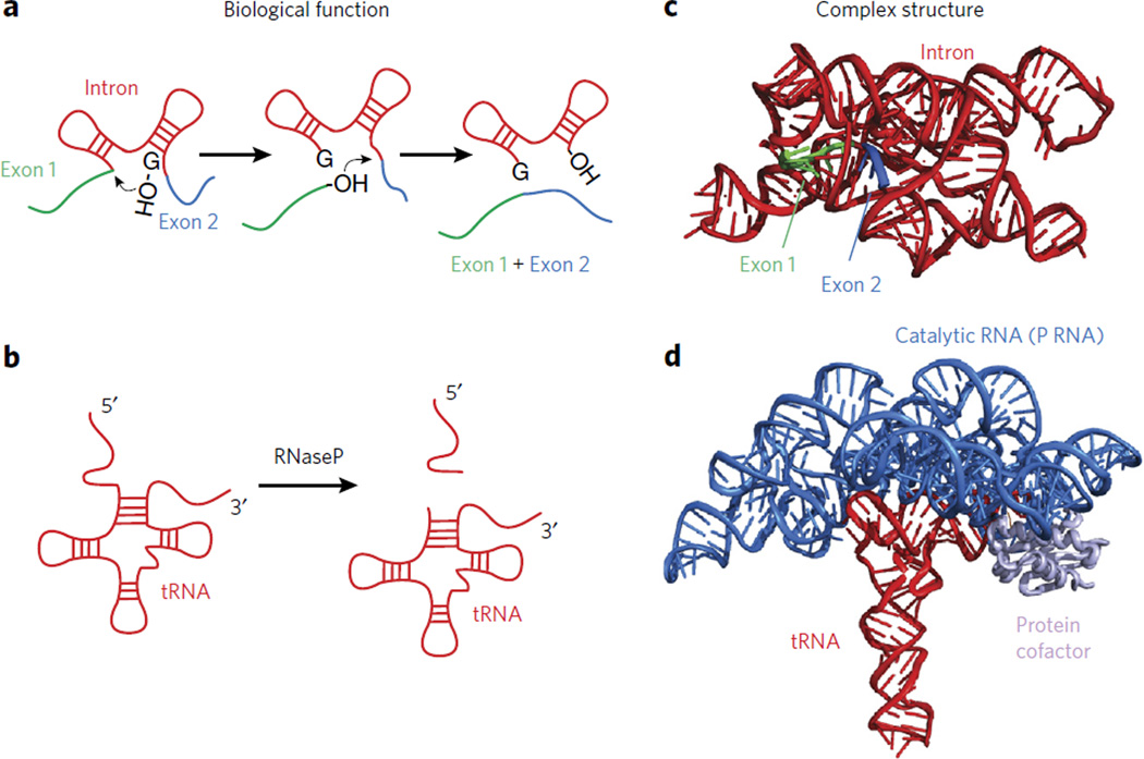
    1. Kruger K, et al. Self-splicing RNA: autoexcision and autocyclization of the ribosomal RNA intervening sequence of Tetrahymena. Cell. 1982;31:147–157. - PubMed
    1. Guerrier-Takada C, Gardiner K, Marsh T, Pace N, Altman S. The RNA moiety of ribonuclease P is the catalytic subunit of the enzyme. Cell. 1983;35:849–857. - PubMed
    1. Noller HF, Hoffarth V, Zimniak L. Unusual resistance of peptidyl transferase to protein extraction procedures. Science. 1992;256:1416–1419. - PubMed

Publication types