

Primary Pulmonary Mucoepidermoid Carcinoma: Histopathological and Moleculargenetic Studies of 26 Cases
- PMID: 26575266
- PMCID: PMC4648574
- DOI: 10.1371/journal.pone.0143169
Primary Pulmonary Mucoepidermoid Carcinoma: Histopathological and Moleculargenetic Studies of 26 Cases
Abstract
Introduction: Pulmonary mucoepidermoid carcinoma (PMEC) is an uncommon neoplasm of the lung and the main salivary gland-type lung carcinoma. The aims of this study were to review the clinicopathological and immunohistochemical features of PMEC and characterize the genetic events in PMEC.
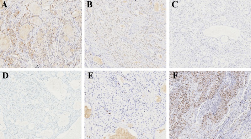
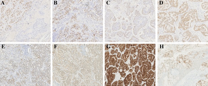
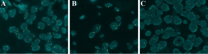
Methods: We reviewed the pathology cases in our hospital and found 34 initially diagnosed PMEC cases, 26 of which were confirmed as PMEC after excluding 8 cases of MEC-like pulmonary carcinoma. The clinicopathological characteristics of the 26 PMEC cases and the 8 cases of MEC-like pulmonary carcinoma were retrospectively reviewed. MAML2 rearrangement was detected by fluorescence In Situ Hybridization (FISH). Immunostains of ALK, calponin, collagen IV, CK7, EGFR, HER2, Ki-67, Muc5Ac, p63, p40, and TTF-1 were performed. DNA was extracted from 23 cases of PMEC. Mutation profiling of the EGFR, KRAS, BRAF, ALK, PIK3CA, PDGFRA, and DDR2 genes were carried out using next-generation sequencing (NGS), Sanger sequencing, and quantitative polymerase chain reaction (QPCR) in 9 successfully amplified cases.
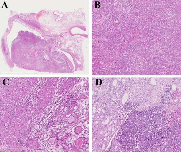
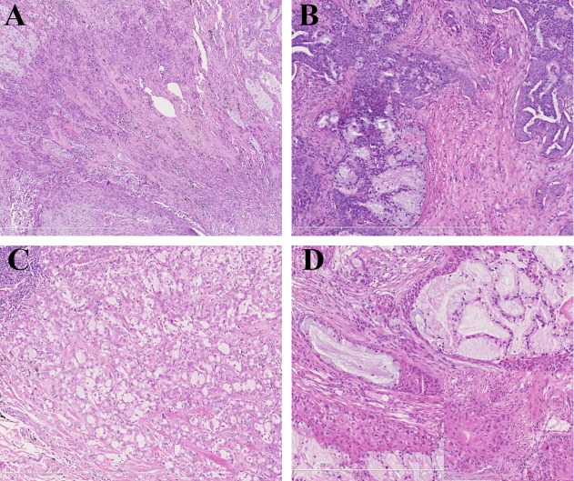
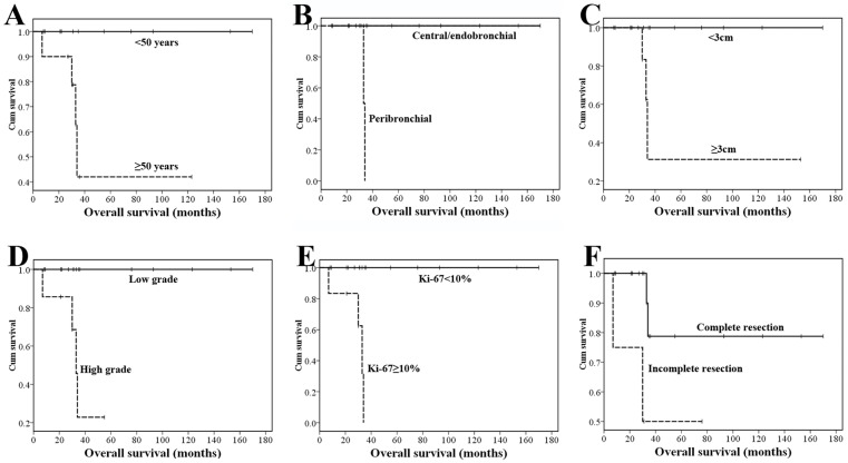
Results: Twenty-six cases of PMEC (18 low-grade, 8 high-grade) included 13 men and 13 women aged 12-79 years. Twenty-two cases had a central/endobronchial growth pattern, and 4 cases had a peribronchial growth pattern. Immunohistochemically, CK7, Muc5Ac, p40, and p63 were positive in all cases (26/26);EGFR was positive in 11 cases (11/26); TTF-1, Calponin, HER2 and ALK were negative in all cases (0/26). MAML2 rearrangement was identified in 12 of 18 PMEC cases. No mutations were detected in any of the 7 genes in the 9 cases that qualified for mutation analysis. Twenty-three PMEC patients had follow-up information with a median interval of 32.6 months. Both the 5- and 10-year overall survival rates (OS) were 72.1%, and a high-grade tumor was an adverse prognostic factor in PMEC. There were 8 cases of MEC-like pulmonary carcinoma aged 36-78 years: 2 cases were located in the bronchus, and 6 cases were located in the lung. p63 and TTF-1 were positive in all cases (8/8), p40 was positive in 5 cases (5/8), and ALK was positive in 5 cases (5/8). No cases of MAML2 rearrangement were detected, but there were 5 cases of ALK rearrangement.
Conclusions: PMEC is a primary malignant pulmonary tumor with a relatively good prognosis that is historically characterized by the presence of mucous cells and a lack of keratinization. There are distinct differences between PMEC and MEC-like pulmonary carcinoma in tumor location preference, immunophenotype, and molecular genetics, and the differential diagnosis is critical due to the therapeutic and prognostic considerations.
Conflict of interest statement
Figures
References
-
- Ishikawa Y, Alvarez-Fernandez E, Aubry MC, Dacic S, Nicholson AG. Mucoepidermoid carcinoma In: Travis WD, Brambilla E, Burke AP, Marx A, Nicholson AG, editors. WHO classification of tumours of the lung, pleura, thymus and heart. Lyon: IARC Press; 2015. pp. 99–100.
-
- Tonon G, Modi S, Wu L, Kubo A, Coxon AB, Komiya T, et al. T (11;9) (q21; p13) translocation in mcoepidermoid carcinoma creates a novel fusion product that disrupts a Notch signaling pathway. Nat Genet. 2003; 33: 08–13. - PubMed
-
- Behboudi A, Enlund F, Winnes M, Andrén Y, Nordkvist A, Leivo I, et al. Molecular classification of mucoepidermoid carcinomas-prognostic significance of the MECT1-MAML2 fusion oncogene. Genes Chromosomes Cancer. 2006; 45: 470–481. - PubMed
MeSH terms
Substances
LinkOut - more resources
Full Text Sources
Other Literature Sources
Medical
Research Materials
Miscellaneous
