. 2015 Dec 8;112(49):15190-5.
doi: 10.1073/pnas.1516992112. Epub 2015 Nov 2.

Evolutionary origins of hepatitis A virus in small mammals

Collaborators, Affiliations

Evolutionary origins of hepatitis A virus in small mammals

Jan Felix Drexler et al. Proc Natl Acad Sci U S A. .

Abstract

Hepatitis A virus (HAV) is an ancient and ubiquitous human pathogen recovered previously only from primates. The sole species of the genus Hepatovirus, existing in both enveloped and nonenveloped forms, and with a capsid structure intermediate between that of insect viruses and mammalian picornaviruses, HAV is enigmatic in its origins. We conducted a targeted search for hepatoviruses in 15,987 specimens collected from 209 small mammal species globally and discovered highly diversified viruses in bats, rodents, hedgehogs, and shrews, which by pairwise sequence distance comprise 13 novel Hepatovirus species. Near-complete genomes from nine of these species show conservation of unique hepatovirus features, including predicted internal ribosome entry site structure, a truncated VP4 capsid protein lacking N-terminal myristoylation, a carboxyl-terminal pX extension of VP1, VP2 late domains involved in membrane envelopment, and a cis-acting replication element within the 3D(pol) sequence. Antibodies in some bat sera immunoprecipitated and neutralized human HAV, suggesting conservation of critical antigenic determinants. Limited phylogenetic cosegregation among hepatoviruses and their hosts and recombination patterns are indicative of major hepatovirus host shifts in the past. Ancestral state reconstructions suggest a Hepatovirus origin in small insectivorous mammals and a rodent origin of human HAV. Patterns of infection in small mammals mimicked those of human HAV in hepatotropism, fecal shedding, acute nature, and extinction of the virus in a closed host population. The evolutionary conservation of hepatovirus structure and pathogenesis provide novel insight into the origins of HAV and highlight the utility of analyzing animal reservoirs for risk assessment of emerging viruses.

Keywords: hepatitis A virus; pathogenesis; small mammals; viral evolution; zoonosis.


Conflict of interest statement

The authors declare no conflict of interest.

Figures
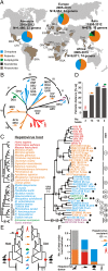
Fig. 1.
Fig. 1.
Hepatovirus evolutionary relationships. (A) Number of sampled host genera, specimens, and dates of collection. Country abbreviations, see Table S1. (B) Hepatovirus VP2 phylogeny (MrBayes, GTR+G+I nucleotide substitution model). (C) Cladogram of hepatovirus hosts (Left) and Hepatovirus phylogeny (Right) as in B. Circled numbers, predicted viral species. Diamonds, viral full genome characterizations. Circles at nodes, posterior probabilities >0.9. (D) Hepatovirus patristic distance per host order; aa, amino acid. (E) Parsimony-based ancestral state reconstructions (ASR) as described previously (3) using 10,000 tree replicates of a VP2 phylogeny (MrBayes, WAG aa substitution model) according to host order (Left) or predominantly insectivorous diet (Right). (F) Averaged number of host switches from ASR shown in E originating from and received by each hepatovirus host order.
Fig. S1.
Fig. S1.
Host relationships, hepatovirus relationships, viral species prediction, and genomic properties. (A) Phylogenetic relationships of mammals included in this study. Relationships of placental mammals according to ref. . Sampled taxa are colored. The superorder Afrotheria contains the sampled order Afrosoricida. (B) Phylogenetic relationships of hepatoviruses in the partial VP2 encoding domain. Values at nodes show support of grouping from Bayesian posterior probabilities. (C) VP2-based species prediction. Pairwise amino acid sequence distances in the translated VP2 were plotted for the complete dataset shown in B. The 7% cutoff separating hepatovirus species in this dataset is highlighted by a slashed line. The distance to the next closely related genus Tremovirus (represented by AEV) is highlighted to the right. (D) Hepatovirus genome architecture. Rodent hepatovirus (RHAV), bat hepatovirus (BtHAV), hedgehog hepatovirus (HgHAV), and shrew hepatovirus (SrHAV). Near-identical HgHAV and SrHAV genomes were represented by one virus only. Genome lengths are given to the right. Domains whose initiation and termination could not be unambiguously identified are shown with curved lines. Annotations and additional genomic features correspond to those in the main article file. The 5′UTR of BtHAVs FO1A-F48 and M32 could not be completely characterized despite repeated trials. The uncharacterized portion is given with a dotted line and the minimum genome size is identified with >. The HAV reference sequence corresponds to the 18f prototype strain. (E) Genomic features of hepatoviruses compared with other picornaviruses. (Top) Percentage G+C content of hepatovirus genomes. (Middle) Relative CpG content of hepatoviruses compared with representative picornaviruses (FMDV, foot-and-mouth disease virus, genus Aphthovirus; AiV, Aichi virus, genus Kobuvirus; SafV, Saffold virus, genus Cardiovirus; PV3, Polio virus 3, HRV89, human rhinovirus 89, both genus Enterovirus; HPeV1, human parechovirus 1, genus Parechovirus; AEV, genus Tremovirus). (Bottom) Effective number of codons. In all panels, dashed lines show minimum and maximum of primate HAV.
Fig. 2.
Fig. 2.
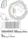
Properties of the genomes of nonprimate hepatoviruses. (A) Hepatovirus genome organization. SrHAV, shrew hepatovirus. (B) Genomic features in primate HAV, nonprimate HAV, and the Picornaviridae. [Scale bars, mean (SD).] (C) IRES folding. RHAV, rodent hepatovirus; BtHAV, bat hepatovirus. (D) 3Dpol cre elements. Gray, conserved AAACG motif. Genomic positions of predicted cre: HAV, 5,945–6,055; RHAV KS11-1230, 5,968–6,078; SrHAV KS12-1289, 6,095–6,181; BtHAV, 5,809–5,870. (E) Phylogenies of hepatovirus domains P1, P2 (only 2C) and P3 (only 3CD; MrBayes, WAG aa substitution model). Circles at nodes, posterior probabilities >0.9. (F) VP2 structure of a SrHAV modeled onto the HAV crystal (8); Box, domain swap. (G) VP2 late domains. (H) aa sequence distance along hepatovirus polyproteins.
Fig. S2.
Fig. S2.
Hepatovirus secondary structures and diversity in different genome regions. (A) Predicted hepatovirus 5′UTR RNA secondary structure of viruses not shown in the main text. Stem-loops (SL) are identified by adjacent numbers. Pseudoknots are given as boxes. Pictograms represent the full 5′UTR from the most 5′ nucleotide to the polyprotein initiation codon at the 3′-end. HgHAV Igel68 represents viruses Igel8 and Igel68; RHAV RMU10-1637 represents the near-identical virus KS11-510 (all these structures were type III IRES-related; SMG-18520 represents Malagasy bat virus; CIV459 represents Ivorian rodent virus). Both viruses contained type IV-like IRES structures including the characteristic pseudoknot preceding the initiation codon. Unpaired sequence between SL I and SL II is represented by dots for graphical reasons. (B) Predicted hepatovirus cis-acting replication elements (cre) in the 3Dpol genomic region. Hosts are shown with pictograms together with virus strains, HAV is included as a reference (genotype Ia, GenBank accession no. AB020564). The conserved AAACG motif in the upper loop structure is highlighted. Rodent virus RMU10-1637 is represented by the near-identical virus KS11-510. Shrew virus KS12-1289 represents the near-identical virus KS12-1232. Hedgehog virus Igel8 represents the near-identical viruses Igel68 and Igel75. (C) Pairwise amino acid sequence distances in the translated VP1, P1, the combined 2C+3CD domains, and the full polyprotein gene plotted for the dataset containing all full genomes used to generate phylogenetic reconstructions shown in the main article. Distances within HAV and those to the next closely related AEV are highlighted. (D) Predicted transmembrane domains of hepatoviruses.
Fig. S3.
Fig. S3.
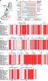
Conservation of hepatovirus VP2 proteins and divergence of hepatovirus 2B proteins. (A) Bayesian phylogeny of the full VP2 using a WAG amino acid substitution model as detailed in SI Materials and Methods. Filled circles at nodes represent Bayesian probabilities above O.9. (B) Identification of structural elements [α-helices, 310-helices (η), β-strands, and strict β-turns (TT)] using the structural models built on the HAV crystal structure (8). Conserved alignment domains are highlighted by red color and boxes. The dot symbol at the C terminus of RHAV CIV459 corresponds to a gap compared with the other sequences. (C) Protein BLAST comparisons of pX and 2B domains. In comparisons of HAV; hepatoviruses were excluded as hits. For each genome segment, the number of residues with identical or biochemically similar residues is indicated per the total number of compared residues. The BLOSUM62 matrix was used for similarity calculations by BLAST according to default parameters.
Fig. 3.
Fig. 3.
Antigenic relatedness of human and nonprimate hepatoviruses. (A) (Upper) immunofluorescence assay (IFA) showing a bat serum reacting with human HAV-infected FRhK-4 cells [red (Cy2); mixed with 50% noninfected cells as internal negative controls]. (Lower) Same cells stained with a monoclonal antibody control (mAb 7E7, 100% infected cells). Blue (DAPI), nuclei. (B) Immunoprecipitation (IP) of HAV by bat sera [IF, IP, neutralization test (NT); red, positive in all assays; gray, discordant assay results; empty, negative in all assays], human sera, and controls. See SI Materials and Methods for details on control sera. Dotted line, threshold precipitation separating positive and negative control sera. (C) aa sequence distance between HAV (genotype Ia, GenBank accession no. AB020564), the Eidolon BtHAV M32, a RHAV (RMU10-1637), and a SrHAV (KS12-1232).
Fig. S4.
Fig. S4.
Hepatovirus epitopes and infection patterns. (A) Amino acid sequence distance along the polyproteins between HAV (Gt Ia) and the nonprimate hepatoviruses. A genomic representation of HAV is given on top. Near-identical viruses from rodents, hedgehogs and shrews are represented by one virus only. (B) Hepatovirus epitopes associated with neutralization according to ref. . Background shading and residue color indicate biochemical properties of residues. AEV was included for comparison. (C) Strand-specific cDNA priming in bat and hedgehog liver and spleen. The number of analyzed animals and biological replicates is indicated on top and above organs, respectively. (D) Partial VP2 neighbor-joining percentage distance phylogeny of the hedgehog viruses in the animal shelter (complete deletion option, 405 nucleotides, GenBank accession nos. KT452748–KT452765). Individual animals are color-coded, sampling number corresponds to increasing time points. The three hedgehog viruses for which the full genomes were characterized were included for orientation. (E) (Upper Left) Geographic location of the roost in Western Germany as detailed in ref. and a picture of female Myotis myotis bats hanging from the roof of the attic forming the roost. (Lower Left) Neighbor-joining amino acid percentage distance phylogeny highlighting the distance between the two hepatovirus lineages detected in the roost in 2008 and 2010 compared with the maximum distance in HAV using the same VP2 fragment. (Right) Detections of lineage 1 during 2008 and detections of lineage 2 during 2010. Height of bars correspond to viral RNA copies per gram of feces; empty spaces correspond to negative samples. The arrow symbolizes the birth of the first pup, after which all pups are born in a short time frame. Height of bars in 2010 was set to 101 to highlight that these specimens were detectable only by nested RT-PCR but could not be quantified using a strain-specific real-time RT-PCR assay due to low RNA concentrations. Data for the year 2009 is not shown because this sample was incomplete as detailed in ref. and no specimen tested positive for hepatoviruses.
Fig. 4.
Fig. 4.
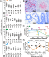
Hepatovirus infection patterns in small mammals. (A) Hepatovirus RNA abundance in solid organs and blood per host taxon determined by qPCR. Li, liver; Sp, spleen; Ki, kidney; Lu, lung; In, intestine; Br, brain; He, heart; Bl, blood. Boxes, median, interquartile range; outliers (circles); extreme values (asterisks). Next to host pictograms, numbers of analyzed animals; Below each organ, ratios of specimens testing positive per specimens available for testing. (B) In situ hybridization of BtHAV RNA in E. helvum liver (Upper Left, 400× magnification, animal M32), spleen (Upper Right, 400×, animal GH297), lung (Lower Left, 200×, animal M32), and intestine (Lower Right, 200×, animal M32). (C) Hepatovirus detections and RNA concentrations determined by qPCR in nine hedgehogs sampled longitudinally. (D) Hepatovirus amplification in a bat maternity roost. Triangles, viral RNA concentrations in pooled fecal specimens over five sampling points. Secondary y axis and orange line, detection rate.

Comment in

References

    1. Luis AD, et al. 2013. A comparison of bats and rodents as reservoirs of zoonotic viruses: Are bats special? Proc R Soc Biol Sci 280(1756):20122753.
    1. Brook CE, Dobson AP. Bats as ‘special’ reservoirs for emerging zoonotic pathogens. Trends Microbiol. 2015;23(3):172–180. - PMC - PubMed
    1. Drexler JF, et al. Bats host major mammalian paramyxoviruses. Nat Commun. 2012;3:796. - PMC - PubMed
    1. Leroy EM, et al. Fruit bats as reservoirs of Ebola virus. Nature. 2005;438(7068):575–576. - PubMed
    1. Han BA, Schmidt JP, Bowden SE, Drake JM. Rodent reservoirs of future zoonotic diseases. Proc Natl Acad Sci USA. 2015;112(22):7039–7044. - PMC - PubMed

Publication types

Associated data

LinkOut - more resources