. 2016 Mar;228(3):443-51.
doi: 10.1111/joa.12419. Epub 2015 Nov 17.

Selective loss of alpha motor neurons with sparing of gamma motor neurons and spinal cord cholinergic neurons in a mouse model of spinal muscular atrophy

Affiliations

Selective loss of alpha motor neurons with sparing of gamma motor neurons and spinal cord cholinergic neurons in a mouse model of spinal muscular atrophy

Rachael A Powis et al. J Anat. 2016 Mar.

Abstract

Spinal muscular atrophy (SMA) is a neuromuscular disease characterised primarily by loss of lower motor neurons from the ventral grey horn of the spinal cord and proximal muscle atrophy. Recent experiments utilising mouse models of SMA have demonstrated that not all motor neurons are equally susceptible to the disease, revealing that other populations of neurons can also be affected. Here, we have extended investigations of selective vulnerability of neuronal populations in the spinal cord of SMA mice to include comparative assessments of alpha motor neuron (α-MN) and gamma motor neuron (γ-MN) pools, as well as other populations of cholinergic neurons. Immunohistochemical analyses of late-symptomatic SMA mouse spinal cord revealed that numbers of α-MNs were significantly reduced at all levels of the spinal cord compared with controls, whereas numbers of γ-MNs remained stable. Likewise, the average size of α-MN cell somata was decreased in SMA mice with no change occurring in γ-MNs. Evaluation of other pools of spinal cord cholinergic neurons revealed that pre-ganglionic sympathetic neurons, central canal cluster interneurons, partition interneurons and preganglionic autonomic dorsal commissural nucleus neuron numbers all remained unaffected in SMA mice. Taken together, these findings indicate that α-MNs are uniquely vulnerable among cholinergic neuron populations in the SMA mouse spinal cord, with γ-MNs and other cholinergic neuronal populations being largely spared.

Keywords: SMA; alpha motor neuron; cholinergic neuron; gamma motor neuron; spinal cord; spinal muscular atrophy.


Figures

Figure 1
Figure 1
Immunohistochemical labelling of cholinergic neuronal populations in the spinal cord of neonatal mice. Representative confocal micrographs showing a transverse section through the upper lumbar spinal cord (L1‐L2; containing both the dorsal commissural nucleus and the intermediolateral nucleus, not present at all levels of the spinal cord) in a postnatal day 9 littermate control mouse, immunohistochemically labelled for NeuN (green) and ChAT (red). Nuclei were labelled with DAPI (blue). In the ChAT section note the presence of motor neurons in the ventral grey horn (white arrow), preganglionic sympathetic neurons in the intermediolateral nucleus (black arrow), partition cells (grey arrow), central canal cluster cells (yellow arrow) and preganglionic autonomic neurons in the dorsal commissural nucleus (blue arrow). Scale bar:  250 μm.
Figure 2
Figure 2
Selective loss of α‐MNs but sparing of γ‐MNs in late‐symptomatic SMA mouse spinal cord. (A,B) Representative low (A) and high (B) power confocal micrographs of the ventral grey horn of the postnatal day 9 mouse spinal cord immunohistochemically labelled for NeuN (green) and ChAT (red) [nuclei were labelled with DAPI (blue)]. Black arrows indicate α‐MNs (ChAT +/NeuN+ cells) and white arrows indicate γ‐MNs (ChAT +/NeuN cells). Scale bars:  50 μm (A) and 25 μm (B). (C,D) Bar charts (mean ± SEM) showing the number of total motor neurons (C) and percentage of α‐MNs and γ‐MNs (D) in the cervical, thoracic and lumbar regions of littermate control mouse spinal cord at P9 (n = 4 mice, mean number of motor neurons counted per control spinal cord region: cervical n = 1146 ± 216; thoracic n = 639 ± 84; lumbar n = 1085 ± 297). (E‐G) Bar charts showing the number of α‐MNs and γ‐MNs in the cervical, thoracic and lumbar regions of spinal cord from littermate control and late‐symptomatic SMA mice at P9 (ns = not significant; *< 0.05, **< 0.01; unpaired two‐tailed Student's t‐test; n = 4 mice per genotype, except for SMA cervical measurements where n = 3).
Figure 3
Figure 3
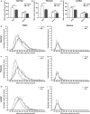
Reduction in the size of the cell somata of α‐MNs but not γ‐MNs in late‐symptomatic SMA mouse spinal cord. (A‐C) Bar charts (mean ± SEM) showing the mean cell soma size for α‐MNs and γ‐MNs in the cervical, thoracic and lumbar regions of spinal cord from littermate control and late‐symptomatic SMA mice at P9 (ns = not significant, *P < 0.05, unpaired two‐tailed Student's t‐test; n = 4 mice per genotype, except for SMA cervical measurements where n = 3). (D‐I) XY graphs showing mean α‐MN (D,F,H) and γ‐MN (E,G,I) cell body size distributions in control (black line) and SMA (grey line) mice at cervical (D & E), thoracic (F & G) and lumbar (H & I) spinal cord levels. α‐MNs and γ‐MN soma measurements were rounded into 50‐μm2 bins and expressed as a percentage of total control motor neuron size distribution measurements. Error bars:  SEM.
Figure 4
Figure 4
Sparing of other cholinergic neuronal populations in the spinal cord from late‐symptomatic SMA mice. (A,B) Representative confocal micrographs showing cholinergic neuronal populations surrounding the central canal (A) and in the intermediolateral nucleus (B) in a transverse section through the upper lumbar spinal cord (L1‐L2) in a postnatal day 9 littermate control mouse, immunohistochemically labelled for NeuN (green) and ChAT (red). Nuclei were labelled with DAPI (blue). Scale bars: 50 μm. (A) In the ‘merged’ panel examples of partition cells (black arrows), central canal cluster cells (white arrows) and preganglionic autonomic neuron in dorsal commissural nucleus (grey arrows) are indicated. (C,D) Bar charts (mean ± SEM) showing the number of ChAT + partition cells (C) and central canal cluster cells per transverse spinal cord section in the cervical, thoracic and lumbar regions in littermate control and late‐symptomatic SMA mice at P9. (E,F) Bar charts (mean ± SEM) showing the number of ChAT + preganglionic autonomic neurons in dorsal commissural nucleus (E; taken from L1‐L2 spinal cord only) and preganglionic sympathetic neurons in intermediolateral nucleus (F; taken from thoracic and upper lumbar levels of spinal cord) in P9 littermate control and late‐symptomatic SMA mice (ns = not significant, unpaired two‐tailed Student's t‐test; n = 4 mice per genotype, except for SMA cervical measurements where n = 3).

References

    1. Arai H, Tanabe Y, Hachiya Y, et al. (2005) Finger cold‐induced vasodilatation, sympathetic skin response, and R‐R interval variation in patients with progressive spinal muscular atrophy. J Child Neurol 20, 871–875. - PubMed
    1. Ashrafi S, Lalancette‐Hebert M, Friese A, et al. (2012) Wnt7A identifies embryonic gamma‐motor neurons and reveals early postnatal dependence of gamma‐motor neurons on a muscle spindle‐derived signal. J Neurosci 32, 8725–8731. - PMC - PubMed
    1. Barber RP, Phelps PE, Houser CR, et al. (1984) The morphology and distribution of neurons containing choline acetyltransferase in the adult rat spinal cord: an immunocytochemical study. J Comp Neurol 229, 329–346. - PubMed
    1. Battaglia G, Princivalle A, Forti F, et al. (1997) Expression of the SMN gene, the spinal muscular atrophy determining gene, in the mammalian central nervous system. Hum Mol Genet 6, 1961–1971. - PubMed
    1. Bertrand SS, Cazalets JR (2011) Cholinergic partition cells and lamina X neurons induce a muscarinic‐dependent short‐term potentiation of commissural glutamatergic inputs in lumbar motoneurons. Front Neural Circuits 5, 15. - PMC - PubMed

Publication types

LinkOut - more resources