. 2016 Jan;60(1):35-46.
doi: 10.1111/1348-0421.12338.

C5a inhibitor protects against ischemia/reperfusion injury in rat small intestine

Affiliations

C5a inhibitor protects against ischemia/reperfusion injury in rat small intestine

Eszter Tuboly et al. Microbiol Immunol. 2016 Jan.

Abstract

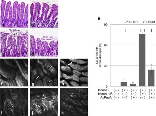
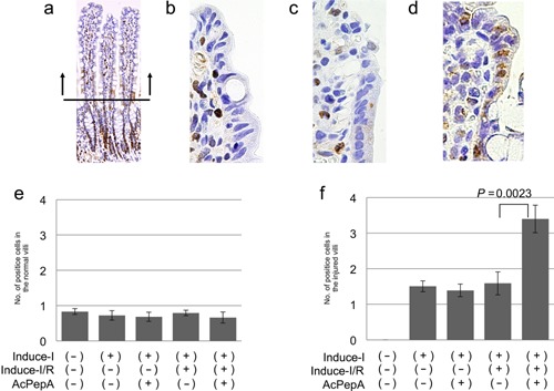
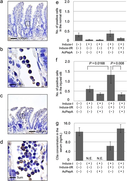
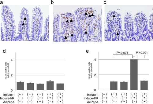
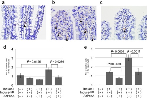
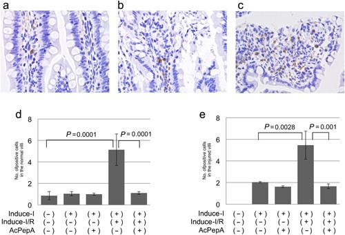
Acute mesenteric ischemia (AMI) is caused by considerable intestinal injury, which is associated with intestinal ischemia followed by reperfusion. To elucidate the mechanisms of ischemia/reperfusion injuries, a C5a inhibitory peptide termed AcPepA was used to examine the role of C5a anaphylatoxin, induction of inflammatory cells, and cell proliferation of the intestinal epithelial cells in an experimental AMI model. In this rat model, the superior mesenteric artery was occluded and subsequently reperfused (Induce-I/R). Other groups were treated with AcPepA before ischemia or reperfusion. Induce-I/R induced injuries in the intestine and AcPepA significantly decreased the proportion of severely injured villi. Induce-I/R induced secondary receptor for C5a-positive polymorphonuclear leukocytes in the vessels and CD204-positive macrophages near the injured site; this was correlated with hypoxia-induced factor 1-alpha-positive cells. Induction of these inflammatory cells was attenuated by AcPepA. In addition, AcPepA increased proliferation of epithelial cells in the villi, possibly preventing further damage. Therefore, Induce-I/R activates C5a followed by the accumulation of polymorphonuclear leukocyte and hypoxia-induced factor 1-alpha-producing macrophages, leading to villus injury. AcPepA, a C5a inhibitory peptide, blocks the deleterious effects of C5a, indicating it has a therapeutic effect on the inflammatory consequences of experimental AMI.

Keywords: complement C5a; hypoxia-induced factor 1-alpha; ischemia; macrophage.


Figures

Figure 1
Figure 1
Effect of AcPepA on the degree of small intestinal injury. Small intestinal injury (×600) was classified as (a) normal, (b) slight, (c) moderate and (d) severe. (e) The percentages of severely injured villi in the variously treated groups. In vivo histology images of the mucosal surface of distal rat ileum recorded under fluorescence confocal endomicroscopy (f, g, h) after i.v. administration of FITC‐dextran and (i, j, k) topical administration of acridine orange. (f) Normal epithelium on the surface of the villi of the control group. (g) Longitudinal fissures on the surface of villi (white arrows) are apparent in the Induce‐I/R group. (h) A few fissures on the surface of villi (thin white arrow) were observed in the Induce‐I/R + AcPepA group. (i) Mucosal vasculature was normal in the control group. (j) Severe dye leakage from vessel lumina was observed 30 min after reperfusion in the Induce‐I/R group. (k) Little dye leakage was observed in the Induce‐I/R + AcPepA group.
Figure 2
Figure 2
Effect of AcPepA on proliferation of small intestinal epithelium. Recovery of the epithelium was evaluated by cell proliferation by counting PCNA‐positive cells (PCNA index). (a) Positive epithelial cells were counted in the villi to avoid the strong proliferative region (below the horizontal bar in [a]) to facilitate detection of small differences in the cell proliferation index. Epithelial cells of injured small intestinal villi were visualized by PCNA staining of tissue from rats in the (b) Induce‐I alone, (c) Induce‐I/R and (d) Induce‐I/R + AcPepA groups. (e) PCNA indices (number of positive cells in each villus) of the epithelium of normal and (f) injured villi are summarized.
Figure 3
Figure 3
Induction of C5L2‐positive cells in villi. (a) Circulating inflammatory cells are often visible in dilated vessels located in the centers of the villi, this being associated with an inflammatory response (Square in 3a). (b) C5L2‐positive cells among inflammatory cells in vessels. (c, d), C5L2‐positive cells are also present in dilated vessels in severely injured villi. (e, f) The number of C5L2‐positive cells in (e) normal and in (f) injured villi. (g) C5a serum concentrations were significantly lower in the Induce‐I/R group than in the Induce‐I/R + AcPepA group.
Figure 4
Figure 4
Induction of macrophages in villi. (a) CD68‐positive macrophages are present in the stromal region outside vessels in the villi (arrowheads). (b) CD68‐positive macrophages are present in the erosion fronts of injured villi of rats in which I/R was induced (arrowheads). (c) Fewer macrophages are present in the Induce‐I/R + AcPepA group. Numbers of the macrophages in (d) normal villi and (e) injured villi.
Figure 5
Figure 5
Induction of CD204‐positive macrophages in villi. (a) CD204‐positive macrophages are present in the stromal region outside vessels in slightly injured villi (arrowheads). (b) CD204‐positive macrophages are present in the erosion fronts of severely injured villi. (c) Fewer CD204‐positive macrophages are present in the Induce‐I/R + AcPepA group than in the Induce‐I/R group. The number of CD204‐positive macrophages in (d) normal and (e) injured villi.
Figure 6
Figure 6
Induction of HIF1‐positive cells. (a, b) HIF1‐α‐positive cells are present in the stromal regions outside vessels in both (a) slightly injured and (b) moderately injured villi. (c) Many HIF1‐α‐positive cells are present in the erosion fronts of severely injured villi. (d, e) Changes in numbers of HIF1‐α‐positive cells in (d) normal and (e) injured villi.

References

    1. Wilson C., Gupta R., Gilmour, D.G. , Imrie, C.W. ( 1987) Acute superior mesenteric ischaemia. Br J Surg 74: 279–81. - PubMed
    1. Oldenburg W.A., Lau L.L., Rodenberg T.J., Edmonds H.J., Burger, C.D. ( 2004) Acute mesenteric ischemia: A clinical review. Arch Intern Med 164: 1054–62. - PubMed
    1. Benjamin E., Oropello, J.M. , Iberti, T.J. ( 1993) Acute mesenteric ischemia: pathophysiology, diagnosis, and treatment. Dis Mon 39: 131–210. - PubMed
    1. Berland T., Oldenburg W.A. ( 2008) Acute mesenteric ischemia. Curr Treat Options Gastroenterol 11: 3–10. - PubMed
    1. Rijke R.P., Van Der Meer‐Fieggen W., Galjaard H. ( 1974) Effect of villus length on cell proliferation and migration in small intestinal epithelium. Cell Tissue Kinet 7: 577–86. - PubMed

Publication types

MeSH terms

LinkOut - more resources