. 2016 Feb;14(2):141-62.
doi: 10.1158/1541-7786.MCR-15-0431. Epub 2015 Nov 17.

Active FOXO1 Is a Key Determinant of Isoform-Specific Progesterone Receptor Transactivation and Senescence Programming

Affiliations

Active FOXO1 Is a Key Determinant of Isoform-Specific Progesterone Receptor Transactivation and Senescence Programming

Caroline H Diep et al. Mol Cancer Res. 2016 Feb.

Abstract

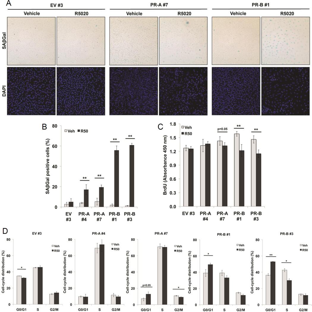
Progesterone promotes differentiation coupled to proliferation and prosurvival in the breast, but inhibits estrogen-driven growth in the reproductive tract and ovaries. Herein, it is demonstrated, using progesterone receptor (PR) isoform-specific ovarian cancer model systems, that PR-A and PR-B promote distinct gene expression profiles that differ from PR-driven genes in breast cancer cells. In ovarian cancer models, PR-A primarily regulates genes independently of progestin, while PR-B is the dominant ligand-dependent isoform. Notably, FOXO1 and the PR/FOXO1 target gene p21 (CDKN1A) are repressed by PR-A, but induced by PR-B. In the presence of progestin, PR-B, but not PR-A, robustly induced cellular senescence via FOXO1-dependent induction of p21 and p15 (CDKN2B). Chromatin immunoprecipitation (ChIP) assays performed on PR isoform-specific cells demonstrated that while each isoform is recruited to the same PRE-containing region of the p21 promoter in response to progestin, only PR-B elicits active chromatin marks. Overexpression of constitutively active FOXO1 in PR-A-expressing cells conferred robust ligand-dependent upregulation of the PR-B target genes GZMA, IGFBP1, and p21, and induced cellular senescence. In the presence of endogenous active FOXO1, PR-A was phosphorylated on Ser294 and transactivated PR-B at PR-B target genes; these events were blocked by the FOXO1 inhibitor (AS1842856). PR isoform-specific regulation of the FOXO1/p21 axis recapitulated in human primary ovarian tumor explants treated with progestin; loss of progestin sensitivity correlated with high AKT activity.

Implications: This study indicates FOXO1 as a critical component for progesterone signaling to promote cellular senescence and reveals a novel mechanism for transcription factor control of hormone sensitivity.


Conflict of interest statement

Conflict of Interest

The authors declare that they have no conflict of interest.

Figures

Figure 1
Figure 1. Stable expression of PR isoforms in ES-2 cells
(A) Inset, Western blot analysis showing total PR expression in ES-2 cell pools expressing GFP-tagged empty vector control (EV), GFP-tagged PR-A (PR-A), or GFP-tagged PR-B (PR-B) and treated without or with R5020 (10 nM) for 24 hr. ES-2 cells expressing EV, PR-A, or PR-B were transiently transfected with a progesterone response element (2X-PRE) containing luciferase reporter gene and treated for 18 hr with R5020 (10 nM). Relative luciferase units (RLU) were normalized to the mean result ± standard deviation (SD) for Renilla luciferase expression (n=3, **p≤0.01). (B) RT-qPCR analysis of HEF1 and BIRC3 mRNA expression after 24 hr R5020 (10 nM) treatment in ES-2 cell pools expressing EV, PR-A or PR-B (n=3, *p≤0.05 **p≤0.01). (C) Western blot analysis of total PR expression in ES-2 cells stably expressing GFP-tagged EV control (clone #3), GFP-tagged PR-A (GFP-PR-A clone #1, #5, #4, #7), or GFP-tagged PR-B (GFP-PR-B clone #1, #3) relative to T47D breast cancer cells stably expressing PR-A-only (YA), PR-B-only (YB), and both endogenous PR isoforms (CO). Actin served as a loading control. (D) Western blot analysis of PR-A and PR-B phosphorylation at Ser294 and Ser190, and total PR protein expression in ES-2 PR-expressing cells treated with R5020 (10 nM) for 1 hr. ◆◆denotes a non-specific band present in the phospho-PR Ser294 blot. Actin served as a loading control.
Figure 2
Figure 2. Gene expression profiling of PR-A and PR-B transcriptional activity in ovarian and breast cancer cells
(A) Heat map highlighting the transcriptional profiles between ES-2 ovarian cancer cells stably expressing EV control (clone #3), PR-A (clone #7), and PR-B (clone #1). Cells were treated with vehicle or R5020 (10 nM) for 24 hr and harvested RNA was subjected to Illumina gene profiling as described in Methods. Genes differentially expressed >2-fold are displayed for each treatment group. The experiment was performed in triplicate. (B) Venn diagrams showing the number of unique genes downregulated or upregulated >2-fold in the absence of R5020 treatment in PR-A− (clone #7) and PR-B-expressing (clone #1) cells. (C) Venn diagrams depicting the number of unique genes downregulated or upregulated >2-fold with R5020 treatment in PR-A− (clone #7) and PR-B-expressing (clone #1) cells. (D) Heat map highlighting the transcriptional profiles between breast cancer cells expressing EV control (T47D Y), PR-A (T47D YA), and PR-B (T47D YB). The cells were treated with vehicle or R5020 (10 nM) for 24 hr and harvested RNA was subjected to Illumina gene profiling as in part A. Genes differentially expressed >2-fold are displayed for each treatment group. The experiment was performed in triplicate. (E) Venn diagrams depicting the number of unique genes downregulated or upregulated >2-fold in the absence of R5020 treatment in T47D YA and YB cells. (F) Venn diagrams depicting the number of unique genes downregulated or upregulated >2-fold with R5020 treatment in T47D YA and YB cells.
Figure 3
Figure 3. Validation of PR isoform-specific gene targets in ovarian cancer cells
RT-qPCR analysis of PR-A selective target gene mRNAs encoding (A) CRISPLD1 and WISP1 or PR-B selective target gene mRNAs encoding (B) BIRC3, GZMA, and PDLIM1 following 24 hr and 96 hr R5020 (10 nM) treatment in ES-2 cells stably expressing EV control (clone #3), PR-A (clone #4, #7), or PR-B (clone #1, #3) (n=3, *p≤0.05, **p≤0.01).
Figure 4
Figure 4. Progestin treatment induces isoform-specific differential expression of cellular senescence mediators in ovarian cancer cells
(A) Venn diagram describing the number of unique genes downregulated in the absence of R5020 treatment in ES-2 cells stably expressing PR-A (clone #7) and upregulated in ES-2 cells stably expressing PR-B (clone #1) with R5020 treatment. Four genes from the shared Venn category are listed. (B) Relative gene expression of FOXO1 and p21 in ES-2 cells stably expressing EV control (clone #3), PR-A (clone #7), or PR-B (clone #1) treated with vehicle or R5020 (10 nM) for 24 hr as determined by Illumina microarray experiments (Figure 2A). RT-qPCR analysis of (C) FOXO1, (D) p21, and (E) p15 mRNA expression following 24 hr and 96 hr R5020 (10 nM) treatment of ES-2 cells stably expressing EV control (clone #3), PR-A (clone #4, #7), PR-B (clone #1, #3) (n=3, *p≤0.05, **p≤0.01). (F) Western blot analysis of PR, FOXO1, and p21 protein expression in ES-2 cells stably expressing PR-A (clone #4, #7) or PR-B (clone #1, #3) after 96 hr treatment of R5020 (10 nM). Actin served as a loading control. (G) ChIP assays showing PR recruitment to a PRE-containing region of the p21 promoter. EV control and PR-A-expressing cells (clone #4, #7) were stimulated with vehicle or R5020 (10 nM) for 1 hr. Fixed lysates were subjected to ChIP assays as described in Methods using specific antibodies targeting PR (or IgG control). Additional data shown are represented as the average fold recruitment (R5020/Vehicle) of PR to a PRE-containing region of the p21 promoter from three separate experiments. (H) ChIP assays demonstrating detection of H3K4me2 at the PRE-containing region of the p21 promoter. EV control, PR-A− (clone #7), and PR-B-expressing (clone #1) cells were stimulated with vehicle or R5020 (10 nM) for 1 hr. Fixed lysates were subjected to ChIP assays as described in Methods using specific antibodies targeting H3K4me2 (or IgG control).
Figure 5
Figure 5. PR isoform expression and activity induces cellular senescence
(A) Representative staining for SAβGal activity in EV control (clone #3), PR-A− (clone #7) or PR-B-expressing (clone #1) ES-2 cells treated with R5020 (10 nM) for 96 hrs (magnification = 100×). Cell samples were mounted onto glass slides using ProLong® Gold Antifade Reagent with DAPI (Invitrogen) for bright-field and fluorescent microscopy. B) Percentage of positive SAβGal cells in EV control (clone #3) and PR-A− (clone #4, #7) and PR-B-expressing (clone #1, #3) cells treated with R5020 (10 nM) for 96 hr was determined by quantifying three fields at 100× magnification. Values were normalized to total nuclei present in each field as determined by DAPI staining (n=3, **p≤0.01). (C) BrdU incorporation analysis of EV control (clone #3), PR-A− (clone #4, #7) or PR-B-expressing (clone #1, #3) cells continuously treated with R5020 (10 nM) for 96 hrs. BrdU was added to the wells, and cells were incubated for 3 hr prior to fixing the cells and denaturing the DNA according to manufacturer’s protocol (n=2, **p≤0.01). (D) Cell cycle analysis by propidium iodide staining of EV control (clone #3) PR-A− (clone #4, #7) or PR-B-expressing (clone #1, #3) cells treated with R5020 (10 nM) for 96 hr (n=2, *p≤0.05 **p≤0.01).
Figure 6
Figure 6. Active FOXO1 restores PR-A sensitivity to progestins and induces cellular senescence
(A) Western blot analysis of total PR, AKT phosphorylation at Ser473, and total AKT protein expression in ES-2 PR-A+ (clone #1) and PR-B+ (clone #1) cells treated with R5020 (10 nM) over a time course of 6 hr. (B) Western blot analysis of PR and FOXO1 protein expression in PR-A-expressing cells (clone #1, #5) stably expressing either EV control or constitutively active FOXO1 (AAA). Actin served as a loading control. (C) RT- qPCR analysis of p21 mRNA expression in cells expressing PR-A (clone #1, #5) and either EV control or constitutively active FOXO1 (AAA) and treated with vehicle or R5020 (10 nM) for 24 hr and 96 hr (n=3, *p≤0.05 **p≤0.01). (D) ChIP assays showing PR and FOXO1 recruitment to the p21 promoter. PR-A-expressing (clone #1) cells stably expressing either EV control or constitutively active FOXO1 (AAA) were stimulated with vehicle or R5020 (10 nM) for 1 hr. Fixed lysates were subjected to ChIP assays as described in Methods using antibodies targeting PR, FOXO1, or IgG control and RT-qPCR was performed on isolated DNA. (E) Representative staining for SAβGal activity in PR-A-expressing cells (clone #1) expressing either EV control or constitutively active FOXO1 (AAA) and treated with vehicle or R5020 (10 nM) for 96 hrs (magnification = 100×). Cell samples were mounted onto glass slides using ProLong® Gold Antifade Reagent with DAPI (Invitrogen) for bright-field and fluorescent microscopy. Percentage of positive SAβGal cells in PR-A-expressing cells (clone #1, #5) expressing either EV control or constitutively active FOXO1 (AAA) and treated with vehicle or R5020 (10 nM, 96 hr) was determined from quantifying three fields at 100× magnification. Values were normalized to total nuclei present in each field from DAPI staining (n=2, **p≤0.01). (F) Western blot analysis of PR phosphorylated on Ser294 or Ser190 and total PR in PR-A+ (clone #1) cells stably expressing either EV control or constitutively active FOXO1 (AAA) treated with either vehicle control, R5020 (R50, 10 nM) or CDB-4124 (CDB, 1 µM) for 1 hr. (G) RT-qPCR analysis of GZMA and IGFBP1 mRNA expression in PR-A-expressing cells (clone #1, #5) co-expressing either EV control or constitutively active FOXO1 (AAA) and treated with vehicle or R5020 (10 nM) for 24 hr and 96 hr (n=3, *p≤0.05 **p≤0.01).
Figure 7
Figure 7. PR-A transactivation of PR-B in PR-B-expressing ovarian cancer cells is dependent on FOXO1 activity
(A) GFP-tagged empty vector (N3-EV) or GFP-tagged PR-A (4 µg) were transiently transfected into PR-B-expressing (clone #1) ES-2 cells with a p21 promoter-driven luciferase reporter construct and treated for 24 hr with R5020 (10 nM). Relative luciferase units (RLU) were normalized to the mean result ± standard deviation (SD) for Renilla luciferase expression (n=2, *p≤0.05, **p≤0.01, ◆◆p≤0.01). RT-qPCR analysis of (B) p21, (C) GZMA, and IGFBP1 mRNA expression in PR-B-expressing (clone #1) cells transiently transfected with either GFP-tagged empty vector (N3-EV) or GFP-tagged PR-A (N3-PRA) (0.5 µg). Cells were treated for 24 hr with R5020 (10 nM) (n=2, **p≤0.01, ◆◆p≤0.01). (D) RT-qPCR analysis of GZMA and IGFBP1 mRNA expression in PR-B+ (clone #1) cells transiently transfected with either GFP-tagged empty vector (N3-EV) or GFP-tagged PR-A (N3-PRA) (0.5 µg) and treated with R5020 (10 nM), AS1842856 (AS, 100 nM), or the combination of AS1842856 and R5020 for 24 hr (n=2, *p≤0.05, **p≤0.01, ◆p≤0.05). (E) Western blot analysis of phospho-Ser294 PR-A and PR-B, total PR, and FOXO1 protein expression in PR-expressing ES-2 cells treated with R5020 (10 nM), AS1842856 (AS, 100 nM), or the combination of AS1842856 and R5020 for 1 hr. Actin served as a loading control.
Figure 8
Figure 8. Progestin mediates p21 and FOXO1 expression ex vivo in PR+ human primary ovarian tumors
(A) Workflow schematic of ex vivo ovarian tumor explant assay as described in Methods. (B) Overview of patient age, final diagnosis, PR expression, and significant regulation of FOXO1 or p21 mRNA levels with R5020 (10 nM) treatment in seven ovarian cancer tumors collected in this study. denotes positive expression of PR-A or PR-B as determined by Western blot analysis (as in C–D). ↓, ↑, or NS denote significant upregulation, downregulation, or no significance, respectively, of R5020-induced changes in the expression of p21 and FOXO1 mRNA levels relative to same-tumor vehicle controls. (C-E) PR protein (Western blots; left) and p21 and FOXO1 mRNA (RT-qPCR; right) expression in human ovarian patient tumor OVC-1, OVC-6, and OVC-7 explants treated without (vehicle) or with R5020 (10 nM) for 48 hrs. In Western blots, PR isoforms expressed in explants are shown relative to T47D CO breast cancer cells (positive control); actin served as a loading control. Data reflect the mean of duplicate samples. (F) Western blot analysis of phospho-Ser473 and total AKT expression in duplicate samples of human ovarian patient tumor explants. Data represented is from the same experimental film exposures, but from separate blots.
Figure 9
Figure 9. Proposed model of PR-A− and PR-B-induced cellular senescence in ovarian cancer cells
PR-A primarily acts to repress expression of p15 or p21 and weakly induce cellular senescence in PR-A+ cells. In contrast, hormone-stimulated PR-B+ cells upregulate FOXO1, p21, and p15 expression. Elevated levels of FOXO1 (a PR-B target gene) then cooperate with PR-B to further upregulate FOXO1, p15, and p21 expression and robustly induce cellular senescence. Expression of PR-A in PR-B+/FOXO1+ significantly enhances progestin-dependent expression of PR-B target genes, p21, GZMA, and IGFBP1 and promotes cellular senescence. In the absence of FOXO1 expression, PR-A and PR-B regulate proliferative and/or pro-survival genes to promote alternate genetic programs.

Similar articles

Cited by

References

    1. Howlader N, Noone AM, Krapcho M, Garshell J, Miller D, Altekruse SF, Kosary CL, Yu M, Ruhl J, Tatalovich Z. SEER Cancer Statistics Review, 1975–2012. National Cancer Institute. 2015
    1. Edmondson RJ, Monaghan JM. The epidemiology of ovarian cancer. International journal of gynecological cancer : official journal of the International Gynecological Cancer Society. 2001;11:423–429. - PubMed
    1. Gabra H, Watson JE, Taylor KJ, Mackay J, Leonard RC, Steel CM, Porteous DJ, Smyth JF. Definition and refinement of a region of loss of heterozygosity at 11q23.3-q24.3 in epithelial ovarian cancer associated with poor prognosis. Cancer Res. 1996;56:950–954. - PubMed
    1. Kelsey JL, Gammon MD, John EM. Reproductive factors and breast cancer. Epidemiologic reviews. 1993;15:36–47. - PubMed
    1. Banks E, Beral V, Reeves G. The epidemiology of epithelial ovarian cancer: a review. International Journal of Gynecological Cancer. 1997;7:425–438.

Publication types

MeSH terms