. 2016 Jan;85(2):193-208.
doi: 10.1111/tpj.13079. Epub 2015 Dec 27.

Hydroxyproline O-arabinosyltransferase mutants oppositely alter tip growth in Arabidopsis thaliana and Physcomitrella patens

Affiliations

Hydroxyproline O-arabinosyltransferase mutants oppositely alter tip growth in Arabidopsis thaliana and Physcomitrella patens

Cora A MacAlister et al. Plant J. 2016 Jan.

Abstract

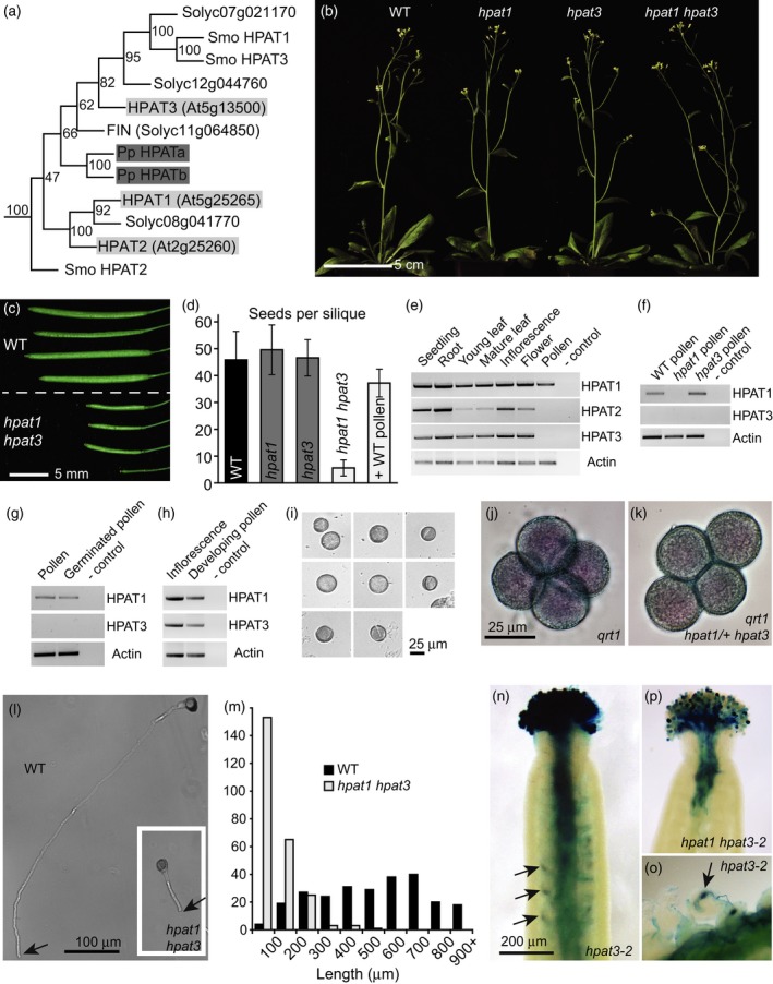
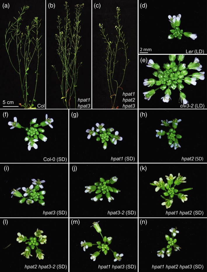
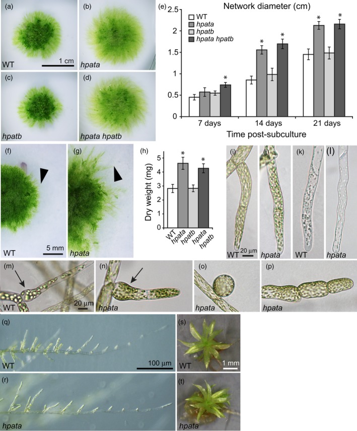
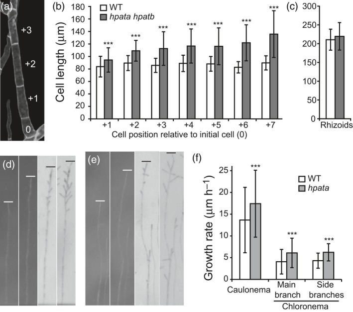
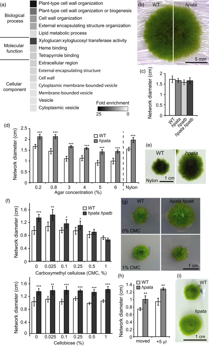
Hydroxyproline O-arabinosyltransferases (HPATs) are members of a small, deeply conserved family of plant-specific glycosyltransferases that add arabinose sugars to diverse proteins including cell wall-associated extensins and small signaling peptides. Recent genetic studies in flowering plants suggest that different HPAT homologs have been co-opted to function in diverse species-specific developmental contexts. However, nothing is known about the roles of HPATs in basal plants. We show that complete loss of HPAT function in Arabidopsis thaliana and the moss Physcomitrella patens results in a shared defect in gametophytic tip cell growth. Arabidopsis hpat1/2/3 triple knockout mutants suffer from a strong male sterility defect as a consequence of pollen tubes that fail to fully elongate following pollination. Knocking out the two HPAT genes of Physcomitrella results in larger multicellular filamentous networks due to increased elongation of protonemal tip cells. Physcomitrella hpat mutants lack cell-wall associated hydroxyproline arabinosides and can be rescued with exogenous cellulose, while global expression profiling shows that cell wall-associated genes are severely misexpressed, implicating a defect in cell wall formation during tip growth. Our findings point to a major role for HPATs in influencing cell elongation during tip growth in plants.

Keywords: Arabidopsis thaliana; Physcomitrella patens; arabinosylation; cell wall; development; extensins; glycosylation; pollination; protonema; tip growth.


Figures

Figure 1
Figure 1
HPAT1 and HPAT3 function redundantly in pollen tube growth.(a) Maximum parsimony phylogenetic tree of the hydroxyproline O‐arabinosyltransferase (HPAT) proteins from Arabidopsis, tomato (Solyc), Physcomitrella patens (Pp) and Selaginella moellendorffii (Smo) with bootstrap support values in nodes. Arabidopsis and Physcomitrella proteins are marked in light and dark grey respectively. (b) Wild type (WT, Columbia‐0), hpat1, hpat3 and hpat1 hpat3 mutant plants all appear morphologically normal. (c) Fully expanded siliques from WT (top) and hpat1 hpat3 double mutants (bottom). (d) Number of seeds per silique (mean ± SD, = 25) for WT (black bar), single mutants (dark grey bars) and hpat1 hpat3 double mutants (light grey bars), with and without pollination using WT pollen. (e)–(h) HPAT expression. Though generally broadly expressed, HPAT3 transcripts are not detected in mature WT pollen (e), in hpat1 pollen (f), or in WT in vitro germinated pollen (g). However, transcripts are detected in a mixture of developing pollen stages (h), like those shown in (i). (j), (k) qrt1 pollen tetrads stained with simplified Alexander's viability stain. Like qrt1 alone (j), all members of a tetrad from qrt1 hpat1/+ hpat3 plants appear morphologically normal (k). (l) Pollen tubes from WT and hpat1 hpat3 genotypes (inset) grown for 8 h in vitro. Arrows mark the tip of the pollen tube. (m) Distribution of pollen tube lengths after 8 h of in vitro pollen tube growth (= 250 tubes per genotype). (n)–(p) hpat3‐2 mutants carry a pollen‐specific GUS reporter within the T‐DNA construct allowing mutant pollen to stain blue. Seven hours after pollination of emasculated WT stigmas with hpat3‐2 (n, o) blue pollen tubes can be seen in the ovary and targeting ovules (arrows) and dissection of the style reveals GUS deposition in the ovule upon fertilization (o). After pollination with hpat1 hpat3‐2/+ pollen, the double mutant pollen tubes penetrate poorly and fail to target ovules (p).
Figure 2
Figure 2
hpat single, double and triple mutants are not fasciated.(a)–(c) Wild type (WT) Columbia‐0 (a), hpat1 hpat3 double mutants (b), and hpat1 hpat2 hpat3 triple mutants (c) show similar growth and morphologies under standard conditions. (d), (e) Compared with the WT background, (Ler, d) clv3‐2 shoots become progressively and severely fasciated due to enlarged meristems, resulting in the initiation of extra floral buds and floral organs in standard long‐day (LD) conditions (e). (f)–(n) Even when flowering and senescence are delayed by growth in short‐day (SD) conditions, hpat single, double and triple mutant inflorescences appear normal and do not initiate extra floral buds or floral organs relative to WT plants (Columbia‐0, f).
Figure 3
Figure 3
Physcomitrella patens hpata mutants show enhanced protonemal growth. (a)–(d) Representative plants 21‐days post‐subculture (dps): (a) wild type (WT, Gransden 2004), (b) hpata, (c) hpatb, (d) hpata hpatb. (e) Diameter of the protonemal network over time (mean ± SD, = 6). Black asterisks mark significant differences from WT at the same time point (P < 0.05) based on Student's t‐tests using the Bonferroni correction for multiple testing. (f), (g) Expanding protonemal networks of WT (f) and hpata mutants (g) showing the extent of filament growth (arrowhead). (h) Dry weight biomass (mg) at 21 dps (mean ± SD, = 6). Black asterisks mark significant differences compared with WT (P < 0.05) based on Bonferroni corrected Student's t‐tests. (i), (j) Chloronemal tip cells from WT (i) and hpata (j). (k), (l) Caulonemal tip cells from WT (k) and hpata (l). (m)–(p) Cell shape abnormalities. Whereas both WT (m) and hpata (n) plants occasionally produce swollen cells at the initiation of a new branch, hpata mutants occasionally also produce more severe abnormalities, including spherical initial cells (o) and swollen filament cells at filament tips (p). (q), (r) Caulonemal filaments initiate branching in both WT (q) and hpata (r). (s), (t) Gametophores from WT (s) and hpata (t) both appear normal.
Figure 4
Figure 4
Protonemal cells of hpata mutants grow longer and faster. (a) Calcoflour white‐stained protonemal filament with the cell positions marked relative to the initial cell (position 0). (b) Cell lengths measured at the indicated position (mean ± SD, = 11–60). Protonemal cells are initially longer in hpata mutants and this effect enhances as filament growth continues. (c) Rhizoid cells, which are tip‐growing and similar in structure to caulonema, but ontogenetically distinct, are not significantly different in length between mutant and wild type (WT) (mean ± SD, = 62). (d), (e) Images of single growing filaments from WT (d) and hpata mutants (e) taken at 24‐h intervals. The two left images are during filament growth in unidirectional red light, and the two right images are after movement to overhead white light. Lines mark the position of the filament tip. (f) Filament growth rates for WT and hpata mutants (mean ± SD,  100). In (b), (c) and (f): ***P < 0.0005 based on Student's t‐tests.
Figure 5
Figure 5
hpata mutants mis‐express cell wall associated genes and can be rescued by exogenous cellulose. (a) Heat map of the fold enrichment for Gene Ontology terms significantly overrepresented among the genes differentially expressed between the wild type (WT) and hpata mutants (Table S5). (b) The appearance of cellophane‐grown networks of WT (left) and hpata (right) plants. (c) The diameter of the protonemal network after 21 days of growth on standard medium overlaid with cellophane shows no significant differences between genotypes, indicating rescue of the hpata mutant phenotype (mean ± SD, = 6). (d) Blocking filament invasion by increasing the agar concentration of the medium or by growing on nylon membrane is not sufficient to rescue the hpata phenotype (mean of network diameter ± SD, = 6). (e) Nylon membrane‐grown WT (left) and hpata (right). (f) Networks grown on medium supplemented with carboxymethyl cellulose (CMC), a soluble cellulose derivative, also show rescue of the hpata at concentrations of 0.5–1% at 21‐days post‐subculture (top). However, plants grown on cellobiose, a disaccharide of β(1→4) linked glucose were not rescued (bottom, mean ± SD, = 6). (g) The appearance of WT (left) and hpata hpatb (right) networks grown on control medium (top) or 1% CMC (bottom). (h) Plants grown for 17 days on cellophane overlay plates that were either physically moved to a virgin position on the cellophane once a day or received a daily addition of 5 μl of liquid growth medium were not rescued by cellophane, suggesting that a stable interaction is necessary for rescue. (i) Networks that were grown on cellophane for 17 days to which 5 μl of liquid growth medium was added directly over the developing network at 24‐h intervals. In (c), (d), (f) and (h): *P < 0.05; **P < 0.005; ***P < 0.0005 based on Student's t‐tests.

Similar articles

Cited by

References

    1. Al‐Shahrour, F. , Díaz‐Uriarte, R. and Dopazo, J. (2004) FatiGO: a web tool for finding significant associations of Gene Ontology terms with groups of genes. Bioinformatics, 20, 578–580. - PubMed
    1. Aoyama, T. , Hiwatashi, Y. , Shigyo, M. , Kofuji, R. , Kubo, M. , Ito, M. and Hasebe, M. (2012) AP2‐type transcription factors determine stem cell identity in the moss Physcomitrella patens . Development, 139, 3120–3129. - PubMed
    1. Baumberger, N. , Ringli, C. and Keller, B. (2001) The chimeric leucine‐rich repeat/extensin cell wall protein LRX1 is required for root hair morphogenesis in Arabidopsis thaliana. Genes Dev. 15, 1128–1139. - PMC - PubMed
    1. Baumberger, N. , Steiner, M. , Ryser, U. , Keller, B. and Ringli, C. (2003a) Synergistic interaction of the two paralogous Arabidopsis genes LRX1 and LRX2 in cell wall formation during root hair development. Plant J. 35, 71–81. - PubMed
    1. Baumberger, N. , Doesseger, B. , Guyot, R. et al (2003b) Whole‐genome comparison of leucine‐rich repeat extensins in Arabidopsis and rice. A conserved family of cell wall proteins form a vegetative and a reproductive clade. Plant Physiol. 131, 1313–1326. - PMC - PubMed

Publication types

MeSH terms