. 2015 Nov 18:5:16763.
doi: 10.1038/srep16763.

High yield extraction of pure spinal motor neurons, astrocytes and microglia from single embryo and adult mouse spinal cord

Affiliations

High yield extraction of pure spinal motor neurons, astrocytes and microglia from single embryo and adult mouse spinal cord

Marie-Josée Beaudet et al. Sci Rep. .

Abstract

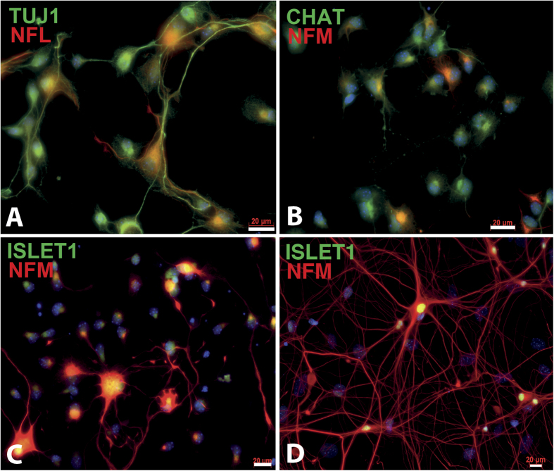
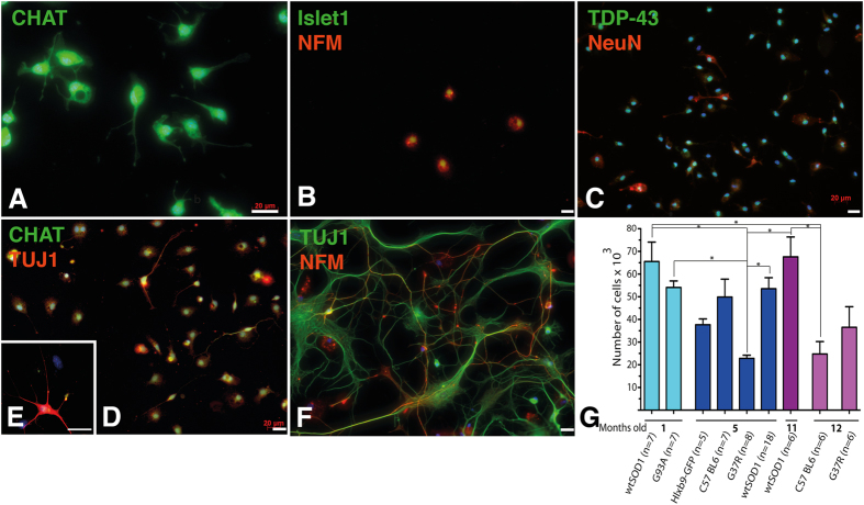
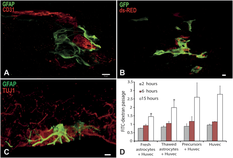
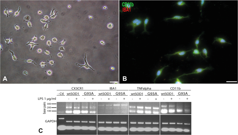
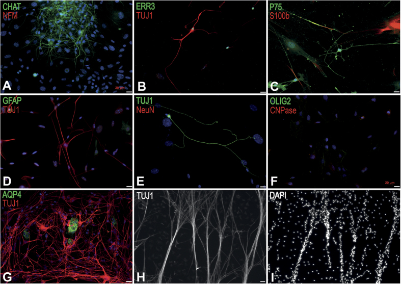
Extraction of mouse spinal motor neurons from transgenic mouse embryos recapitulating some aspects of neurodegenerative diseases like amyotrophic lateral sclerosis has met with limited success. Furthermore, extraction and long-term culture of adult mouse spinal motor neurons and glia remain also challenging. We present here a protocol designed to extract and purify high yields of motor neurons and glia from individual spinal cords collected on embryos and adult (5-month-old) normal or transgenic mice. This method is based on mild digestion of tissue followed by gradient density separation allowing to obtain two millions motor neurons over 92% pure from one E14.5 single embryo and more than 30,000 from an adult mouse. These cells can be cultured more than 14 days in vitro at a density of 100,000 cells/cm(2) to maintain optimal viability. Functional astrocytes and microglia and small gamma motor neurons can be purified at the same time. This protocol will be a powerful and reliable method to obtain motor neurons and glia to better understand mechanisms underlying spinal cord diseases.


Figures

Figure 1
Figure 1. Characterization of purified MNs obtained from E14.5 embryo spinal cord of CD-1 mice.
MNs were extracted from E14.5 embryos and characterized by immunofluorescence staining using the TUJ1 (A) in green), NFL ((A), in red) and NFM ((B–D) in red) neuronal markers and the CHAT (B) in green) and Islet1 ((C,D) in green) specific MN markers after 2 days of culture on a poly-D-lysine coated cover glass (200,000 cells/cm2). NFM (in red) and Islet1 (in green) positive cells with long neurites can be observed after cultivating MNs for 14 days (D). Scale bars 20 μm.
Figure 2
Figure 2. Characterisation of purified MNs obtained from adult spinal cord.
MNs (with a 90% purity) were extracted from 5-month-old SOD1G93A mice at disease onset, and cultured for one day (A–D) and 15 days (E,F), and characterized for expression of CHAT (in green, (A,D,E)), Islet1 (in green, (B)) and NFM (in red, (B,F)), TDP-43 (in green, (C)) and NeuN (in red, (C)), TUJ1 (in red, (E,D), and in green, (F)) and NFM (in red, (F)) (200,000 cells/cm2). The yield of MNs obtained from different mouse lineages 1, 5, 11 and 12 months after birth was quantified and showed significant variations from 19 000 to 65 000 MNs (*p < 0.05) (G). Scale bars 20 μm.
Figure 3
Figure 3. Characterisation of purified astrocytes obtained from adult spinal cord of 5-month-old SOD1G93A mice.
Phase contrast microscopy of extracted cells after 7 days of culture showing astrocytes, microglia and small MNs (A,D). Identification of purified astrocytes after 14 days of in vitro maturation by immunofluorescence staining using GFAP (In green; (B)), EAAT2 (in green) and GS (in red) (C), aquaporin-4 (AQP4) in green and TUJ1 in red (D), Vimentin in red (E), and O4 in red (F). Cells were negative for O4 marker excluding them as oligodendrocytes. Scale bar 20 μm.
Figure 4
Figure 4. Characterisation of astrocyte functionality obtained from adult spinal cord of 5-month-old wtSOD1mice.
Astrocytes were cocultured with endothelial cells and fibroblasts in a three-dimensional culture system in which capillary-like tubes form (A,B). Astrocytes were characterized by GFAP expression in green (A,C), or transduced to express ds-Red (B). Endothelial cells were detected by CD31 expression in red (A) or transduced to express GFP in green (B). Astrocytes were cocultured with MNs in a three-dimensional culture system promoting axonal migration. Astrocytes were identified using GFAP expression in green, and MNs using TUJ1 expression in red (C). The capacity of astrocytes to decrease endothelial cell permeability was assessed by quantification of FITC conjugated dextran diffusion through an endothelial cell monolayer, compared to spinal cord-derived oligodendrocyte progenitor cells as a control. Astrocytes significantly decreased FITC-dextran diffusion through endothelial cell monolayer after 15 hours of contact compared to endothelial cells alone or endothelial cells cocultured with oligodendrocyte progenitor cells (p < 0.005; n = 3). Scale bar 10 μm.
Figure 5
Figure 5. Characterisation of microglia purified from adult spinal cord of 5-month-old mice.
Phase contrast microscopy of purified microglia cells after 14 days of culture (20,000 cells/cm2) (A). Immunocytochemical characterization of purified microglia using CD11b in green and IBA1 in red (B). Scale bar 20 μm. Qualitative PCR showing expression by microglia from wtSOD1 and SOD1G93A mice of specific markers such as CX3CR1, IBA1, TNFalpha and CD11b, after activation or not with LPS (C). Urothelial cells were used as negative control.
Figure 6
Figure 6. Characterisation of small gamma MNs from adult spinal cord from 5-month-old wtSOD1mice.
(A) Identification of extracted small gamma MNs by immunofluorescence staining after 21 days of culture using CHAT in green and NFM in red (A), ERR3 in green and TUJ1 in red (B), P75 in green and S100b in red (C) (50,000 cells/cm2). The TUJ1-positive MNs did not express GFAP (in green, (D)), NeuN (in red, (E)), AQP4 (in green, (G)), OLIG2 (in green) and CNPase in red (F). Nuclei were stained in blue with DAPI (A,D,E,I). The TUJ1-positive MNs formed aligned nerve bundles after 3 months in culture (H,I). Scale bar 20 μm.

References

    1. Bunton-Stasyshyn R. K., Saccon R. A., Fratta P. & Fisher E. M. SOD1 Function and Its Implications for Amyotrophic Lateral Sclerosis Pathology: New and Renascent Themes. Neuroscientist 21, 519–29 (2014). - PubMed
    1. Dion P. A., Daoud H. & Rouleau G. A. Genetics of motor neuron disorders: new insights into pathogenic mechanisms. Nature reviews. Genetics 10, 769–782 (2009). - PubMed
    1. Berthod F. & Gros-Louis F. in Amyotrophic Lateral Sclerosis. (ed. Maurer M. ) 81–124 (InTech, Rijeka, 2012).
    1. Graham J. M. Isolation of a mouse motoneuron-enriched fraction from mouse spinal cord on a density barrier. Sci. World J. 2, 1544–1546 (2002). - PMC - PubMed
    1. Gingras M., Gagnon V., Minotti S., Durham H. D. & Berthod F. Optimized protocols for isolation of primary motor neurons, astrocytes and microglia from embryonic mouse spinal cord. J. Neurosci. Methods 163, 111–118 (2007). - PubMed

Publication types

LinkOut - more resources