

Test-retest reliability of fMRI experiments during robot-assisted active and passive stepping
- PMID: 26577598
- PMCID: PMC4647500
- DOI: 10.1186/s12984-015-0097-2
Test-retest reliability of fMRI experiments during robot-assisted active and passive stepping
Abstract
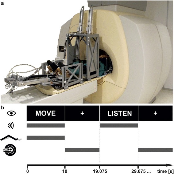
Background: Brain activity has been shown to undergo cortical and sub-cortical functional reorganisation over the course of gait rehabilitation in patients suffering from a spinal cord injury or a stroke. These changes however, have not been completely elucidated by neuroimaging to date, mainly due to the scarcity of long-term, follow-up investigations. The magnetic resonance imaging (MRI) compatible stepper MARCOS was specifically developed to enable the investigation of the supraspinal adaptations in paretic patients undergoing gait-rehabilitation in a controlled and repeatable manner. In view of future clinical research, the present study aims at examining the test-retest reliability of functional MRI (fMRI) experiments using MARCOS.
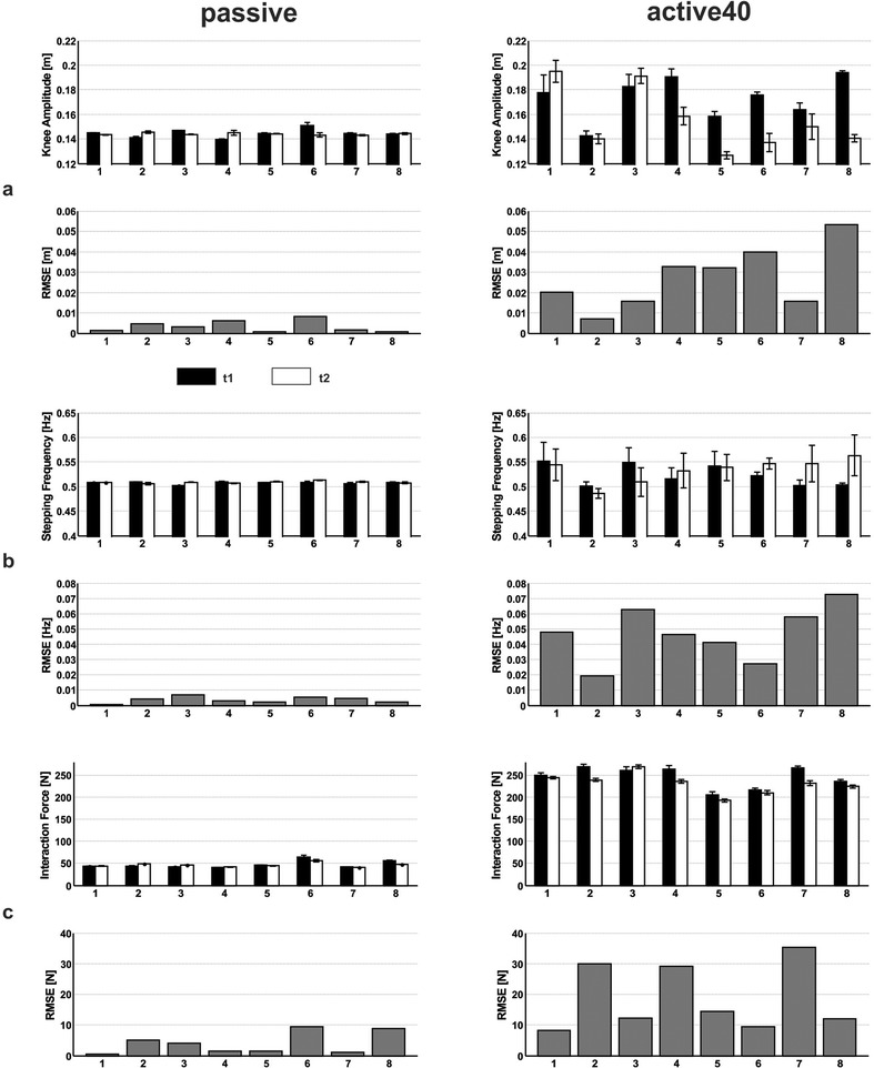
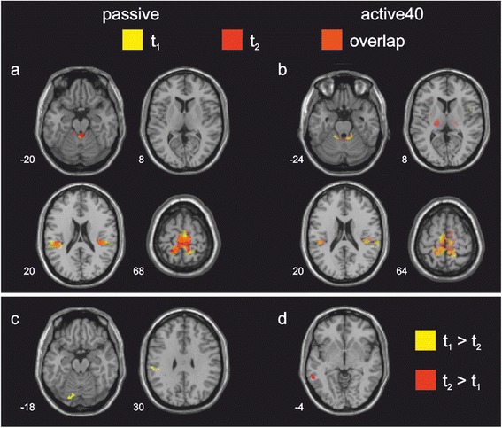
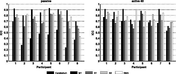
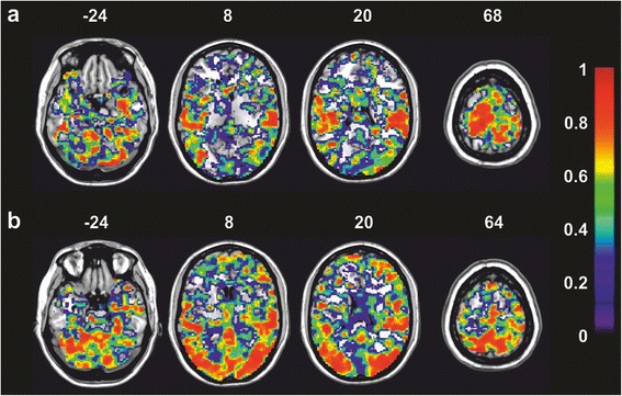
Methods: The effect of repeated active and passive stepping movements on brain activity was investigated in 16 healthy participants from fMRI data collected in two separate imaging sessions six weeks apart. Root mean square errors (RMSE) were calculated for the metrics of motor performance. Regional overlap of brain activation between sessions, as well as an intra-class correlation coefficient (ICC) was computed from the single-subject and group activation maps for five regions of interest (ROI).
Results: Data from eight participants had to be excluded due to excessive head motion. Reliability of motor performance was higher during passive than active movements, as seen in 4.5- to 13-fold lower RMSE for passive movements. In contrast, ICC ranged from 0.48 to 0.72 during passive movements and from 0.77 to 0.85 during active movements. Regional overlap of activations was also higher during active than during passive movements.
Conclusion: These findings imply that an increased variability of motor performance during active movements of healthy participants may be associated with a stable neuronal activation pattern across repeated measurements. In contrast, a stable motor performance during passive movements may be accompanied by a confined reliability of brain activation across repeated measurements.
Figures
Similar articles
-
Within-session and between-session reproducibility of cerebral sensorimotor activation: a test--retest effect evidenced with functional magnetic resonance imaging.J Cereb Blood Flow Metab. 2001 May;21(5):592-607. doi: 10.1097/00004647-200105000-00014. J Cereb Blood Flow Metab. 2001. PMID: 11333370
-
A reliability study on brain activation during active and passive arm movements supported by an MRI-compatible robot.Brain Topogr. 2014 Nov;27(6):731-46. doi: 10.1007/s10548-014-0355-9. Epub 2014 Apr 10. Brain Topogr. 2014. PMID: 24718725
-
Brain activation associated with active and passive lower limb stepping.Front Hum Neurosci. 2014 Oct 28;8:828. doi: 10.3389/fnhum.2014.00828. eCollection 2014. Front Hum Neurosci. 2014. PMID: 25389396 Free PMC article.
-
Intersession reliability of fMRI activation for heat pain and motor tasks.Neuroimage Clin. 2014 Jul 22;5:309-21. doi: 10.1016/j.nicl.2014.07.005. eCollection 2014. Neuroimage Clin. 2014. PMID: 25161897 Free PMC article.
-
Activation likelihood estimation meta-analysis of motor-related neural activity after stroke.Neuroimage. 2012 Feb 1;59(3):2771-82. doi: 10.1016/j.neuroimage.2011.10.023. Epub 2011 Oct 17. Neuroimage. 2012. PMID: 22023742 Review.
Cited by
-
Characterizing the supraspinal sensorimotor control of walking using MRI-compatible system: a systematic review.J Neuroeng Rehabil. 2024 Mar 5;21(1):34. doi: 10.1186/s12984-024-01323-y. J Neuroeng Rehabil. 2024. PMID: 38443983 Free PMC article.
-
There is No test-retest reliability of brain activation induced by robotic passive hand movement: A functional NIRS study.Brain Behav. 2020 Oct;10(10):e01788. doi: 10.1002/brb3.1788. Epub 2020 Aug 13. Brain Behav. 2020. PMID: 32794359 Free PMC article.
-
Lower extremity Interlimb coordination associated brain activity in young female athletes: A biomechanically instrumented neuroimaging study.Psychophysiology. 2023 Apr;60(4):e14221. doi: 10.1111/psyp.14221. Epub 2022 Nov 23. Psychophysiology. 2023. PMID: 36416574 Free PMC article.
-
VALIDITY OF AN MRI-COMPATIBLE MOTION CAPTURE SYSTEM FOR USE WITH LOWER EXTREMITY NEUROIMAGING PARADIGMS.Int J Sports Phys Ther. 2020 Dec;15(6):936-946. doi: 10.26603/ijspt20200936. Int J Sports Phys Ther. 2020. PMID: 33344010 Free PMC article.
-
A Novel Approach to Evaluate Brain Activation for Lower Extremity Motor Control.J Neuroimaging. 2019 Sep;29(5):580-588. doi: 10.1111/jon.12645. Epub 2019 Jul 3. J Neuroimaging. 2019. PMID: 31270890 Free PMC article.
References
-
- Newton JM, Dong Y, Hidler J, Plummer-D'Amato P, Marehbian J, Albistegui-DuBois RM, et al. Reliable assessment of lower limb motor representations with fMRI: Use of a novel MR compatible device for real-time monitoring of ankle, knee and hip torques. Neuroimage. 2008;43(1):136–46. doi: 10.1016/j.neuroimage.2008.07.001. - DOI - PMC - PubMed
Publication types
MeSH terms
LinkOut - more resources
Full Text Sources
Other Literature Sources
Medical
